 |
| About Bioline | All Journals | Testimonials | Membership | News |
|
||||||
|
||||||
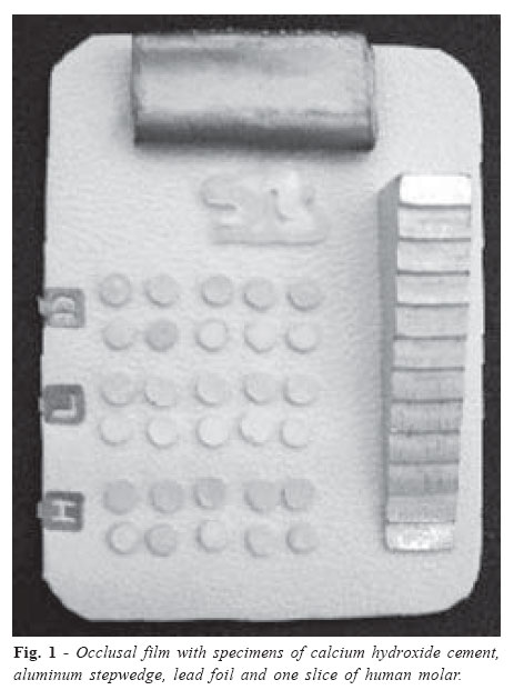
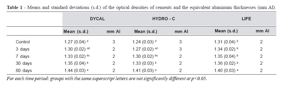
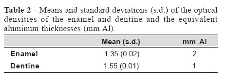
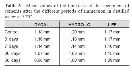
Brazilian Journal of Oral Sciences, Vol. 5, No. 16, Jan - March, 2006, pp. 958-962 Effect of the storage in water on theradiopacity of calcium hydroxide cements Karina Lopes Devito1 Ana Isabel Ortega2 Abstract Francisco Haiter-Neto3 1PhD, Professor, Department of Radiology, Juiz
de Fora Dental School, UFJF, Juiz de Fora,
Brazil. Received for publication: February 11, 2004 Code Number: os06005 Abstract In extensive cavities usually calcium hydroxide cements are used in order to protect the pulp. These materials can present a reduction in the radiopacity due solubility resulting from contact with oral fluids. This study investigated the radiopacity of calcium hydroxide cements stored in distilled water at 37o C, with the aim to verify the alteration of radiographic density in function of solubility. Samples of Dycal, Hydro-C and Life cements were submitted to immersion for periods of 3, 7, 30 and 60 days. After each of these periods, the samples were radiographed and their optical densities obtained. The radiopacity values were expressed in equivalent thickness of aluminum and were compared with the values of enamel and dentine. ANOVA was used to determine if there was a significant difference between the radiopacity values of the cements submitted to different immersion times. The results showed that Dycal and Hydro-C cements presented a significant radiopacity reduction after one week of immersion, while Life had its radiopacity significantly reduced only after 2 months. However, the three tested cements kept similar radiopacity values to those of enamel. Key Words: calcium hydroxide cement, radiopacity, solubility Introduction The materials added to teeth should present a radiopacity similar to or higher than that of enamel1-5. This characteristic is essential to detect secondary caries and excess of materials at approximal surfaces; to evaluate approximal contour; to distinguish base materials from the restorative materials and from surrounding dental structures, to detect voids in the restorations and to locate some material that has been accidentally aspired, inhaled or can eventually be in soft tissues1, 4, 6-9. Usually, in extensive cavities, base materials are used beneath the restorative material, with the function to help the affected pulp and to protect it against several types of aggressions to which it may be submitted. A variety of materials that has been used as base, among which the hydroxide calcium cement should be pointed out10. Hydroxide calcium cement has the property to allow remineralization of decalcified dentine and induces formation of repaired dentine, possessing antibacterial properties due to its high pH11. It is the material of choice to be used in resin restorations, since it does not influence the polymerization of these materials. However, the hydroxide calcium cement presents the characteristic to be hydrolitically unstable, is dissolved when in contact with oral fluid, and may reach total disappearance, and thus the restorations remain without support and with formation of voids, that can induce an erroneous radiographic diagnosis of secondary caries12-14. This study analyzed the radiopacity of hydroxide calcium cement stored in water, with the objective to verify the influence of its solubility in the radiographic density. Material and Methods Ten specimens, with a thickness of 1 mm (± 0.2), of three commercially available calcium hydroxide cements: Hydro-C (Dentsply/York Division Co., USA), Dycal (Dentsply/York Division Co.) and Life (Kerr, Oregon, USA) were made. Thickness was determined using a pachymeter (Mitutoyo, Japan). For the preparation of samples, elastomer molds with a 4-mm diameter opening and 1-mm thickness were used to contain the material. The cements were manipulated following the manufacture’s recommendations. The samples were kept at 37oC for one hour before the removal of the mold. All specimens of calcium hydroxide cement; an aluminum stepwedge with thickness varying from 1 to 12 mm (with increments of 1 mm); a lead foil (to determine the base density and fog); one slice of human molar of 1 mm thickness, in the mesiodistal direction, were placed on the same occlusal film (Eastman Kodak Co., NY, USA) to minimize the possible variable regarding film and radiographic processing (Figure 1). The radiographs were obtained using a dental X-ray machine (General Electric Co., USA), model 1000, 70 kVp, 10 mA and 10 impulses of exposure time. The standardization of the focus-film distance was obtained with the use of a device that provided an incidence of the radiation beam perpendicular to the plan where the film and the objects to be radiographed were placed. With the aim to evaluate the solubility and its influence on the radiopacity, the samples were stored in distilled water at 37oC, in a culture stove, model 002 CB (Fanem Ltda, Brazil). For the control group, eight radiographs of the specimens were obtained soon after their preparation. The specimens were then stored in water for 3, 7, 30 and 60 days. After each of the periods, eight radiographs were obtained. For the verification of the possible thickness reduction due to solubility, the samples were measured with a pachymeter. These measurements were obtained before each cycle of radiographs. The radiographic processing was manual (20oC/4min) with processing solutions (Eastman Kodak Co.) without previous use. After development, densitometric measurements of the images of each cement specimen, of all the steps of the stepwedge, of the lead foil and of the enamel and dentine were obtained, using a digital densitometer (MRA, SP, Brazil) with a diaphragm opening of 1.5-mm diameter. The radiopacity values of the calcium hydroxide cements, dentine and enamel were expressed in equivalent aluminum thickness (mm Al). Then, the radiopacities of these materials were compared with those of the dental structures. A variance analysis (ANOVA) was performed to determine if there was a statistically significant difference between the radiopacity values of the calcium hydroxide samples submitted to the different immersion times. Results Tables 1 and 2 show optical densities and equivalent aluminum thicknesses for the tested cements, enamel and dentine. After seven days of immersion in distilled water, Dycal and Hydro-C cements presented statistically significant differences (p<0.01) regarding radiopacity values when compared with the control group, showing a reduction of these values. Differently, Life cement only presented a significant reduction in its radiopacity after two months of immersion. However, in any of the immersion times (3, 7, 30 and 60 days) the samples presented mean densities in the range equivalent to 2 or 3 mm aluminum. Evaluating the enamel and dentine radiopacities in equivalent aluminum thickness, we will have 2 and 1 mm, respectively. Therefore, despite the solubility, the samples still present similar or higher radiopacity values when they are compared with that of the enamel. The occurred dissolution was determined by the measurement of the dimensions of the samples, presenting a mean loss of 0.22, 0.20 and 0.08 mm thickness of the Dycal, Hydro-C and Life cement samples, respectively. Table 3 presents the mean values of the thickness of the samples for each of the period of immersion in water. Discussion The results obtained in this study showed a gradual reduction of the radiopacity of the three tested cements in function of the immersion time in water. This reduction in radiopacity values occurred due the solubility of the material that can be evidenced by the reduction in the dimensions of the samples. The disintegration or total disappearance of the calcium hydroxide bases was noticed by some clinical studies when amalgam restorations were placed directly on the calcium hydroxide bases15. The justification of the solubility of the calcium hydroxide is related to the correct proportion and manipulation of the pastes and to the specifical components of each cement. Basically, they consist of calcium hydroxide, an alkaline salicylate and a plasticizer. Depending on the nature of the plasticizer, water diffusion is facilitated, allowing the complete disintegration of the cements. However, if the water difusion is made difficult, there will be less release of hydroxyl and calcium ions, resulting in less soluble material, but with lower antibacterial properties16-17 . An additional factor to be considered in the dissolution of the base is the presence of bacteria associated with microleakage on the material17 . Some in vitro studies also reached the same results of clinical evaluations, for example, Gourley and Rose18 , assessing the solubility of materials used under amalgam restorations, concluded that Dycal cement experienced disintegration and presented high solubility when stored in distilled water at 37o C. Prosser et al.16 studied the effect of the storage in water, for 3 days, of four commercial brands of calcium hydroxide cements and concluded that all cements are dissolved to varied degrees, and are considered hydrolitically unstable. Dycal presented one of the greater solubility values. In 1983, McComb11 , evaluating the solubility in distilled water, for 24 hours, of five calcium hydroxide cements, observed that four of then presented reasonably high solubility values, especially Dycal that presented a significantly higher value than that of Life. Driscoll et al.19 studied the solubility of Life and Dycal in water at 37o C and observed that the tested cements dissolved during the whole period of study (3 months), with Dycal cement presenting higher solubility values than those of Life. However, Hwas and Sandrik20 who studied four commercial brands of calcium hydroxide for 24 hours, observed that Dycal presented the least solubility value in water, followed by Procal, Life and Renew cements. Pardini et al.21 evaluated in vitro the solubility and disintegration of calcium hydroxide cements under amalgam restorations kept for 29 days in distilled water at 37°C. The results also showed Dycal with the least solubility value, however, when this value was compared with the solubility value of Life cement, no statistically significant difference was found. The differences in the results of the several studies can be justified by inherent variations in the methodology of each research. Moreover, the presence of possible variations in the composition of one same cement should not be discarded, a problem that has been already mentioned by other authors17 . The results of this study are similar to those of most studies, where Life cement was the least soluble. Dycal and Hydro-C cements presented higher solubility values and these values were similar for both. The solubility values found in this study were higher than those that would be found in a clinical situation, where an entire restoration over a calcium hydroxide base helps to protect it from the contact with saliva. Moreover, the dissolution caused by dentinal fluid circulation in the tubules does not come close to that resulting from in vitro solubility tests. However, the joint action of dentinal fluid with the marginal infiltration may result in partial solubility or total disappearance of the calcium hydroxide bases14 . None of the mentioned studies on solubility of calcium hydroxide cements concomitantly evaluates the influence of the dissolution on the radiopacity of these materials. According to the International Standard Organization (ISO 4049)22 , a radiopacity of a material must be higher than that of a same thickness of aluminum in order to be considered radiopaque. Moreover, most studies state that the materials must present an equal or higher radiopacity than that of enamel in order not to be misinterpreted as secondary caries1-5 . The evaluation of the means of the radiopacity values indicated that Life cement was the least radiopaque material, however, it was also less soluble, presenting a significant reduction in radiopacity only after 2 months of immersion. This can be explained by the fact that the lowest the percentages of substances added to the materials to give radiopacity, the lowest the solubility values23 . Despite the gradual reduction in the radiopacity for the whole period of study (60 days), the three cements presented higher values than those of the same aluminum thickness, in agreement with the specifications of ISO and with a radiopacity value equivalent to that of enamel. Thus, the radiopacity reduction found for 60 days of storage in distilled water at 37°C, probably is not important for the radiographic interpretation. Tests with longer immersion times are necessary to complement this study, emphasizing, once more, that the behaviour of cements in the oral cavity is quite different from that resulting from in vitro tests. The conclusions are: 1-Significant radiopacity reduction occurred after 7 days of immersion of Dycal and Hydro-C cements in distilled water at 37°C; 2-Life presented a significant radiopacity reduction after 60 days of immersion in water distilled at 37°C; 3-After 60 days of immersion, despite the radiopacity reduction, all tested cements presented a thickness equivalent to 2 mm aluminum, with similar radiopacity values to those of the enamel. References
Copyright 2006 - Piracicaba Dental School - UNICAMP São Paulo - Brazil The following images related to this document are available:Photo images[os06005t2.jpg] [os06005f1.jpg] [os06005t3.jpg] [os06005t1.jpg] |
| |||||||||
