 |
| About Bioline | All Journals | Testimonials | Membership | News |
|
||||||
|
||||||
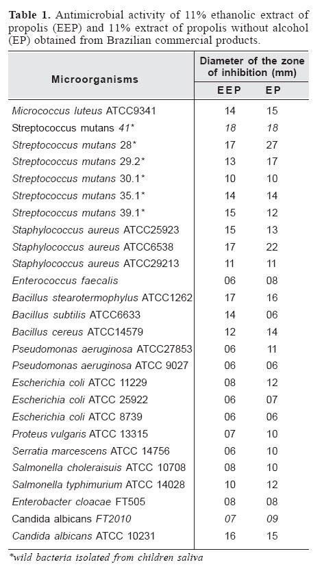
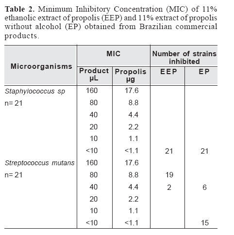
Brazilian Journal of Oral Sciences, Vol. 5, No. 16, Jan - March, 2006, pp. 967-970 Antimicrobial activity of two brazilian commercial propolis extracts Giovanna Pires da Silva Ribeiro de Rezende1 Fabiana Cristina Pimenta2 Luciane Ribeiro de Rezende Sucasas da Costa3 1Health Sciences Program sponsored by University of Brasilia, DF, Brazil 2Tropical Pathology and Public Health Institute, Federal University of Goias, GO, Brazil 3Faculty of Dentistry, Federal University of Goias, GO, Brazil Received for publication: August 05, 2005 Code Number: os06007 Abstract The antimicrobial activity of propolis extracts is well documented, but little is known about the antimicrobial properties of commercial products containing propolis, since these vary according to the geographical region in which the propolis is obtained. This study evaluated the antimicrobial activity of two samples of commercial propolis on 26 species of microorganisms obtained from ATCC and some wild strains: Gram-positive cocci and bacilli, and Gram-negative rods and yeasts. The tested products were two samples of Brazilian commercial propolis from Apis Flora™: 11.0% ethanolic extract of propolis (EEP) and Propomax™ 11.0% extract of propolis without alcohol (EP). Antimicrobial activity was determined by the agar diffusion technique, well method. MIC was determined for Staphylococcus sp. and Streptococcus mutans using the method of broth dilution with the propolis extract in serial concentrations. EEP and EP showed antimicrobial activity against all tested bacteria and yeasts, having a more pronounced action against Gram-positive bacteria and Candida albicans ATCC 10231, and a less evident activity against Gram-negative and Candida albicans FT2010. For S. mutans , the EEP MIC ranged from 8.8 to 4.4 mL of propolis, and the EP MIC, from 4.4 to <1.1 mL. For Staphylococcus sp., the MIC of both extracts was <1.1. Key Words: microorganisms, propolis, antimicrobial activity Introduction The antimicrobial activity of propolis against Gram-positive bacteria and yeasts is well documented1. However, this antimicrobial activity depends on the chemical composition of propolis2, which in turn seems to vary depending on the geographical region where it is extracted3-5. The main source of Brazilian propolis is Baccharis dracunculifolia DC, but its antimicrobial efficiency is controversial; that is, Brazilian propolis may promote better or worse effects than that from other countries6. Thus, while commercial products containing propolis from various regions of Brazil are sold, their efficacy is not clear. The aim of this study was to evaluate the antimicrobial activity of two samples of commercial propolis on different groups of microorganisms including oral pathogens. Material and Methods Twenty-six sample species of microorganisms obtained from the American Type Culture Collection (ATCC) and some wild strains from the Laboratory of Medical Bacteriology, Tropical Pathology and Public Health Institute, Federal University of Goias, Brazil, were selected. They included Gram-positive cocci and bacilli, and Gram-negative rods and yeasts (Table 1). The tested products were two samples of Brazilian commercial propolis from Apis Flora™: 11.0% ethanolic extract of propolis (EEP) and Propomax™ 11.0% extract of propolis without alcohol (EP). The antimicrobial activity was determined using the agar diffusion technique, well method7. Sterile Mueller Hinton (MH) agar or Brain Heart Infusion, according to the requirements of the microorganisms, was poured into 20 (20 mL) Petri dishes and left to set. Then, 10 mL of agar was inoculated with 1 mL of the microorganism inoculum poured on top. The inoculum was prepared with an overnight culture of test microorganism and the size was adjusted to 0.5 McFarland standard turbidity, approximately 108 colony forming units (CFU/mL). Equidistant wells of 5 mm in diameter and 4 mm in depth were bored into the agar using a sterile cork borer and the wells were completely filled with the tested products. The plates were left at room temperature for two hours and then incubated at 37oC for 24 hours. Antimicrobial activity was determined by measuring the diameters of the zone of inhibition of EEP and EP. Controls were maintained with methanol and penicillin G 10 mg/mL (for Gram-positive) and erythromycin 15 mg/mL (for Gram-negative). The Minimal Inhibitory Concentration (MIC) was determined for EEP and EP by the agar dilution method in MH agar medium (NCCLS 2003). Staphylococcus sp (21 strains) were grown on MH agar plates (DifcoÒ) and suspended in MH broth (DifcoÒ). Streptococcus mutans (21 strains) were grown on brain heart infusion (BHI) broth (DifcoÒ) and the assay done with BHI agar. The inoculum suspensions were prepared with an overnight culture of test microrganism and approximately 108 colony forming units (CFU/mL). Serial 10-fold dilutions were made that furnished a concentration range from 1.1 to 17.6 mg/mL for EEP and EP. Before gelling, the MH agar was added to each of the Petri dishes containing the dilutions and swirled carefully until the agar began to set. The bacterial suspensions were inoculated using a Steers replicator on the Muller Hinton agar surface and incubated at 37oC for 24 hours. The MIC was defined as the lowest concentration able to inhibit any visible bacterial growth. Control cultures, containing only the MH/BHI agar, were also prepared. Tests were performed in duplicate. Results and Discussion In the present study, commercial propolis with or without ethanol showed in vitro antimicrobial activity against bacteria and yeasts (Table 1). The standard strains tested were chosen according to a screening protocol including Gram-positive cocci and bacilli, Gram-negative bacteria and yeasts. In the first step of an antimicrobial activity screening, the product should be tested against these standard strains, which represent microorganisms associated with important infections. Commercial propolis products had a more pronounced activity against Gram-positive bacteria and Candida albicans FT2010, and a less evident action against Gram-negative bacilli and Candida albicans ATCC 10231. The controversial result concerning Candida albicans FT2010 and Candida albicans ATCC 10231 could be explained by the inherent virulence of each strain. That is one reason to employ different microbial strains of a same species. This efficient antimicrobial action, mainly towards Gram-positive bacteria, was also observed in other studies which tested non-commercial extract of propolis8-10. Probably, the antibacterial activity of propolis is greater on Grampositive bacteria due to flavonoids, acids and aromatic esters found in the resin, which would act on the cell wall through an unknown mechanism11. This study is in accordance with Sforcin et al.12, who verified that the growth of Gram-positive bacteria is inhibited by low propolis concentrations (0.4%) whereas Gram-negative bacteria were less susceptible to this substance, with the MIC ranging from 4.5 to 8.0%. Drago et al.8 also observed that in low concentrations propolis shows bacteriostatic rather than bactericidal activity. Among the yeasts, this study showed that the C. albicans was more susceptible to propolis than other species. This result is supported by Ota et al.13, who found the following order of susceptibility to hydroalcoholic propolis: C. albicans > C. tropicalis > C. krusei > C. guilliermondii. Another study has shown that a commercial 20% ethanol propolis extract inhibited Candida albicans strains collected from HIV-positive patients with oral candidiasis14. The results of this study have to be interpreted carefully as far as its methodological procedures are concerned. It is reported that the best microbiological method to evaluate the activity of propolis extracts against species of Candida is agar dilution in plates9. Otherwise, serial dilution in tubes is the best method for the evaluation of the bactericidal activity of propolis samples. However, agar plate diffusion tests are strongly influenced by the solubility of the components of propolis in agar, leading to incorrect results. This method should not be used for the comparison of samples of different hydro-solubility nor for the evaluation of poorly hydro-soluble samples9. After the evidence of in vitro antimicrobial activity against all tested strains in the screening diffusion test, the minimum inhibitory concentration (MIC) was established using the agar serial dilution method for 21 Staphylococcus sp and 21 Streptococcus mutans isolated from saliva. Both products tested contained 11.0% of propolis (ethanolic extract of propolis-EEP and Propomax™-EP). The products were measured in µL and the propolis in µg. Table 2 illustrates the MIC obtained for S. mutans and Staphylococcus sp., showing an extremely low concentration, especially against Staphylococcus sp. The EEP MIC for Streptococcus mutans ranged from 80 to 40mL (8.8mg to 4.4mg of propolis) and the EP MIC for Streptococcus mutans ranged from 40 to <10mL (4.4 to <1.1mg of propolis). For Staphylococcus sp, the MIC of the two extracts ranged from <10mL (<1.1mg of propolis). On the other hand, Gebara et al.2 showed a greater MIC for propolis ethanolic extract (14 µg/mL) against S. aureus. However, it should be borne in mind that the determination of MIC values depends on technical details that may vary between laboratories and the bacteria’s inherent virulence and susceptibility. The results of this study do not corroborate the statement that one of the limitations to propolis use is its variability in composition and action as a consequence of variations in the flora of the region where it is produced, since the commercial propolis studied consist of a mixture of various propolis collected in Brazil. Future in vitro and in vivo research must be conducted to analyze the biological effects and the viability of using different propolis formulations in various oral infections. It is important to remember that in vitro tests do not reflect the real conditions found in clinical infections, because they do not take into account biofilm formation. Therefore, this finding can hypothetically permit a more comprehensive clinical use of propolis after further in vivo studies prove its efficacy in the treatment of oral infections, since preliminary antimicrobial propolis activity against cariogenic microorganisms3 and periodontopathogens15 has already been demonstrated. Another potential field for propolis research is endodontics. References
Copyright 2006 - Piracicaba Dental School - UNICAMP São Paulo - Brazil The following images related to this document are available:Photo images[os06007t1.jpg] [os06007t2.jpg] |
| |||||||||
