 |
| About Bioline | All Journals | Testimonials | Membership | News |
|
||||||
|
||||||
Brazilian Journal of Oral Sciences, Vol. 5, No. 16, Jan - March, 2006, pp. 971-976 The effects of anti-oxidant agents as neutralizers of bleaching agents on enamel bond strength Carlos Rocha Gomes Torres* Alexandre Fuzuko Koga* Alessandra Bühler Borges*
* Department of Restorative Dentistry - São José dos Campos - School of Dentistry - São
Paulo State University - SP, Brazil
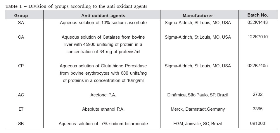
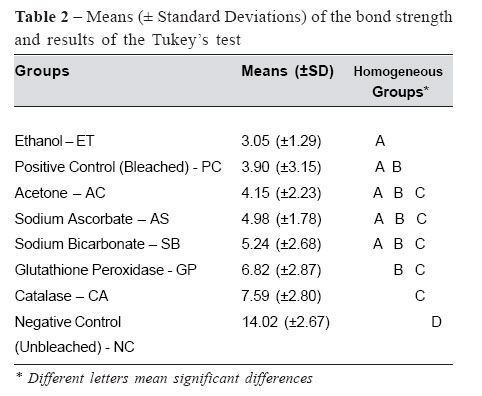
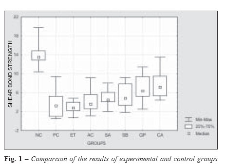
Received for publication: March 23, 2005 Code Number: os06008 Abstract The aim of this study was to investigate the neutralizer effect of antioxidant agents on the bond strength of bleached enamel. Buccal enamel surfaces of 80 bovine incisors were divided into eight groups. The negative control group (NC) received no bleaching treatment and the other groups were bleached with 35% hydrogen peroxide gel for 30min., activated with a photocuring unit. Ten specimens were selected for the positive control group (PC) and received no anti-oxidant agents. The remaining bleached specimens received one of the six anti-oxidant agents, for 20min (10% Sodium Ascorbate–SA; Catalase–CA; Glutathione Peroxidase–GP; Acetone–AC; Ethanol–ET; 7% Sodium Bicarbonate– SB). Bonds were formed and shear bond test was carried out after 24h. The data were analyzed by one-way ANOVA and Tukey’s tests (α =5%). The PC group showed significantly lower bond strength mean than the NC group. All of the experimental groups showed bond strength means significantly lower than the NC group. The CA group showed significantly higher bond strength mean than the bleached PC group and the groups SA, GP, AC, ET and SB showed no significant differences compared to PC group. It was concluded that only the Catalase application resulted in significant increase of bond strengths in relation to the PC group and that none of the treatments was able to completely neutralize the deleterious effects of bleaching on bond strength. Key Words: dental bleaching, enamel, adhesion, antioxidants Introduction With the increasing interest in esthetic dentistry, vital bleaching has become widespread. Previous studies have shown that hydrogen peroxide (HP) and carbamide peroxide (CP) used as bleaching agents affect the bond strength of composites to acid etched enamel when bonding is performed immediately after the bleaching treatment1-3.It is recommended delays in bonding of 1 to 3 weeks following the bleaching procedure for the enamel to return to conditions that lead to normal bond strengths4-6, rendering the immediate reestablishment of esthetics impossible. The lower bond strengths of bleached enamel and dentin are a result of the oxidative process caused by the bleaching agents7-11. Some authors assert that the oxygen remains in the dental structure after bleaching and can interfere with the polymerization of adhesive monomers5,11. Nevertheless, it was observed that the use of anti-oxidant agents before the bonding process can reverse the compromised bonding to bleached enamel3,11. Thus, the restorative procedures could be performed immediately after the bleaching procedures, reducing the total time of the complete esthetic treatment. The present study was therefore designed to evaluate the neutralization effect of various anti-oxidant agents on the bond strength of bleached enamel. Material and Methods Eighty 3-year-old bovine incisors erupted and intact were extracted immediately after slaughter. The teeth were then cleaned and the roots were sectioned at the middle level with a low speed diamond saw and the pulp was removed using endodontic instruments. The pulp chamber was rinsed with distilled water and the apical region filled with utility wax to avoid penetration of embedding media. The teeth were embedded in self-curing acrylic resin (Jet, Clássico, São Paulo, SP, Brazil) using a heavy-body silicon mould (Rodhorsil, Clássico, São Paulo, SP, Brazil) to expose the buccal area. The enamel surface was polished with wet 600grit silicon carbide abrasive paper on a polishing machine (DP 10, Panambra, São Paulo, SP, Brazil) for 60 seconds to create a flat enamel surface. Ten specimens were randomly selected for the negative control group (NC). They were stored in distilled water and received no bleaching treatment. In the seventy remaining specimens, the 35% HP bleaching gel was applied (Whitness HP, FGM, Joinville, SC, Brazil). The bleaching procedures consisted of 3 applications of 10 minutes. In each application, the gel was light activated four times for 40 seconds with a photocuring unit (Optilight 600, 600mW/cm2 -Gnatus, Ribeirão Preto, SP, Brasil). The gel was agitated with a dental explorer to displace bubbles after light activations. At the end of the bleaching procedures, the surfaces were rinsed with tap water for 1 minute to remove the bleaching gel. A special Scotchtape Mold (Barbi Ind. Metalúrgica, Itu, SP, Brazil) with a standard central hole, 3mm in diameter, was placed on each specimen. Ten bleached specimens were randomly selected for the positive control group (PC) and received no anti-oxidant agents. The remaining sixty bleached specimens were randomly divided into six groups (n=10) to receive the anti-oxidant agents for 20 min, as observed in Table 1. For the groups Sodium Ascorbate (SA), Catalase (CA), Glutathione Peroxidase (GP) and Sodium Bicarbonate (SB), 1 ml of the respective solutions was applied on the delimitated area. To allow the application during the determined time, the acetone and ethanol solutions were applied with a cotton pellet on the surfaces, which were keep moist regularly, due to their quick evaporation. The surfaces were then rinsed with tap water for 30 seconds and air-dried for 20 seconds. All the specimens were acid etched with 37% phosphoric acid (Dentsply, Petrópolis, RJ, Brazil, Batch No. 42859) for 15 seconds, rinsed for 30 seconds and air dried for 20 seconds. A thin layer of adhesive (Magic Bond, Vigodent, Rio de Janeiro, RJ, Brazil, Batch No. 006/03) was applied on the etched enamel, gently spread with compressed air and light-cured for 10 seconds. This adhesive is composed essentially by hydrophobic monomers (BisGMA and methacrylic acid esther), with no solvent. The embedded specimens were mounted in an apparatus containing a split teflon mould with a circular hole 3mm in diameter and 4mm high. Two increments of a composite resin (Z250 - 3M, St Paul, MN, USA, Batch No. 2PW) were inserted into the opening of the split mould and each one was light cured for 40 seconds. After curing, the split mold was removed and the composite cylinder was additionally light cured for 40 seconds in the two opposite sides, to ensure the maximum polymerization of the resin, enabling an homogeneous stress distribution to the bonding interface during the shear test. The specimens were identified and stored in distilled water at 37oC for 24 hours prior to testing. Each specimen was locked in a special device that was seated on the universal testing machine (EMIC, São José dos Pinhais, PR, Brazil). A shear load was applied to the base of the composite cylinder with a 1mm thick knife-edge rod, at a crosshead speed of 1 mm/min. The results were expressed in MPa and were subjected to one-way analysis of variance (ANOVA) and Tukey’s test at the 5% level of significance. Results The ANOVA test showed significant differences in the tested groups (p=0.00; F=19.32 to 7 degree of freedom). The means and standard deviations of the bond strength values for the groups and the results of the Tukey test are listed in Table 2. The PC group showed significant lower bond strength mean than the NC group. All of the experimental groups (SA, CA, GP, AC, ET and SB) showed bond strength means significantly lower than the NC group. The CA group showed significantly higher bond strength mean than the bleached PC group and the groups SA, GP, AC, ET and SB showed no significant differences compared to PC group (Table 2). The Figure 1 compares the mean bond strength values of the different groups. A significant reduction of the bond strength mean for the bleached groups compared to the unbleached group is evident. At the same time, a trend of increase of the bond strength values for the groups that received the neutralizer agents is noted. Nevertheless, there was no reversal of the compromised bonding for the bleached groups compared to the NC group. Discussion In the present study, it was observed that the bleaching procedure resulted in a significant decrease of bond strength values compared to the unbleached group (Table 2 , Figure 1). The bleaching agents release free radicals as the nascent oxygen and hydroxyl or peri-hydroxyl ions when they are applied to the dental structure. Free radical is any molecule that has one unpaired electron, providing it high reactivity. These molecules are able to react with the electron-rich regions of the pigments inside the dental structure, breaking down large pigmented molecules into smaller, less pigmented ones2. On the other hand, this property could be deleterious to the bonding of resinous materials. One theory proposed to explain the influence of bleaching agents on bonding suggests that peroxides and their byproducts present inside the dental structure are capable to interfere with the polymerization process of the adhesive material4-5,12-13. Some eletronic and optical microscopic studies showed that the resin tags in the bleached enamel were sparse, short, poorly defined, structurally incomplete and, in some areas, completely absent5,12-14. It is supposed that the bonding agent applied on the etched enamel is not completely polymerized, impairing the mechanical retention and decreasing the bond strength5. Bubble-like structures and voids were observed inside the adhesive layer, suggesting that the oxygen released by the hydrogen peroxide is trapped within the adhesive during light-activation4,11-12. According to McGuckin et al.4, the void areas present at the interface decrease in bonds formed at longer time intervals after bleaching cessation, indicating a gradual elimination of HP and its byproducts with consequently increase of the bond strength. According to Titley et al.15, the dentin and dentinal fluid can act as a peroxide and oxygen reservoir. The reservoirs of gaseous or dissolved oxygen products could persist until removed by pulpal microcirculation and diffusion from the external surface. A greater surface diffusion would be expected based on a reduced pulpal microcirculation16. Thus, levels of peroxide or oxygen higher than normal may be present in bonding interface, inhibiting the polymerization reaction and reducing bond strengths4. Various anti-oxidant agents were tested in this study. It is possible that, reestablishing the altered redox potential of the oxidized bonding substrate, the anti-oxidants allow free radicals polymerization of the adhesive resin without its premature termination10. Free radicals are deficient electrons and the neutralization process results in oxidation of the neutralizer agent7. They are classified in three categories: the full-time prevention, the active detoxication against an oxidative stress and the passive detoxication. The role of the full-time prevention is to prevent the overproduction of free radicals by inactivating molecules, which are responsible for their generation. The active detoxication is based on three enzymes that compose the basic system of anti-oxidant defense system. They are superoxyde dismutase, catalase and glutathione peroxidase and can be found in cells where the oxidative stress is greater, as cytosol, mitochondria and peroxysomes17. To control the HP, the human body uses two enzymes. The catalase accelerates the transformation of HP into O2 and water17-18. The amount of peroxide decomposed is directly proportional to the concentration of enzyme. The rate of this reaction is rapid and little energy is involved as catalase accelerates the decomposition of HP by more than 100.000.000 fold. Rotstein8 investigated the efficacy of catalase applied following intracoronal bleaching and observed the total elimination of HP after 3 minutes. The role of glutathione peroxidase is also to reduce HP. When two molecules of glutathione react between each other, they produce two hydrogen ions (H+) which react with HP and result in two water molecules19. In the present study, it was observed that the application of both catalase and glutathione peroxidase resulted in greater bond strength means than the positive control group (Table 2, Figure 1). However, there was significant difference only when catalase was applied, showing its better activity in comparison to glutathione peroxidase. This result is probably related to the catalase mechanism of action, which needs a reduced number of molecules. Nevertheless, both enzymes were unable to completely neutralize the pH, because the bond strengths were significant lower than the NC group. It can be supposed that if more concentrated solutions were used, the results would probably be more expressive. Another line of defense of the human body, the passive detoxication, allows the reduction of free radicals which could have survived the two other lines of defense. As examples, we have vitamin E, vitamin C or ascorbic acid, flavonoid and beta-carotene18,20-21. The ascorbic acid is a reducing agent capable to donate two high-energy electrons to scavenge the free radicals22. Previous studies showed that the application of 10% sodium ascorbate was effective to reverse the compromised bonding to oxidized dentin and enamel3,10-11,23. The use of the sodium ascorbate, instead of the ascorbic acid, is recommended to avoid the potential double-etching effect of this mild acid on etched teeth10. The pH of the solution used in the present study was around 7.76, with no demineralizing effect. Since vitamin C and its salts are nontoxic and are commonly used in the food industry as antioxidants, it is unlikely that their intra-oral use will result in adverse biological effects or clinical hazard10-11. In this study, the sodium ascorbate application increased the bond strength means compared to the positive control group, but the difference was not significant. According to Lai et al.11, 10% sodium ascorbate needs to be applied for at least onethird of the bleaching time and sometimes, this may not be clinically acceptable. In this study, it is probably that more consistent effects could be observed with the increase of the application time. Some previous studies showed that the use of adhesive systems containing organic solvents like ethanol24 or acetone9 can reverse the negative effects of bleaching on bonding. It was also observed that the alcohol application on bleached enamel increased the bond strengths, although the values did not return to the levels of the non-bleached group9,13,25. These effects may be due to two factors. The first one is related to the fact that the bleached enamel is more porous and therefore, has more water containing oxygen. The high pressure solvents associated to resin monomers are able to displace the water and to improve the impregnation of the dental substrate13,26. By the other hand, it was supposed that the solvent could interact with the free radicals present in enamel and inactivate them7, 13. In the present study, we aimed to test the anti-oxidant ability of ethanol and acetone, so they were applied on enamel and then the surface was rinsed to remove residues. Thus, all of the groups were similarly hydrated and the anti-oxidant effects of the different treatments could be individualized. It is known that enamel, on the contrary of dentine, can be completely air-dried and the dehydrate action of solvents is unnecessary. The adhesive used is essentially composed by resinous monomers without the addition of solvents, enabling the independent study of the effects of the tested treatments. It can be observed in Table 2 that the application of solvents had no significant effect on bond strength, showing its inability to neutralize free radicals. The aqueous solution of 7% sodium bicarbonate has been indicated by the manufacturer to neutralize the damaging action of the bleaching gel on soft tissues. According to them, its action is based on the fact that the high pH of the sodium bicarbonate solution (around 8.67) can destabilize the peroxide molecule and result in its decomposition and inactivation. The treatment of bleached enamel with sodium bicarbonate resulted in a mild increase, non-significant effect on bond strength means compared to the positive control group (Table 1, Figure 1). Since a great number of human teeth are difficult to be obtained, bovine enamel was used as substitute to bond strength tests. Although there are differences in density and porosity of human and bovine enamel27, the mechanism of acid etching is similar28. According to Nakamichi et al.28, bovine teeth can be used as substitute to human teeth in bond strength tests. Nevertheless, if the presence of peroxide in the interprismatic spaces is the explanation for the adverse influence on adhesion, the effect in bovine enamel may be greater compared to human enamel, due to their inherent differences of structure and size of interprismatic areas27. While using bovine teeth does not lead to results that are identical with those obtained when human teeth are used, they produce results that are comparable and certainly useful in evaluating the influence of various treatments on enamel bond strengths1. In this study, it was observed that the catalase resulted in the most efficient treatment, although within the tested parameters, it was not able to completely reverse the deleterious effects of bleaching on bonding. Nevertheless, this substance is very unstable, loosing quickly its activity, and is high costly. Thus, the preparation of a commercially disposable product would be hardly viable. Therefore, it is still recommendable to wait a period of around two weeks after the bleaching treatment to proceed the bonding process. Additional studies are needed to test higher concentrations of anti-oxidant agents, reducing the time needed to an efficient neutralizing action. It was concluded that only the catalase application resulted in significant increase of bond strengths, in relation to the positive control group and none of the treatments was able to completely neutralize the deleterious effects of bleaching on bond strength. Acknowledgements The authors thank FUNDUNESP for the financial help and the above mentioned manufacturer for supplying the bleaching agent. The authors have no financial interest in the companies whose products are mentioned in this article. References
Copyright 2006 - Piracicaba Dental School - UNICAMP São Paulo - Brazil The following images related to this document are available:Photo images[os06008f1.jpg] [os06008t2.jpg] [os06008t1.jpg] |
| |||||||||
