 |
| About Bioline | All Journals | Testimonials | Membership | News |
|
||||||
|
||||||
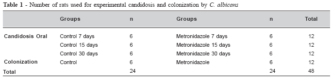
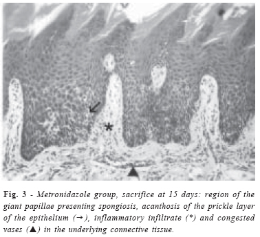
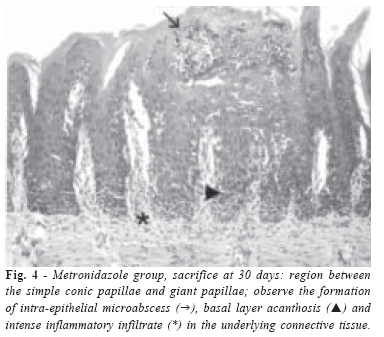
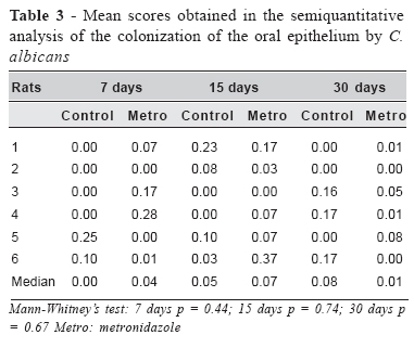
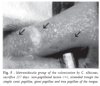
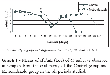
Brazilian Journal of Oral Sciences, Vol. 5, No. 17, Apr-June, 2006, pp. 1041-1047 The role of metronidazole on the establishment and persistence of oralcandidosis Veronica Quispe Yujra1 Alexandre Prado Scherma2 Juliana Campos Junqueira3 Antônio Olavo Cardoso Jorge4 Rosilene Fernandes da Rocha5 1Undergraduate student - School of Dentistry of São José dos Campos, São
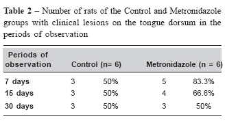
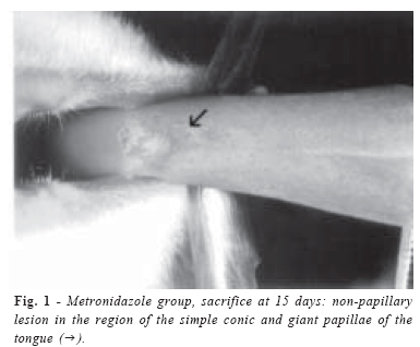
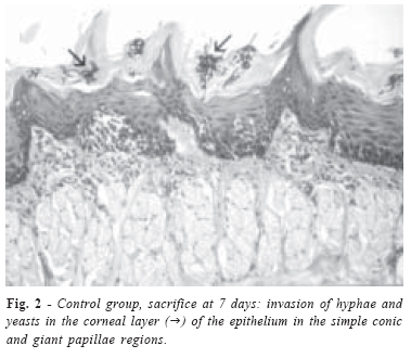
Paulo State University (UNESP), SP, Brazil Received for publication: February 03, 2006 Accepted: April 18, 2006 Code Number: os06021 Abstract The aim of this study was to study the effects of metronidazole on the establishment of oral candidosis and Candida albicans colonization in the oral cavity of rats. Forty-eight male rats, negative for yeasts in the oral cavity, were used in the study. The rats were inoculated with a suspension of Candida albicans and treated with metronidazole or plain water (control group). The rats of the candidosis experimental group were sacrificed 7, 15, or 30 days after inoculation and their tongues were analyzed by light microscopy. Colonization by Candida albicans was evaluated 1, 2, 5 and 7 days after inoculation and progressively at 15-day intervals, with a total of 18 collections. The results demonstrated the development of candidosis on the tongue dorsum was similar between the Control and Metronidazole groups for each sacrifice period. However, the colonization results showed that yeasts were recovered in the Metronidazole group in greater numbers than in the Control group after the 37th day of the experiment (6th collection). According this, the long term metronidazole therapy favored the colonization of C. albicans in the oral cavity of rats. Key Words: Candida albicans, candidosis, metronidazole, rats Introduction The long-term use of antibiotics can lead to an imbalance in the oral microbiota, which can increase yeast colonization in the oral cavity, including Candida albicans1-3. A majority of researchers have observed an exacerbation and persistence of clinical and histological lesions on the tongue dorsum of rats treated with tetracycline, after the inoculation of C. albicans1,4-5. Russell and Jones3 studied the effect of both tetracycline-laced drinking water and a carbohydrate-rich diet (CRD) on candidal infection. The persistence of Candida in the mouth of each rat was examined by swabbing the tongue and mucosal surfaces throughout the experimental period. The oral yeast carriage was monitored semi-quantitatively by counting the number of oral swabs positive for C. albicans. Tetracycline administration resulted in the oral persistence of C. albicans in all rats over a period of 24 days. The prolonged carriage induced by a tetracycline-laced diet was far superior to that achieved by feeding with CRD alone. Furthermore, an increased frequency and severity of infection were seen in tetracycline-fed rats compared with the control. The relation between oral candidosis and the use of antibiotics is explained through the imbalance in the ecosystem that the medicament can cause. Fungi present in the normal microbiota are not destroyed by antibacterial drugs, but their competitors are. Therefore, the survivors can undergo an increase in their population and become opportunistic pathogens, to the point of originating a superinfection. Some researches have shown that the oral microbiota can show alterations within the first 24 hours of antibiotic therapy6-8. Metronidazole, a broad spectrum antibiotic with bactericidal action, is a synthetic 5-nitroimidazole. The mechanism of metronidazole activity and other 5-nitroimidazoles, appears to depend on the ferredoxin-mediated reduction of their nitro group, with the generation of a reactive metabolite or metabolites which interact with DNA, leading to the subsequent inhibition of nucleic acid and protein synthesis9. It is active only against obligate anaerobic bacteria; however, showing little or no effect against most aerobic bacteria. It is also recommended for the treatment of trichomoniasis, vaginitis by Gardenerella vaginalis, giardiasis and amebiasis10-11. In the area of dentistry, recent studies have suggested the use of metronidazole, specially combined with amoxicillin, for the treatment of patients with juvenile and refractive periodontitis, where it is an effective adjunct to conventional mechanical therapy10,12. During the systemic use of metronidazole, it reaches high concentration in the gingival fluid. Its long-term use is commonly associated to the development of pilous tongue and stomatitis, although superinfections by Candida are rarely reported2,13. The evidence of the many works performed to study experimental candidosis in rats treated with tetracycline, makes it clear that few studies have been realized to verify the role of metronidazole in this process. Therefore, the purpose of the present work was to evaluate the effects of metronidazole in the development of oral candidosis on the tongue dorsum of rats and in the colonization of C. albicans in the oral cavity. Materials and Methods Animals: Fifty-four male rats (Rattus norvegicus, Albinus, Wistar) initially weighing 250g, at 12 weeks old, were used. This study was approved by the Research Ethics Committee of the School of Dentistry of São José dos Campos/UNESP, under protocol number 002/2003-PA/CEP. Research on natural colonization by Candida spp.: All the rats were submitted to an examination to verify previous natural colonization by Candida spp. For this purpose, material was collected from the oral cavity with a sterile swab, which was then grown on Sabouraud dextrose agar plates (Difco, Detroit, USA), with chloramphenicol (0.1mg/mL, Itafarma, SP, Brazil) in duplicate. The plates were incubated for 48 hours at 37ºC. Colonies of Candida were isolated and identified according to Williams and Lewis14: germinative tube formation, production of chlamydoconidium and hyphae in microculture, and the fermentation and assimilation of carbohydrates. Out of the fifty-four rats analyzed, six animals presented natural colonization by yeasts in the oral cavity and were excluded from the study. Only forty-eight rats, negative for Candida spp., were used in the subsequent experiments, thirty-six for the experimental candidosis study and twelve for colonization by C. albicans (Table 1). Inoculation: A suspension of C. albicans, containing 5 x 10⁸ viable cells/mL was prepared according to Reed et al.15 (1990). A strain of C. albicans originating from the Microbiology Laboratory of the School of Dentistry of São José dos Campos/UNESP was used, isolated from a patient who presented stomatitis due to prosthesis use. This strain presented high lethality for mice and produces proteinase, hyaluronidase, chondroitin sulfatase and phosphatase16. For the inoculation, the rats were sedated with a Rompun (Bayer, SP, Brazil) and Dopalen (Vetbrands, SP, Brazil) solution in the proportion of 1/0.5mL, at a dose of 0.05mL/100g of body weight, intramuscularly. The suspension of C. albicans (0.2mL) was dripped into the rat‘s mouths with the aid of an insulin syringe. The material was uniformly spread on the mucosa of the tongue dorsum with a sterile swab soaked in the suspension. This procedure was repeated for three consecutive days. Treatment with metronidazole: The metronidazole (Aventis, SP, Brazil) was administrated at a dose of 22.5mg/Kg/day11 in the rat‘s drinking water. The rats of the experimental candidosis began treatment with metronidazole on the first day of C. albicans inoculation, and were treated for 7, 15 or 30 days. The rats used for the study of C. albicans colonization began treatment with metronidazole four days after the first inoculation of the yeast, and were treated for a total of one hundred fifty seven days. Experimental candidosis: After 7, 15, or 30 days of treatment with metronidazole, the rats were sacrificed and their tongues were removed and submitted for macroscopic analysis using a stereoscopic microscope (Zeiss, Germany). For the histopathological examine, the tongues were fixed in 10% formalin for 24 hours and sectioned in two parts in direction of the sagittal. After being paraffinated, a series of cuts of 7µm thickness in microtome were obtained, which were stained with Hematoxylin Eosin (HE) and Periodic Acid Schiff (PAS). To determine the degree of C. albicans colonization, a semiquantitative analysis was performed based on the methodology proposed by Junqueira et al.17. Twenty-eight histological fields of the tongue dorsum from each cut were analyzed, in anteroposterior direction , at 400X magnification. Each histological field was attributed a score (score 0: absence of colonization; score 1: one to five hyphae; score 2: six to fifteen hyphae; score 3: sixteen to fifty hyphae; and score 4: over fifty hyphae). Thus, 28 scores were attributed to each histological cut. From each rat, two randomly chosen histological cuts were analyzed, totaling fifty-six scores per rat. For the realization of statistical analysis, the mean between the fifty-six scores was determined. Colonization by C. albicans: Oral samples were collected on days 1, 2, 5, and 7 after the last inoculation of C. albicans and after that, every fifteen days until eighteen samples were collected. The oral samples were collected from the oral cavity by using a swab for 60 seconds, immersing it in a tube with 0.95mL sterile physiologic solution and agitating for 60 seconds (one hundred times diluted) according to Jorge et al.4, Freire Garabal et al.18 and Junqueira et al.19. 100µL samples from this suspension were dropped in duplicate, after serial four fold dilution, on Sabouraud agar plates containing chloramphenicol (0.1mg/mL, Itafarma, SP, Brazil). The plates were incubated for 48 hours at 37°C. After the incubation period, the colony forming units per milliliter of saliva (cfu/ mL) were calculated. Statistical analysis: The results were submitted to the Mann-Whitney test and Student´s t test at a significance level of 5%. Results Research on natural colonization by Candida spp.: Out of the fifty-four rats analyzed at the beginning of the study, six (11.1%) presented Candida spp. in their oral cavity and were excluded. In relation to their identification, three samples were C. guilliermondii (50%); two were C. tropicalis (33.3%) and one was C. lusitaniae (16.6%). Experimental Candidosis: The macroscopic analysis showed that 21 (58.33%) out of the 36 animals in the experimental candidosis study, presented clinically visible lesions on the tongue dorsum (Table 2). The clinical lesions were erythematous and nonpapillated (Figure 1), well delimited and situated in the simple conic papillae, near the giant papillae. The true papillae were the least affected. The following characteristics were observed in the microscopic analysis: Control Group: out of the 18 rats analyzed, 9 (50%) presented lesions constituted by yeasts and hyphae inside the corneal layer, predominantly in the region of simple conic papillae, near the giant papillae (Figure 2). The epithelium presented acanthosis and discrete hydrophic degeneration. In the subjacent connective tissue congested vases were observed, as well as the presence of a local discrete inflammatory infiltrate. In only one of the histological cuts of this group, an extensive candidosis lesion and the formation of intraepithelial microabscesses were observed. Metronidazole Group: out of the 18 rats studied, 13 (71%) presented foci of hyphae and yeasts invasion, most being well situated affecting some simple and giant conic papillae. The epithelium displayed acanthosis of the prickle layer, exocytosis, spongiosis and epithelial disorganization. The subjacent connective tissue showed many congested vases and discrete inflammatory infiltrate (Figure 3). Some cuts of the 30-day period presented hyperparakeratosis, hydropic degeneration, epithelium hyperplasia, loss of the basal cell layer stratification, in one of them papillary destruction with the formation of intraepithelial microabscesses were observed (Figure 4). In relation to the semiquantitative analysis of epithelium colonization by Candida, there was no statistically significant difference between the Control and Metronidazole groups (Table 3). Colonization by C. albicans: Yeasts were recovered in all rats of the Control group in the periods of 1, 5, 7, 22 and 37 days after inoculation. After that, the number of rats positive for yeasts decreased to 4 (66.66%) rats after 52 days and to 2 (33.33%) rats after 67 days. On the 142nd and 157th days, C. albicans was no observed. In the metronidazole group, the C. albicans colonization occurred in all rats in the periods of 1, 5, 7, 22, 37 and 52 days after inoculation. After the 67th day, the number of rats positive for Candida dropped to 3 (50%), remaining that way up to the 172nd day. On the 187th day of C. albicans colonization, another rat was negative, and on the 202nd day C. albicans was recovered from only one rat; this result was repeated up to the 217th day of recovery, when it was sacrificed. The candidosis observed in this rat was nonpapillated and extended through the simple conic papillae, giant papillae and true papillae, very similar to median rhomboid glossitis (Figure 5). The logarithm of the cfu/mL of C. albicans colonization from the oral cavity of the rats up to the 22nd day was similar between the Control and Metronidazole groups. After the 37th day, the mean log of cfu/ml was higher in the Metronidazole group then in the Control group (Graph 1). Discussion Out of the fifty-four animals examined, six rats (11.1%) were positive for the Candida spp. in the oral cavity. This result was inferior to those reported by Jorge et al.4 (17%) and Totti et al.20 (42%). On the other hand, this result is similar to that reported by Junqueira17, who found 9.48% of rats colonized by Candida at the beginning of their work. These isolates were identified as C. guilliermondii (50%), C. tropicalis (33.3%) and C. lusitaniae (16.6%). These data corroborated with the results of Junqueira17, who found 44.4% of C. guillermondii, 5.55% of C. tropicalis and 38.8% of C. lusitaniae. These species were also found by Jorge et al.21 in the following percentages 2.5%, 8.13%, and 1.25%, respectively. In relation to C. albicans colonization, out of the six control rats, 100% were positive up to the 37th day of observation. These results are superior to those found by Jorge et al.4, who recovered Candidain only 50% of the control rats 35 days after inoculation and to the results of Junqueira et al.19, who observed colonization by yeasts in 80% of the control animals up to the 37th day of recovery. These data demonstrate that the inoculations of C. albicans realized in the current work were able to induce a strong Candida infection in the oral cavity of rats. Therapy and prophylaxis with broad spectrum antibiotics are known to be major risk factors for the development of fungal infections22. Oral administration of antibiotics may induce alterations in the normal oropharyngeal, intestinal and vaginal microbiota. These alterations depend on the dosage and schedule, the spectrum of activity and the pharmacokinetic properties of the antimicrobial agent used23. Up until the present, numerous studies have been developed to investigate the effect of tetracycline in oral candidosis in rats, demonstrating that this antibiotic predisposes the host to C. albicans colonization and the development of persistent infections1,3-4. However, many other antibacterial medications are prescribed in clinical odontology and their potential effects on the establishment of candidosis need to be studied. Several studies have documented that drugs active against anaerobic microbiota such as vancomycin, clindamycin or metronidazole are significantly more associated with fungal superinfections than are other antimicrobials22. For these reasons, this work studied the effects of metronidazole in the development of candidosis and C. albicans colonization in the oral cavity of rats. In the experimental candidosis study the control rats developed candidosis lesions on the tongue dorsum similar to the treated group with metronidazole in all periods of observation (7, 15, and 30 days). In the study of C. albicans colonization, the number of yeasts recovered from the oral cavity of the rats up to the 22nd day (5th collection) was similar between the control and metronidazole groups. However, after the 37th day (6th collection), the number of cfu/ml was higher in the metronidazole group than in the control group. These results show that the metronidazole influenced the oral colonization by Candida when administered for long periods (over 37 days). However, Pultz et al.24 verified an increase in intestinal colonization by Candida glabrata in mice after subcutaneous administration of metronidazole for 8 days. The differences between the current results and the results of Pultz et al.24 are probably due to a combination of factors; the way metronidazole was administered, the species of Candida studied, the colonization site in the organism and experimental animal model. Besides Pultz et al. (2005), other authors have also studied the influence of metronidazole on C. albicans colonization of the gastrointestinal tract of mice25-26. Samonis et al.25 reported that mice fed on a diet containing C. albicans (concentration of C. albicans in the food was 7.48 log10 cfu/ g) and treated with antibiotics for ten days presented substantially higher Candida counts in their stools than control mice fed C. albicans and treated with saline. The concentrations of Candida in the stools of mice treated with tetracycline were much higher when compared to those of mice treated with metronidazole and norfloxacin. However, Kinsman and Pitblado26 verified that the treatment with metronidazole, gentamicin and vancomycin for three days did not affect the gastrointestinal Candida levels. Only ceftizoxime, augmentin and cefoperazone promoted Candida overgrowth. The lower predisposition of metronidazole to the development of candidosis in relation to other antimicrobials could be due to the slight inhibition of anaerobic microbiota induced by this medication. This can be explained by the fact that the in vivo susceptibility of anaerobic microorganisms to metronidazole is considerably less than the in vitro susceptibility, and that high doses of the drug are needed to suppress oropharyngeal and intestinal microbiota23. The effects of metronidazole on candidosis have already been studied in humans23,27. Maraki et al.23 demonstrated that treatment with metronidazole for 10 days increased human gastrointestinal, oropharyngeal and vaginal colonization by the Candida species, though not significantly. Trenschel et al.27 found that the combined use of ciprofloxacin, metronidazole and fluconazole, as an antifungal prophylaxis, increased intestinal yeast colonization when compared to ciprofloxacin and fluconazole alone in patients undergoing allogenic bone marrow transplantation. Although an evaluation of experimental candidosis for longer periods was not performed, a higher incidence of yeasts in the oral cavity of rats in the metronidazole group was observed in the study of C. albicans colonization after the 37th day (6th collection), indicating that the long-term use of metronidazole favored the C. albicans colonization in the oral cavity of treated rats. References
Copyright 2006 - Piracicaba Dental School - UNICAMP São Paulo - Brazil The following images related to this document are available:Photo images[os06021t2.jpg] [os06021f2.jpg] [os06021t1.jpg] [os06021f3.jpg] [os06021f4.jpg] [os06021t3.jpg] [os06021f1.jpg] [os06021f5.jpg] [os06021g1.jpg] |
| |||||||||
