 |
| About Bioline | All Journals | Testimonials | Membership | News |
|
||||||
|
||||||
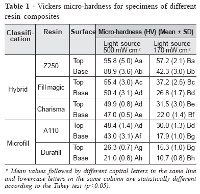
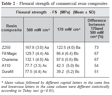
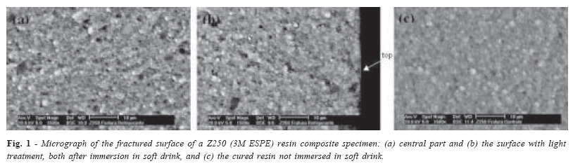
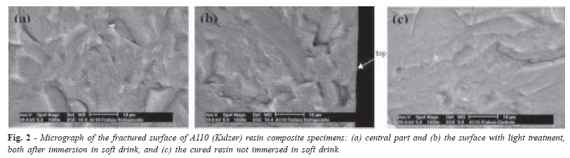
Brazilian Journal of Oral Sciences, Vol. 5, No. 17, Apr-June, 2006, pp. 1048-1053 Influence of light intensity on mechanical properties of the resin composite Betsy Kilian Martins Luiz1 Luiz Henrique Maykot Prates2 José Roberto Bertolino3 Alfredo Tibúrcio Nunes Pires3 1Research Group of Polymeric Materials -POLIMAT and Graduate Program of Science and Material Engineering - UFSC Received for publication: February 18, 2006 Accepted: May 29, 2006 Code Number: os06022 Abstract The aim of this study was to evaluate Vickers micro-hardness and flexural strength, of five commercial resin composites cured by two light curing units (LCU) with different intensities. The inorganic filling content of the composite was evaluated by thermogravimetric analysis (TGA) and the morphology of the surface was analyzed by scanning electron microscopy (SEM), in composites cured by different light intensities and immersed into a commercial soft drink or distilled water. The results show an improvement in the mechanical properties when the highest intensity of the halogen light-curing unit was used. At the same intensity of LCU, the highest values for the mechanical properties were found in specimens with a higher percentage by weight of filler. The better values were observed for micro-hardness and flexural strength in hybrid resin composites than in microfill resin composites. When the material was stored in the soft drink, changes occurred in the composite structure. The SEM images show a fractured specimen immersed in the soft drink compared with the control group, maintained in distilled water. The images show the low level of light beam penetration into resin composites, since the removal of resin components at the central part of the specimen was higher than at the surface near the light incidence. Key Words: resin composites, micro-hardness, microscopy, flexural strength, light curing Introduction Due to a growing demand for light-cured resins with good properties in aesthetic dentistry, an efficient way to increase the curing degree and the development of a new formulation was studied. Changes in size, type and percentage of inorganic particles and composition of organic matrix have been proposed. Visible light-curing units (LCU) are commonly used to initiate the polymerization process of light-sensitive materials in dentistry, such as resin composites, modified glass resins, pit and fissure sealants, bonding systems, bases, liners and resin luting agents. The degree of cure in photopolymerizable materials depends on the intensity, wavelength, and exposure time of the light-curing units, which are directly related to changes in mechanical properties1-2. The consequences of a decrease in the degree of cure may be color instability and water sorption, as well as the eventual appearance of holes, pulp damage, and a reduction in the restoration life3. It has been observed that a high intensity of the light source will favor the resin reticulation, resulting in superior chemical and mechanical properties those achieved by the use of a light source of low intensity4. Halogen dental curing lights have been commonly used in dentistry in recent decades. Despite some inherent limitations, such as an intensity decrease with use, due to degradation of the halogen bulb and its reflector, blistering and cracking of the internal filter, and damage to the fiber optic tips used to focus the light on the restorative material5. Studies have shown that the majority of light sources in use have inadequate technical maintenance, and a substantial number delivers less than the minimum power density required to adequately polymerize light-cured restoratives6-7. Resin composites must have high wear resistance, good adaptation to the dental surface, and a micro-hardness with the same characteristics as those of enamel and dentin. Other requirements for the use of resin composites are dimensional stability and resistance to occlusion stress, allowing a simple technique for durable restoration, and acceptable aesthetics in terms of color8. Many factors such as resin composition, specimen geometry, concentration of the photo initiator, intensity and exposure time of the light beam and temperature of the curing process are related to the cure degree9. The purpose of this study was to evaluate Vickers microhardness and flexural strength of five commercial resin composites cured using two light curing units with different intensities. Inorganic filler percentage of the resins and the surface morphology of the specimens immersed in a commercial soft drink and distilled water were analyzed. Material and Methods The materials used were three hybrid resin composites: Z250 (3M ESPE Dental Products, St. Paul, Minnesota, USA), Charisma (Heraeus Kulzer, Hanau, Germany), Fill Magic (Vigodent, Rio de Janeiro, Brasil) and two microfill resin composites: A110 (3M ESPE Dental Products, St. Paul, Minnesota, USA) and Durafill (Heraeus Kulzer, Hanau, Germany). All resin composites used in this investigation were shade A2. The curing units investigated were two conventional halogen lights: Primelite (Dentsply Company), with 170 mW/cm2 of light intensity and Curing light 2500 (3M ESPE Dental Products), with 500 mW/cm2; verified with a Curing Radiometer 100 Demetron power meter (Demetron Research Corporation, Oregon, USA). Vickers micro-hardness testCylindrical stainless steel molds, 6 mm in diameter and 1 mm in height, were packed with resin composites, and a Mylar strip (0.06 mm thick) and a glass slide (3mm) were placed on the top of the mould. One of surfaces was treated with a light beam for 40 s. The glass slide thickness standardized the distance from the light source to the resin composite and provided a smooth, non-air-inhibited surface for subsequent hardness testing10. After, the specimens were ground with a sequence of abrasive papers (240 to 1200 grit). Vickers microhardness (Shimadzu HMV–2000) is based on the resistance of the material to penetration by a diamond pyramid (square base and angle of 136o between the faces), when applying a load of 100 g for 10 s. The specimens were stored in distilled water for 24 h before analysis, and at least three hardness measurements taken on a light beam treated surface (top) and also on a non-treated surface (base) of each commercial resin composite, according to American Dental Association (ADA) specifications11. Flexural strength test The specimen dimension was 25 mm X 2 mm X 2 mm and to carry out the curing process a mechanical support was adapted to move half of the light beam diameter to cover the entire length of the specimen. The same procedure was carried out on each side of the specimen. The specimens were maintained in distilled water at 37 oC for 24 h, ground on 240 grit abrasive paper to remove shavings and measured with an electronic digital caliper (Starrett 727) with an accuracy of 0.01 mm. The flexural strength (three-point loading) test was carried out at least twelve times, with 20 mm between supports. A load of 2000 N and a crosshead speed of 0.75 mm min-1 were applied using an Instron Universal Testing Machine (Instron 4444). The measurements were carried out according to the International Standards Organization (ISO)12. Data from the mechanical tests were analyzed statistically using a three-factor design in the STATISTIC 6.0 program (p< 0.05). Thermal analysis (inorganic filler percentage) The resin degradation of the specimens was analyzed by thermogravimetry (TGA) with a Shimadzu 50 thermal gravimetric analyzer under a nitrogen atmosphere. Nonisothermal experiments were performed in the temperature range of 25 to 800 oC, at a heating rate of 20oC min-1. Nitrogen flow was maintained at 50 cm3 min-1 and samples of ca. 12 mg were used for all the experiments. Scanning electron microscopy (SEM) The cured specimens (6 X 1 mm) of five commercial resins (500 mW/cm2 light intensity), were cryogenically fractured and kept in a commercial soft drink (Coca-Cola Company) or distilled water for 24 h, and then morphologically analyzed by scanning electron microscopy. The specimens were covered by a thin gold layer in a D2 metalizer of a Diode Sputtering System, ISI (International Scientific Instruments). A Phillips XL 30 scanning electron microscope with a tungsten electron source and a secondary electron detector was also used. Results By thermogravimetric curves were obtained 80, 81 and 78% of inorganic filler for Fill Magic, Z250 and Charisma (hybrid resins) and 61 and 56% for A110 and Durafill (microfill resins), respectively. The Vickers micro-hardness values for different resin specimens for both sides (top and base) using two light beam intensities are shown in Table 1. On comparing the micro-hardness of the top and base of the specimens, it was seen that the values for the base were around 10% lower than those for the top, for the photopolymerization with a light intensity of 500 mW/cm2. For the 170mW/cm2 light curing unit the base values were around 30 % lower than those for the top. The flexural strength values are shown in Table 2. The percentage difference in flexural strength of the resin specimens cured using LCU intensities of 500 and 170 mW/ cm-2 was lower for the micro-fill resins. The micrograph images showed similar characteristics for all hybrid and all micro-fill resins, so only the images for one hybrid (Z250) and one micro-fill (A110) resin are given as examples. Figure 1 shows a micrograph of a fractured specimen of the Z250 (hybrid resin), where (a) is the central part and (b) is the surface with light treatment, both after immersion in soft drink, and (c) is the cured resin not immersed in soft drink. Figure 2 shows a micrograph of a fractured specimen of A110 resin (microfill), cured with 500 mW/cm2 light intensity: (a) central part and (b) is the surface with light treatment, both after being immersed in soft drink, and (c) the cured resin not immersed in soft drink. Discussion Due to the influence of light beam intensity on the curing of light-activated dental materials, the LCU is an important tool in dental offices. The proper performance of these units, i.e., their ability to provide adequate intensity, is crucial to optimizing the physical properties of light-activated materials. An inadequate cure degree has been associated with low physical properties, solubility, retention failures and adverse pulpal responses, due to the residual unpolymerized monomers10. Continuous use of LCU in dental offices results in the degradation of their components in a short period of time. This is a common cause of diminished light beam intensity in dental practices, which has adverse consequences for the restorations and for the tooth. The performance of a halogen light is known to diminish over time if the bulb, reflector, fiber-optic tips, and filter are not properly technically maintained. The literature showed similar studies related to the effects of the light beam intensity, as in Pilo et al.13 who carried out an investigation measuring 130 photopolymerization light sources. They found that 45 % of the LCU were of appropriate intensity (> 300 mW cm-2), 22 % were of inappropriate intensity (200 - 299 mW/cm-2) and required a longer light source exposure time, and 33 % were inappropriate for use (< 199 mW/cm-2). It is also known that dentists are often unaware of this degradation and continue to use poorly performing lights, which leads to inadequate polymerization. Higher Vickers micro-hardness values for both sides of specimens treated with 500 mW/cm2 of light intensity, when compared to 170 mW/cm2, suggest that LCU intensity is directly related to the penetration of the light beam and resin composite curing across the entire specimen. For all specimens, LCU with an intensity of 170 mW/cm-1 resulted in low values for the micro-hardness of both surfaces14-16. The hardness values for the base of specimens treated with 500 mW/cm-1 were higher than values for the top of specimens treated with 170 mW/cm-1, showing that the light source with low intensity does not cure the specimen even at the top. Regarding micro-hardness, the ANOVA revealed that the resin composite and light beam intensity have a significant effect on micro-hardness, and the light source intensity is related to the depth of penetration of the light beam into the specimen. Results for Vickers micro-hardness showed that the LCU influences the mechanical properties regardless of the type of commercial resin used, probably changing the cure degree of the organic component of the resins. Based on the percentage of inorganic particles present in the resin composites, the higher values for flexural strength and micro-hardness obtained for the hybrid when compared to the microfill resins may be due to the particle size of the material structure and the higher percentage by weight of inorganic particles in the composition of these resins. However, in a comparative study between condensable composites and hybrid resins, Adabo et al.17 did not observe a relationship between volume of inorganic filler particles and flexural strength. Observations gained from the micrograph of the fractured specimen immersed in a soft drink are consistent with the fact that the cure degree is greater near the light-treated surface and lower at depth, even using the 500 mW/cm-2 LCU. This is observed from the homogeneity of the material near the surface treated with the light beam and the removal of components in the central part of the fracture. It can be assumed that the porous resin is that which was not properly cured and was partially removed by the soft drink. This difference results from the penetration of the light beam into resin composites, resulting in variations in the degree of cure in different parts of the resin. An inadequate degree of polymerization can result in lower values for the mechanical properties with the removal of resin components by agents of the everyday diet. Appropriate light beam intensities and exposure times induce a higher degree of polymerization at the surface, reducing the removal of resin components by chemical agents present in foods and drinks. In this regard, modifications in the composition of soft drinks have been suggested in order to minimize dental erosion, including that of restorations18. The texture homogeneity of the whole fractured surface showed in the micrograph for A110 resin specimen immersed in soft drink and its similarity with that of the specimen not immersed suggest similar cure degree. It indicates that the efficiency of the curing process is related with the size of the inorganic particles when the light intensity and the light source are not changed. Microfill resin composites allow a higher degree of cure not only because of the smaller particle size but also due to the lower percentage of inorganic components, and probably to better penetration of the light beam into the resin specimen. These results agree with those of previous studies by Yoon et al.19 on the degree of cure of resin composites treated with different LCU, and those of Rueggeberg et al.1 who showed that even an LCU with relatively low intensity can cure the microfill resin matrix to an extent almost equal to that when high intensity light was used. However, as light passes through the bulk of the restorative material its intensity decreased, greatly reducing the degree of cure. Park and Lee showed that the degree of cure of a resin composite diminished as the distance from the light source increased. They observed that for the top surface, only the exposure time was a significant factor contributing to monomer conversion, while exposure time and intensity became significant factors in curing a 1 mm-depth of the composite20. Intensity of light-curing units is directly related to the mechanical properties, as we can see from the results for the Vickers micro-hardness and flexural strength properties, due to differences in the penetration of the light beam into the resin composite specimen, and the percentage of the organic component. The lower cure can affect the durability of the restoration in the mouth, and the risk of its degradation in a short period of time. In this study light-curing units with intensities of 170 and 500 mW/cm-2 were used to compare mechanical properties and analyze the behavior of the material, during the light beam penetration. It is generally reported in dental science literature that an intensity of 300 to 400 mW/cm-2 and wavelength between 380 and 520 nm are necessary for adequately curing a 2 mm thick composite specimen1. Barghi6 studied the efficiency of the light source in relation to the reticulation degree of a resin composite, using a wavelength in the range of 400 – 520 nm, recommending an intensity of 400 mW/cm-2 for the cure of the material. Dentists should check periodically the photopolymerization light source, and use appropriate treatment times for the curing of resin composites in order to guarantee the required clinical success and durability of the restorations. The effect of drinks on the resin depends not only on factors related to the light source but also the composition of the resin composites, which can lead to different dissolution patterns. The effect of the light beam intensity on the polymerization of resin composites is extremely important; it is directly related to the degree of cure of the material, consequently affecting the mechanical properties. For similar light beam intensity, the effects of filler particle size and percentage by weight are directly related to micro-hardness and flexural strength. Acknowledgments This study was supported by CNPq (Conselho Nacional de Desenvolvimento Científico e Tecnológico). References
Copyright 2006 - Piracicaba Dental School - UNICAMP São Paulo - Brazil The following images related to this document are available:Photo images[os06022t2.jpg] [os06022f2.jpg] [os06022t1.jpg] [os06022f1.jpg] |
| |||||||||
