 |
| About Bioline | All Journals | Testimonials | Membership | News |
|
||||||
|
||||||
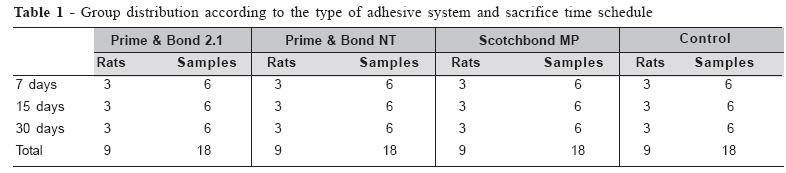
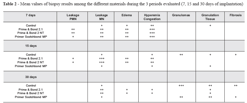
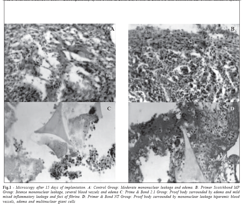
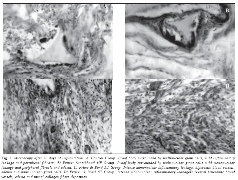
Brazilian Journal of Oral Sciences, Vol. 5, No. 18, July-September 2006, pp. 1079-1084 Biocompatibility of the Prime & Bond 2.1, Prime & Bond NT and Scothbond MP Primer adhesive systems Marcos Ribeiro Moysés1 William Luis Lopes2 Alessandro Antônio Costa Pereira1 José Carlos Rabelo Ribeiro1 Sérgio Candido Dias1 Luciano José Pereira1 1Professor Clinical Dentistry Post-Graduation Program, Vale do Rio Verde University – UNINCOR, Três Corações – MG – Brazil Received for publication: May 05, 2006 Code Number: os06027 Abstract The objective of this study was to verify the biocompatibility of dental adhesives (Prime & Bond 2.1; Prime & Bond NT and Scotchbond MP). Thirty-six male rats (Rattus norvegicus) received artificial sponges containing the adhesive materials in their back, and they were polymerized before suture. As a control, one group had sponges containing water. Each group consisted of 9 rats with two implants, resulting in 18 samples per group. Three animals of each group were killed after 7, 15 and 30 days and the tissue was analyzed. At sevenday period, inflammation was acute and severe in all other groups, except water. After 15 days, Prime & Bond 2.1 and Prime & Bond NT groups showed a more severe inflammatory infiltrate with foci of neutrophils inside the cavity border when compared with the Scotchbond Primer. After 30 days, in the control group the cavity was shown to be filled by well organized granulomas next to the sponge samples. Prime & Bond 2.1 and Prime & Bond NT groups maintained intense inflammatory process when compared with the Scotchbond Primer MP group. The present results showed that Scotchbond MP Primer Group demonstrated better biocompatibility than the other two groups. Key Words: biocompatible materials, dentin-bonding agents Introduction The objective of an adhesive system is to establish a strong mechanical bond between the restorative material and the dental structure. The area of dentin in contact with the adhesive is of great importance. However, components commonly used in adhesive system compositions have proven citotoxic effects1-4. In accordance with this statement, adhesive biocompatibility is still a cause of controversy. Usually, dentin adhesives are selected on the basis of mechanical experiments and marginal leakage tests, in which the bond capacity and tooth-restoration sealing effectiveness are evaluated. Nevertheless, as dentin is considered a living tissue, despite its relationship with the pulp, adhesive system biocompatibility is of interest at the time of deciding the choice of this material. It has long been demonstrated that different resinous material components can be released in an adjacent aqueous phase5. Thus, when applied to a wet surface, such as dentin, unpolymerized free monomers released from resin-based materials may diffuse across dentinal tubules to reach the pulpal space6. Some investigations have shown that released monomers cause chemical damage to cultured cells7-8. In addition, many in vivo studies have shown that unpolymerized resin components that reach the pulpal space cause noticeable chronic inflammatory response and inner dentinal resorption9. The acid conditioning of dentin, particularly in the pulpal wall of deep cavities seems to increase dentin permeability6. In this clinical situation, it has been demonstrated that bonding agent application may result in inward residual monomer movement10 due to a reversal in dentinal fluid .flow caused by the light-activation step11. Once these residual monomers reach the pulp tissue, a foreign body reaction is elicited. Macrophages engulfing these leached resin components have been found as long as 300 days after the restorative procedure6 had been done, triggering a persistent chronic inflammatory pulpal response6,9,12. Thus, the objective of the present research was to compare the biocompatibility of three dentin adhesives (Prime & Bond 2.1; Prime & Bond NT and Scotchbond MP) implanted under the subcutaneous tissue of rats. The inflammatory reaction induced by these systems after polymerization was evaluated, to simulate the same condition found “in vivo”. The results of this experiment aim to facilitate adhesive system selection for clinical use. Material and Methods For the experiments, 36 male rats (Rattus norvegicus, Holtzman) from the UNINCOR Laboratory were used. The protocol was approved by the Ethics Committee of the same institution. The animals were standardized according to their weight (200 to 300 grams) and state of health. All the procedures were performed in an aseptic environment and all the surgical instruments were sterilized in autoclave. Group distribution The rats were distributed into 4 groups according to the adhesive (Prime & Bond 2.1; Prime & Bond NT and Scotchbond MP) or water as a control. Each rat received two polyvinyl (PVA) sponge implants containing the same substance; consequently each group consisted of 9 rats with two implants, resulting in 18 samples per group. Three animals of each group (6 samples) were, however, killed after 7, 15 and 30 days and the tissue containing the sponges and adhesive was analyzed by microscopy (Table 1). Adhesive Implantation All the animals received (ketamine 50 mg/kg; xylazine 10 mg/ kg;) intra-peritoneal anesthesia. Afterwards, the animals had their backs shaved (4x4cm) and after a small incision, the sponges were implanted in the subcutaneous tissue, in accordance with the animals’ groups. Finally, the incision was sutured and the animals received analgesic injection (Sodium dipyrone – 0.3mL/100g weight). The animals were killed according to the pre-determined time table, in accordance with each group, and biopsies were conducted to evaluate the tissue response to the adhesive systems. Antisepsis was performed with digluconate of chlorexidine 4%. The incision location was delimited taking into account the medium line and the distance between the rat’s head and tail. The two incisions were made with surgical blade number 15 and each site was approximately 18 mm deep. Using scissors, the subcutaneous tissue was separated laterally and a space was created to implant the sponges. The dentin adhesives (Prime & Bond 2.1; Prime & Bond NT and Scotchbond MP) were prepared in accordance with the manufacturer’s instructions. Polyvinyl sponges (PVA) 6.0mm in diameter and 2.0mm high were soaked with 2 drops of adhesive and were then put into the cavities. Before closing the hollow space, the adhesives were photopolymerized using the Optilight 600 equipment (Gnatus-Brazil). The time for the adhesives was established in accordance with the manufacturer’s recommendations. After each 5 procedures, the light intensity was measured by radiometry. After completing polymerization, the cavities were sutured with 4.0 (Johnson & Johnson) needled line. The rats were kept in their cages and were fed normally. After 7, 15 and 30 days the rats were anesthetized and the biopsy materials were taken by one examiner. This procedure ended with the animal being sacrificed by the cervical displacement technique. The tissues were analyzed at the Pathology Department – UNINCOR. Next they were included in paraffin and cut into 6µm samples. The HE (Hematoxilin -Eosin) method was applied. Using optical visualization, the inflammatory reaction was compared among groups. The reactions were classified according to a severity scale: mild, moderate and severe inflammation. Biocompatibility was determined in accordance with the evaluation procedures described by ISO 10993-313. Results Microscopic evaluation showed that the cavities were filled with foreign bodies and inflammatory infiltration, in addition to edema and blood vessels. Bordering the cavity, an inflammatory process with multinuclear giant cells, sometimes intensely acute and sometimes chronic was observed, depending on the type of material tested and the time interval of the experimental period. Data relative to the 3 different periods of evaluation for the biopsy samples are given in Table 2. In the seven-day period, inflammation was moderate and chronic in the control group and acute and intense in all other groups, with no significant variation among groups. After 15 days, organization of granulomas with multinuclear giant cells was observed in the control group. In groups containing Prime & Bond 2.1 and Prime & Bond NT implants, a more intense inflammatory infiltrate was observed, with foci of neutrophils inside and in the cavity border when compared with Scotchbond Primer (Figure 1). After 30 days, the cavity in the control group was shown to be filled by granulation tissue and well organized granulomas next to the sponge samples. At this period of time, groups Prime & Bond 2.1 and Prime & Bond NT maintained a more intense inflammatory process when compared with the Scotchbond Primer MP group, which presented more organized granulomas around the samples and discrete granulation tissue bordering the cavity (Figure 2). Discussion Biocompatibility evaluation by means of dentin adhesive implants under rat dorsal subcutaneous tissues is of great importance. The tissue response is similar to that expected when the same material is applied to mechanically exposed dental pulp in human teeth. These procedures avoid the use of humans as volunteers14. Several studies have investigated the biocompatibility of dental materials1-2,4,6,9,15-18 however, there are discrepancies among the methodologies used by many authors. Most of the studies on subcutaneous adhesive implants in rats use polyethylene tubes with prepolymerized adhesives. Clinically, however, the adhesive is placed on the dentin surface or dental pulp still in a liquid state, and is only polymerized afterwards. In the present study, polyvinyl sponges (PVA) were used; they were impregnated with the adhesives and were then put into the subcutaneous cavities. The adhesives were polymerized in contact with the tissues, in an attempt to simulate the real clinical process. Costa et al.3 used polyvinyl sponges, but the adhesives were not polymerized when in contact with the live tissues. Thus, the adhesive and its monomers were in deep contact with the rats’ subcutaneous tissues. As a result, the authors mentioned that despite the citotoxic effects of the adhesives systems and the persistent inflammatory reactions deflagrated by the resin components, these materials did not seem to be adequate for direct application on conjunctive tissue. Cox et al.18 reported that inflammatory pulp reactions are caused exclusively by bacterial microleakage through toothrestoration material spaces. There was a large correlation between bacterial presence and inflammatory pulp response. This was corroborated by Akimoto et al.19 in a sample of monkeys, and they concluded that adhesive systems are not toxic when delivered directly or indirectly on the dental pulp. On the other hand, there are several researchers who do not agree with this theory2-3,15,20-21. According to Consolaro et al.22, cellular necrosis can be determined by the action of physical agents in the same way as radiation and traumatisms, chemical substances such as alkalis and acids, and by biological organisms such as bacteria, virus and fungi. This evidence is also reinforced by Santos and Barbosa2 who suggested that pulp aggression is histologically independent of bacterial contact with the pulp, and that adhesive systems do have irritant properties. The citotoxicity of the adhesive components has been stated by several authors4,23-25. The divergent results presented by the literature can be explained by the different compositions of the adhesive systems tested, different application methods and methodological discrepancies1. Various research protocols must be developed to evaluate the biological behavior and clinical indications of dental materials. Biocompatibility is indispensable to make an adhesive system successful and thus, biological tests are encouraged to verify this question. These tests should provide preliminary biological responses related to specific materials14. The most studied method to test biocompatibility in vivo is inflammatory evaluation. Inflammatory cells and substances cross the endothelial walls from the blood and diffuse into the harmed tissues. The extracellular matrix is replaced by exudates and dead cells or results in necrosis, to which leucocytes and inflammation mediators are added. The last phase of inflammation is local area repair with granulation tissue before complete healing takes place22. The inflammatory response begins with a more intense reaction to the surgical procedures and the implanted foreign body, which is nonspecific for each adhesive system most of the time, and therefore the first hours after the material is implanted are normally not considered. After 7 days, is expected that a better organized inflammatory reaction will appear, which should be related to the aggression agent properties and not to the surgical procedure. In the present experiment, a mild inflammatory reaction against the sponge containing water was shown, and a moderate to severe reaction to the sponges containing the adhesives. After 15 and 30 days, a more intense difference in the degrees of inflammatory reaction was expected to become visible, and this could be interpreted as better or worse biocompatibility between the tested materials. Usually, a small area with necrosis, edema and dilated vessels surrounded by a well organized fibroid structure is observed, with chronic inflammation and decreased number of blood vessels. Multinuclear giant cells could also be seen, suggesting a granuloma formation originated by the presence of the sponge and adhesive material. The formation of granulomas associated with the occurrence of cells and lack of vascular reactions are described as a good biocompatibility response caused by the material. In the present study, the group containing the adhesive Primer Scotchbond MP was the one with most of the mentioned characteristics after 30 days implantation. Using the current methods and analyzing the inflammatory and healing characteristics, it was possible to classify the experimental groups compared with the controls. The results suggested that the group containing the Primer Scotchbond MP showed better tolerance by the tissues. This material demonstrated the formation of a chronic inflammatory reaction, with healing characteristics at the border as well as the occurrence of multinuclear giant cells, in a shorter experimental period than in the other two adhesive systems tested. In accordance with the methods used and its limitations, the present results suggested that the group with Primer Scotchbond MP showed a better biocompatibility in comparison with the Prime & Bond 2.1 and Primer & Bond NT. References
© Copyright 2006 - Piracicaba Dental School - UNICAMP São Paulo - Brazil The following images related to this document are available:Photo images[os06027f2.jpg] [os06027t2.jpg] [os06027t1.jpg] [os06027f1.jpg] |
| |||||||||
