 |
| About Bioline | All Journals | Testimonials | Membership | News |
|
||||||
|
||||||
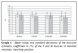
Brazilian Journal of Oral Sciences, Vol. 6, No. 20, January - March 2007, pp. 1269 - 1273 Electromyographyc evaluation of complete denture wearers using the balancing ramps concept Daniel Filgueiras Ferreira1 ,Marcelo Ferraz Mesquita 2 ,Frederico Augusto Peixoto Silva3 ,Wagner Araújo de Negreiros4 ,Rafael Leonardo Xediek Consani5 ,Guilherme Elias Pessanha Henriques6 School of Dentistry of Piracicaba, Department
of Prosthodontics and Periodontology, University
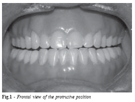
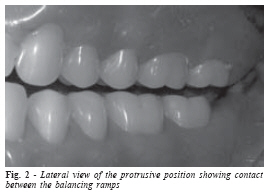
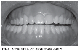
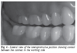
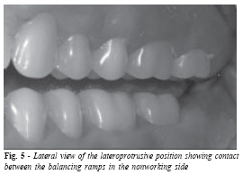
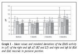
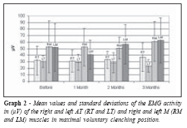
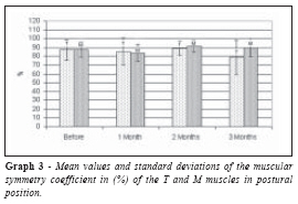
of Campinas, Piracicaba, Brazil. Received for publication: December 12, 2006 Code Number: os07005 Abstract This study verified through electromyography the masticatory muscle behavior resulting of the balancing ramps technique to obtain balance in double complete dentures. Eleven totally edentulous patients that used their complete dentures for 5 years at least, had new other ones made and balanced by the balancing ramp technique. The electromyographyc (EMG) activity pattern was registered in anterior temporal (T) and masseter (M) muscles in the rest position (R) and in the maximal voluntary contraction in the intercuspal position (MVCIP), using Myosystem I® (Prosecon Ltda., Uberlândia, MG, Brazil), in four experimental moments: T0 before the prostheses installation; T1 1 month after installation; T2 2 months after installation; T3 3 months after installation. Results showed a symmetry reduction between T muscles and a little increasing between M muscles in the R position. In the MVCIP position, it there was a reduction for both T and M muscles. The F test showed no statistical significant differences (p<0.05). Authors concluded that the employed technique promote no balance of the masticatory muscles in the tested experimental moments. Key Words: Complete denture, electromyography, balanced occlusion, balancing ramps, masticatory muscles Introduction In the last three decades, a little scientific progress could be seen in relation to the occlusal aspects in complete dentures. However the clinical experience has guided dentists to establish a simpler occlusal pattern that attend the majority of patient complaints1. Understanding the functional behavior of masticatory muscles of complete denture wearers may be important for diagnosing and planning the clinical treatment2. Among the several occlusal concepts existent, the more accepted one is the bilateral balanced occlusion introduced by Bonwill3 and studied by other known authors4-6. Basis extension, flange area, cusp height, width of occlusal table, and the lost of periodontal sensibility are aspects that promote a reduction of the masticatory efficiency in dentures wearers. In fact the occlusal pattern seems to have a great influence on the increasing of balance between the prosthesis7. Balancing ramps may promote denture balance through the smooth sliding between the prosthesis5,8. Besides the excellent esthetics due to the anterior overjet, it is possible to have more stability during the food bolus interposition than in the classical balanced bilateral occlusion pattern 9. Masticatory function may be analyzed using both objective and subjective methods. Electromyographic activity evaluation is an objective method that provides the masticatory muscle behavior in many situations during the function. That technique is based on capturing electric potentials from the muscles in action during natural and voluntary movements10. According to Tallgren et al.2 alterations in occlusal relation due to residual ridge reabsorption and complete denture seating would affect the EMG activity of masticatory muscles. However, there are few eletromyographic (EMG) studies on the effects of tooth loss and complete denture use2. Considering the evidences about the masticatory muscle behavior, electromyographyc evaluations seem to be an important auxiliary instrument employed in the clinical diagnosis of the sthomatognatic system function11. The aim of this study was to evaluate through electromyography the muscular behavior resulting of use of the balancing ramps technique between the pared masticatory muscles. Material and Methods Eleven volunteers (10 female and 1 male) were selected from Department of Prosthodontics and Periodontology of Piracicaba Dental School respecting the following inclusion criteria: totally edentulous subjects aging 58-79 years with mean age of 66,3 , wearers of complete dentures for 5 years at least, free of signs and symptoms of temporomandibular disorders (TMD) and systemic diseases. Informed consent was obtained from all volunteers, using a written form approved by Ethical Committee of the Piracicaba Dental School (nº 032/2005). New pair of complete dentures was made for each subject using the balancing ramps technique5. Anterior temporal (T) and masseter (M) muscles were evaluated in both postural (P) and maximal voluntary contraction in the intercuspal position (MVCIP) during 4 experimental moments: T0 before the prostheses installation; T1-1 month; T2-2 months; T3-3 months after installation. Occlusal balance was obtained through sliding contacts between balancing ramps. Dentatus ARL semi-adjustable articulator (Dentatus AB, Hagersten, Sweden) was used with individualized measures for Bennett angle and condylar guidance obtained by modified Gysi intraoral tracing. The posterior teeth were then mounted in a straight occlusal plane determined by an anterior point (canine position) and a posterior point (retromolar papillae). After superior teeth arrangement, the articulator was individualized and stabilized in a protrusive position so that the inferior teeth could be mounted. After teeth mounting, a mutual protected occlusion was obtained. Balancing ramps were fabricated following the posterior guidance inclination that was transferred to the superior cast. The balance pattern was obtained through contacts of anterior teeth and bilaterally between the ramps during protrusion (Figures 1 and 2). In the lateroprotrusive position the balance pattern (Figure 3) was obtained through the contact of the working side canines (Figure 4) and the contact between the ramps at the nonworking side (Figure 5). Electromyographyc evaluations were recorded at the Electromyography Laboratory - Department of Morphology of the Piracicaba Dental School, using a 12 channel Myosystem I® (Prosecon Ltda., Uberlandia, Minas Gerais, Brazil). Skin surface active-electrodes (Lynx Electronic Technology Ltda., São Jose dos Campos, SP, Brazil) of parallel bars of pure silver (Ag) were used in the experiment. Evaluations were recorded inside the Faraday cage. During the recording procedures, each subject was seated in a dental chair with the head unsupported, in natural balance, and Frankfort plane parallel to the floor. The subject's skin was carefully cleaned with 70% alcohol before electrode placement. The electrodes were positioned in relation to the muscle fiber length with the silver bar positioned perpendicularly in order to maximize signal capture and to minimize noise interference. Muscle function test12 was used to position the electrodes over the evaluated muscles. The EMG activity of T and M muscles was recorded at the P and MVCIP positions 3 times during 5 seconds. Analysis of EMG signals was accomplished by root mean square (RMS) considering their amplitude. A Myosystem I® software version 2.12 was used to visualize and process the EMG signal. The individual EMG activity value obtained was the result of the mean calculation of the three repetitions performed during each position. Evaluation of the muscular behavior was performed through the analysis of the EMG activity pattern between the pared masticatory muscles using a muscular symmetry coefficient introduced by Ferrario et al.13, presented as follow: CSM = [1 -∑ | right muscle left muscle| / ∑ (right muscle + left muscle)] x 100 Where: CSM = coefficient of muscular symmetry % ∑ = Sum. Right muscle = EMG activity of right muscle Left muscle = EMG activity of left muscle The symmetry degree of the EMG activity of AT and M muscles was evaluated analyzing their behavior at each time. It was evaluated the correlation pattern of data through the Pearson correlation coefficient and F Test (p<0.05) to verify possible differences in the symmetry pattern of the EMG activity among the evaluated times. Results Mean and standard deviation values of the EMG activity amplitude in microvolts (µV), at the P and MVCIP positions are presented at Graphs 1 and 2. Mean and standard deviation values of the EMG activity symmetry, in percentage, between pared muscles are presented at Graphs 3 and 4. Discussion The necessity of the bilateral balanced occlusion for complete denture balance and the relationship between occlusion and neurofunctional behavior of the masticatory muscles may evidence the importance of occlusal aspects, although this complete understanding has not been properly elucidated14-15. Electromyographyc studies compared two occlusal concepts utilized in complete denture, canine guidance pattern and bilateral balanced occlusion, evaluating T and M muscles in P and MVCIP positions, lateral excursions and protrusive movement. Canine guidance group obtained lesser EMG activity values during lateral excursions and protrusive movement in both evaluated muscles16-17. In another EMG activity study, with the same methodology, 9 denture wearers presented lesser values for both T and M muscles when utilizing canine guidance scheme. The theory in which inputs originated from stimulated receptors could stimulate motor neurons in the masticatory muscles try to explain these results18. Balancing ramps did not balance the EMG activity between pared masticatory muscles in this study, despite of permitting a smooth sliding between the prosthesis, what possibly promote a lesser effort in the physiologic muscle behavior8-9. However the symmetry degree in the submaximal clenching levels was implicated with occlusal aspects such as difference of occlusal support between right and left sides, occlusal interferences, and lateral and anterior mandibular sliding of eccentric to centric positions19. A lesser difference in the EMG activity between pared muscles was observed on the sixth month of evaluation, showing a temporal character of the symmetry pattern in the masticatory muscle20. A study showed that early alterations in mandible and in the occlusal aspect, followed by bone loss and difficult control in the prostheses use may affect the muscular activity, and temporal muscle seems to be more sensible in this situation21. That phenomenon may be explained by the maintenance of the central mechanism of neurophysiologic masticatory process even after tooth loss17. However it was demonstrated edentulism really influence on the sensorial and motor aspects of masticatory process22. Adaptation of neuromuscular system takes a long time and may be a determinant factor in influencing EMG activity, and this aspect can change the results20. Results verified in this study did not show a direct relationship between the functional behavior of the evaluated technique and EMG activity, confirming the theory proposed by Veyrune and Mioche22. The EMG activity could have been affected by alteration in the central mechanism of masticatory process, influenced by different stimulus originated from receptors still present on the bucal mucosa, muscular and tendon tissues. It should be also considered the mechanic aspect of the balance obtained during the protrusion in which 3 contacts exist between the dentures. However only 2 contacts exist in lateral movements, fact that promotes denture instability around an axis formed by canine and balancing ramps in the nonworking side. An important functional characteristic is that the alimentary bolus function as a third contact point in the working side, increasing the articular balance during the mastication. Therefore the results showed not great influence of occlusal scheme on the muscle behavior, in contrast what is seen in natural dentition in which the occlusal pattern may modify EMG activity. A longer time of evaluation could better show the effect of this therapy or an adapting process could occur. Within of limitations of this study, it may be affirmed that balancing ramps technique had no influence on the symmetry muscular pattern of EMG activity in the evaluated experimental moments. This study may support the theory in which there are important alterations in sensorial and motor functions in denture wearers. Acknowledgements Financial Support provided by FAPESB, Bahia State Agency for Research, Bahia, Brazil. References
© Copyright 2007 - Piracicaba Dental School - UNICAMP São Paulo - Brazil The following images related to this document are available:Photo images[os07005f1.jpg] [os07005f3.jpg] [os07005g3.jpg] [os07005g4.jpg] [os07005g1.jpg] [os07005f4.jpg] [os07005g2.jpg] [os07005f5.jpg] [os07005f2.jpg] |
| |||||||||
