 |
| About Bioline | All Journals | Testimonials | Membership | News |
|
||||||
|
||||||
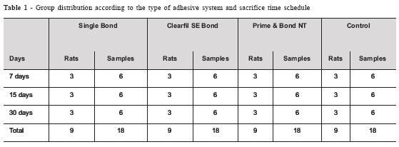
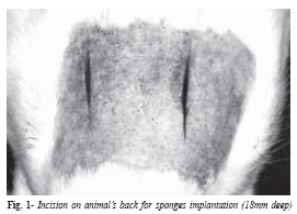
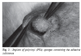
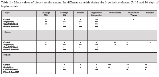
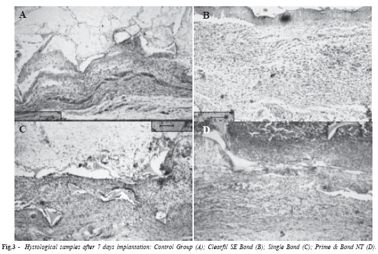
Study of dentinal adhesives compatibility using histological analysis Neusa Pereira Machado 1 , Marcos Ribeiro Moysés 2* , Alessandro Antônio Costa Pereira 3* , Luciano José Pereira4* , José Carlos Rabelo Ribeiro 2* , Sérgio Candido Dias 5* 1 DDS, Undergraduate student Três
Corações Dental School, Vale do Rio Verde University
UNINCOR Três Corações MG Brazil Received for publication: October 11, 2006 Code Number: os07009 Abstract The objective of this study was to verify the biocompatibility of three different dental adhesives (Single Bond, Clearfil SE Bond and Prime & Bond NT). Thirty-six male rats (Rattus norvegicus, Holtzman) received two subcutaneous implants in the region of their backs. Artificial sponges containing the adhesive material were placed in the cavities and they were polymerized before the surgical site was sutured. As a control, one group was implanted with sponges containing water. Each rat received two implants containing the same adhesive; consequently each group consisted of 9 rats with two implants, resulting in 18 samples per group. After 7, 15 and 30 days, 3 animals of each group were killed and the tissues containing the sponges were analyzed by microscopy. The results showed a great similarity between the control group and the Single Bond implanted tissues, followed by the Clearfil SE Bond and Prime & Bond NT. The Prime & Bond NT adhesive system appeared to show less biocompatibility when compared with the other two tested materials. Key Words: adhesive systems, biocompatibility Introduction A great effort has been made over the last few years to develop new adhesives that can effectively bond to dentin. Adhesives need to establish a strong mechanical bond between the restorative material and dental surface. To achieve this in adhesives for dentin surfaces, a large surface has to be in contact with adhesive. However, compared to enamel, dentin has a complex structure and odontoblastic derived from the pulp tissue cells are present in its tubules1. Several studies have demonstrated that some of the substances present in the adhesive systems, like Bis-GMA, TEGMA, HEMA and UDMA, show cytotoxic effects2-5. In many previous studies, it has been observed that different components of resinous material can be released in an aqueous phase6 and ,when applied to a wet surface such as dentin, uncured free monomers released from resin-based materials may diffuse across dentinal tubules to reach the pulpal space7. It was shown that these released monomers can induce chemical damage to cultured cells in vitro and8-9 can also cause noticeable chronic inflammatory response and inner dentinal resorption if they reach the pulpal space in vivo10. The acid conditioning of dentin, particularly on the pulpal wall of deep cavities seems to increase the dentin permeability7. In this clinical situation, light-activation step may cause a reversal in dentinal fluid flow and due to this the application of bonding agents may result in inward residual monomer movement11-12. Once these residual monomers reach the pulp tissue, a foreign body reaction is elicited. Macrophages engulfing these leached resin components which were found 300 days after the restorative procedure7, triggering a persistent chronic inflammatory pulpal response7,10,13. Because of the mentioned factors, the biocompatibility of the adhesive systems is still controversial. Clinicians usually consider the mechanical properties of the adhesive, its retention and amount of leakage to choose an adhesive. However, dentin is a live tissue and has a close relationship with the pulp. Thus, the choice of an adhesive material should also take its biocompatibility into consideration13. The objective of the present research is to compare the biocompatibility of three different dentinal adhesives (Single Bond, Clearfil SE Bond and Prime & Bond NT) implanted under the subcutaneous tissue of rats14. The inflammatory reaction induced by these systems after polymerization was evaluated, in order to simulate the same condition found "in vivo". Material and Methods In this study, 36 male rats (Rattus norvegicus, Holtzman) from the UNINCOR Laboratory were used. The protocol was approved by the Ethics Committee of the same institution. The animals were standardized according to their weight (200 to 300 grams) and state of health. All the procedures were performed in an aseptic environment and all the surgical instruments were sterilized in autoclave. Group distribution The rats were distributed into 4 groups. For three groups adhesives (Single Bond, Clearfil SE Bond and Prime & Bond NT) were used and for one group water was used as control. Each rat received two implants of polyvinyl (PVA) sponges containing the same substance; consequently each group consisted of 9 rats with two implants, resulting in 18 samples per group. After 7, 15 and 30 days, 3 animals of each group (6 samples) were killed, and the tissues containing the sponges and adhesives were analyzed by microscopy (Table 1). Adhesive Implantation All the animals received (ketamine 50 mg/kg; xylazine 10 mg/kg) intra-peritoneal anesthesia. Afterwards, the animals had their backs shaved (4x4cm), small incisions were made, and the sponges were implanted in the subcutaneous tissue, in accordance with the animals groups. Finally, the incisions were sutured and the animals received analgesic injection (Sodium dipyrone 0.3mL/100g weight). The animals were killed according to the pre-determined time table for each group, and then biopsies were obtained to evaluate the tissue response to the adhesive systems. Antisepsis was carried out with 4% digluconate of chlorexidine. Considering the medium line and the distance between the rat's head and tail, the incision place was delimited. The two incisions were made using surgical blade number 15 and each site was approximately 18 mm deep (Figure 1). Using scissors, the subcutaneous tissue was separated laterally and the space created to place the sponges. The dentin adhesives (Single Bond, Clearfil SE Bond and Prime & Bond NT) were prepared in accordance with the manufacturers' instructions. Polyvinyl sponges (PVA) 6.0mm in diameter and 2.0mm high were soaked with 2 drops of adhesive and then put into the cavities (Figure 2). Before closing the wound, the adhesives were photopolymerized using Optilight 600 equipment (Gnatus-Brazil) in the time recommended by the manufacturers. After each 5 procedures, the light intensity was measured using a radiometer. When polymerization was completed, the cavities were sutured with 4.0 (Johnson & Johnson) needled line. The rats were kept inside their cages and were fed normally. After 7, 15 and 30 days the rats were anesthetized and the biopsy materials were removed by one examiner. This procedure was performed by sacrificing the animal with cervical displacement technique. The tissues were analyzed at the Pathology Department UNINCOR and the tissues were included in paraffin and cut into 6µm samples. The HE (Hematoxylin -Eosin) method was applied. Using optical visualization the inflammatory reaction was compared among groups. The reactions were classified according to a severity scale: mild, moderate and severe inflammation. Biocompatibility was determined in accordance with the evaluation procedures described by ISO 10993-315. ResultsThe results of this study has shown that 7 days after implantation, there was a large mono and polymorphonuclear inflammatory infiltration along with several blood vessel and circulatory alterations (dilatation and edema) in the 3 adhesive groups. In the control group there was a mild response, demonstrating a less intense inflammatory effect (Table 2, Figure 3). For all adhesive systems, there was less inflammatory response after 15 days than after 7 days period. Among these groups, it was possible to note a more intense inflammatory effect in the Prime & Bond NT group, with more eminent vascular phenomena and the presence of neutrophils. In the control group, multinuclear giant cells and granulomatous organization were noted. After 30 days of implantation, the Single Bond material showed less inflammatory response compared with the other two adhesives, with granulomatous organization and multinuclear giant cells as well. Although there were some giant multinuclear cells, the SE Bond showed a more intense inflammatory response than the Single Bond group. The Prime & Bond NT group, however, showed a different aspect from the other two groups, with intense mononuclear inflammatory infiltrate, dilated and hyperemic blood vessels and foci with neutrophils (Table 2). Thus, it was considered the least biocompatible material out of the three tested in this protocol. Discussion Several studies have evaluated the biocompatibility of adhesive systems2-5,10,16-19. But there are discrepancies among methodologies as well as the results. In most of the studies evaluating the biocompatibility of dentinal adhesives in rat subcutaneous tissue, polyethylene tubes containing polymerized materials were used. In dental practice, however, the adhesive is placed on the dental tissue in the liquid state, and it is polymerized afterwards. Due to this, in this study, polyvinyl sponges immersed in the liquid form of the adhesive were used, and the polymerization procedure was performed with the material in contact with the live tissue. So it is expected that the obtained results from this study would be more accurate and relevant to the usage of adhesive in clinical practice. Only in one study conducted by Costa et al.4, polyvinyl sponges were used. In this study, the sponges were immersed in the adhesive material which had not undergone polymerization and the adhesive and its monomers had been in contact with the connective tissue. As a result, the authors concluded that because of the cytotoxic effects of the tested adhesives and the long inflammatory response, these adhesives are not appropriate for direct delivery on the connective tissue. Moreover, phosphoric acid which is used in the process of bonding opens the dentinal tubes and facilitates bacterial penetration and fluid flow1. Some studies indicate that baterial microleakage is the only cause of pulpal inflammation but other studies do not corroborate these findings3-4,18,20-22, as cellular necrosis can be elicited by physical agents, such as radiation and traumatism, or chemical agents, such as alkalis and acids or biological agents, such as bacteria, virus and fungi. Cox et al.17 reported that pulpal inflammatory responses are caused only by bacterial microleakage as a result of a restoration fault. The authors demonstrated a significant correlation between the presence of bacteria and inflammatory response. This is also supported by Akimoto23, who demonstrated in a research with monkeys that the adhesive systems are not toxic when applied direct or indirectly on the pulp. Other studies Reinforcing this theory, Santos and Barbosa3 stated that pulp injury is histological evidence independent of bacteria in contact with the tissue, and that adhesive systems are pulp irritants. Several studies corroborate these findings5,24-26 by showing the cytotoxic effects of adhesive components. The discrepancies in the literature can be justified by research methods or materials with different components and means of applying the adhesive2. Thus, it is clear that the selection criteria for an adhesive system should take the biocompatibility of the material into consideration. Further research is needed on the evaluation of behaviour and Clinical indications of dental materials. Biocompatibility is indispensable to make an adhesive system successful and thus, biological tests are encouraged to verify this question. These tests should provide preliminary biological responses related to specific materials14. The most studied method for biocompatibility tests in vivo is inflammatory evaluation. The inflammatory cells and substances cross the endothelial walls from the blood and diffuse into the harmed tissues. The extracellular matrix is replaced by exudates and dead cells, or by necrosis, which are added to leucocytes and inflammation mediators. The last phase of inflammation is the local area repair with granulation tissue before complete healing20. The decrease in inflammatory response during the periods of 7, 15 and 30 days is due to the body's defense system. This system limits the injury action of the adhesive components. In present current research, it was possible to differentiate the inflammatory intensity among groups, with a more intense response being shown to the Prime & Bond NT than to the other two materials. The results of the 3 adhesives were compared with a control group, enabling conclusions to be drawn about the biocompatibility of the tested materials. The Single Bond group presented the highest tolerance by the tissues, by showing the healing phenomena at the borders of the tissue in a shorter period of time, and also by the presence of multinuclear giant cells surrounding the test specimen. In accordance with the present methods and results, it was possible to conclude that the adhesive system Single Bond showed the best biocompatibility followed by the Clearfil SE Bond and the Prime & Bond NT. References
© Copyright 2007 - Piracicaba Dental School - UNICAMP São Paulo - Brazil The following images related to this document are available:Photo images[os07009t1.jpg] [os07009t2.jpg] [os07009f2.jpg] [os07009f1.jpg] [os07009f3.jpg] |
| |||||||||
