 |
| About Bioline | All Journals | Testimonials | Membership | News |
|
||||||
|
||||||
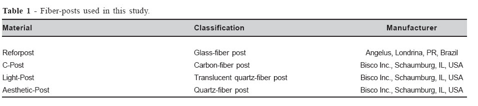
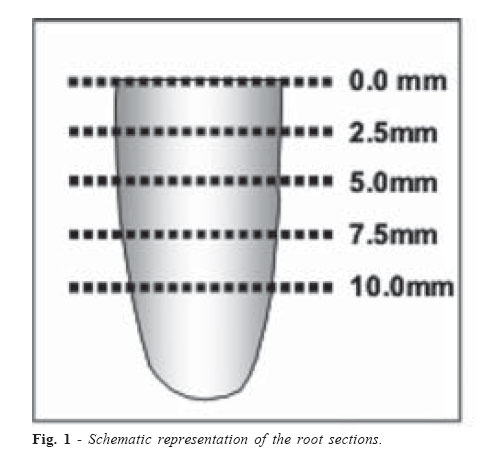
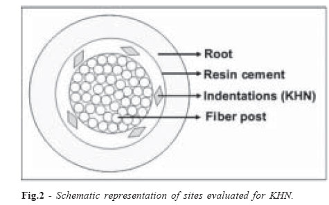
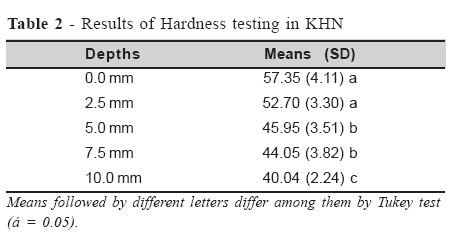
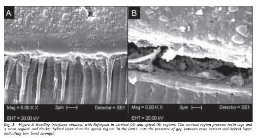
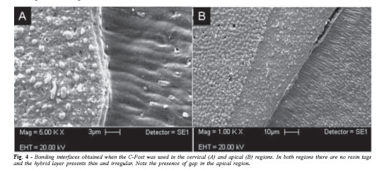
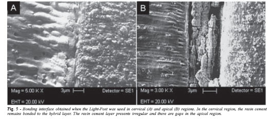
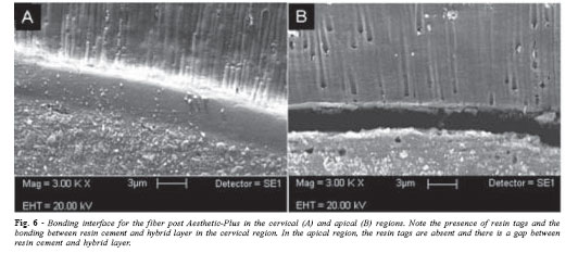
Brazilian Journal of Oral Sciences, Vol. 6, No. 21, April - June 2007, pp. 1337 - 1343 Influence of root depth and the posttype on Knoop hardness of a dualcured resin cement Murilo de Sousa Menezes1, Adeliana Garcia Veríssimo2, Rodrigo Borges Fonseca1, André Luís Faria e Silva1, Luís Roberto Marcondes Martins1, Carlos José Soares3 1Department of Restorative Dentistry, Piracicaba Dental School, State University of Campinas, Piracicaba, SP, Brazil. Correspondence to: Carlos José Soares Universidade Federal de Uberlândia Faculdade de Odontologia -Departamento de Dentística e Materiais Odontológicos Av. Pará, 1720, Bloco 2B, Sala 24, Campus Umuarama Uberlândia - Minas Gerais - CEP. 38400-902 Phone.: +55 34 3218 2255 Fax.: +55 34 3218 2279 E-mail: carlosjsoares@umuarama.ufu.br Received for publication: February 12, 2007 Code Number: os07017 Abstract Fiber posts are usually luted to the root canal with dual-cured resin cements. However, some of these cements require light-activation in order to reach optimal degree of cure. The aim of this study was to evaluate the effect of the fiber post type on microhardness of a dualcured resin cement used for luting these posts. After endodontic treatment, four fiber post types were luted to the root canal of bovine incisors with RelyX ARC. The samples were sectioned in order to obtain four slices, representing different depths (0.0; 2.5; 5.0; 7.5 and 10.0mm). Knoop microhardness testing was performed in each slice. The data were submitted to split-plot ANOVA and Tukey post-hoc tests. An additional sample per fiber post type was used to analyze bonding interface by SEM. At the depths of 0 and 2.5 mm, the resin cement presented the highest hardness values. The lowest values were observed at the 10 mm depth, and the resin cement presented intermediary hardness means at the depths of 5 mm and 7.5 mm. By means of scanning electronic microscopy, it was possible to see the more homogeneous hybrid layer in the cervical region of the root. Key Words: fiber post, Knoop hardness, polymerization depth, dual-cured resin cement IntroductionFiber posts are commonly used to restore endodontically treated teeth when their remaining coronal tissue can not provide adequate support and retention for the restoration1. The similarity of the fiber post modulus of elasticity to that of resin cement and dentin was perceived to be advantageous for producing a stress field similar to that of natural teeth and resulting in reduced root fractures2. To achieve adequate fiber post retention to canal, resin cements are routinely used in combination with adhesive systems. Resin-based luting agents provide significantly increased fiber-post retention when compared with conventional cements, such as zinc phosphate and glass ionomer3. This may be attributed to the greater compressive and bond strength of resin cements than conventional cements. However, the mechanical properties of the resin cements depend of the degree of cure4-5. Most clinicians generally use dual-cure resin cement to lute fiber-posts. Dual-cure cements were developed to conciliate the favorable characteristics of self-cured and light-cured cements6. The rationale was to have a material with extended working time, and capable of reaching a high degree of conversion either in the presence or absence of light. Nevertheless, some dual-cure resin cements are over dependent on light-activation and its absence may lead to an inadequate degree of conversion5. During post fixation, exposed marginal areas can benefit largely from both selfand light-curing, as they are readily accessible to the curing light. As light is irradiated, a significant reduction in intensity occurs due to light scattering within the resin cement and shadowing produced by both tooth structure and post7. The use of the light-transmitting translucent fiber post was reported to increase the depth of resin cure in studies that used light-cured resin composites8-9. No study has, however, evaluated the ability of the translucent fiber posts to enhance degree of conversion of dual-cured resin cements. Thus, the aim of this study was to evaluate the effect of the fiber-post type on the hardness of one dual-cure resin cement at different depths. The null hypothesis tested was that there was no difference in the resin cement Knoop hardness values when it is compared different types of fiber-posts and different root depths. Material and Methods The fiber-posts, with respective classification, shape and manufacturers, used in this study are listed in Table 1. Twenty four bovine incisors with mature apices and roots without curves were selected for this study. The teeth were cleaned with periodontal scalers and stored in a 0.2% thymol solution. The crowns were removed above the cementumenamel junction with a low-speed diamond saw under irrigation in order to obtain a 17 mm long remaining root. For the endodontic treatment, a step-back preparation technique was used with stainless-steel K-files (Malleiffer, Ballaigues, Switzerland) and Gates-Glidden drills #2 to 4 (Malleiffer, Ballaigues, Switzerland). All-enlargement procedures were followed by irrigation with 1.0 % sodium hypochlorite. The prepared root canals were obturated with gutta-percha cones using the lateral condensation technique and a sealer containing calcium hydroxide (Sealer 26, Dentsply, Petrópolis, Brazil). After endodontic treatment was completed, the samples were stored in 100% humidity for at least 72 hours. Post-holes were prepared by standardizing the length at 12 mm, leaving 5 mm of gutta-percha in the canal space after post preparation. Preparation was performed with drills specifically for each fiber-post system. After preparation, the bonding procedure was carried out as it follows: the canal walls were etched with 35% phosphoric acid (Dentsply, Petrópolis, Brazil) for 15 seconds, rinsed for 15 seconds and gently air dried. Excess water was removed from the post space with absorbent paper tips. Two coats of an one-bottle adhesive system (Single Bond, 3M-ESPE, Saint Paul, USA) were applied in the root canal with a microbrush of compatible size. Excess adhesive was removed with an absorbent paper tip (Dentsply, Petrópolis, Brazil), the remaining material was gently air dried and light-cured for 20 seconds. The fiber posts were cleaned with ethanol and treated with a silane coupling agent (Ceramic Primer, 3M ESPE, Saint Paul, USA). After completing the adhesive application, the base and catalyst pastes of the dual-cured resin cement RelyX ARC (3M ESPE, Saint Paul, USA) were mixed and the cement was inserted in the root canal with a #40 lentulo spiral. Afterwards, the post was put into the root canal with light pressure, and excess luting material was removed. Light curing was performed through the cervical portion of the root for 40 seconds from buccal and lingual surfaces with halogen light unit with 800 mW/cm2 (XL 3000, 3M ESPE, Saint Paul, USA). Five samples were made for each fiber post. The roots with their cemented posts were stored in distilled water at 37º C for 24 hours. After the storage period, the samples were fixed with sticky wax on an acrylic resin table adapted to a precision cutting machine (Isomet 1100- Buehler, Lake Bluff, IL, USA). The posts were kept parallel to the table and perpendicular to the saw blade. The roots were sectioned using a low speed diamond saw (4"x 0.12 x 0.12, Extec, Enfield, CT, USA) under constant water cooling. Five slabs with approximately 2.0 mm in thickness were obtained per root (Figure 1). The slabs were embedded in polystyrene resin cylinders to facilitate handling. The included samples were finished with # 1000, 1200 and 1500-grit SiC papers (Norton, Campinas, SP, Brazil) under water and then polished with 6, 3, 1 and 0.5 µm diamond paste (Arotec, São Paulo, SP, Brazil) using a polishing cloth (Arotec, São Paulo, SP, Brazil). Microhardness was measured by means of a Knoop indenter under 25 grams load and 30 seconds dwell time (HMV - 2000, Shimadzu, Japan). Indentations were made at five positions in each specimen (Figure 2) and the hardness readings were obtained in Knoop Hardness (KHN). Statistical analysis was performed by applying split-plot ANOVA followed by Tukey’s post-hoc test at the 95% confidence level. The factors tested in this study were the fiber post (four levels) and depths (five levels). The other four bovine incisor roots (one per fiber-post type) were used to analyze the bond interface. The fiberposts were luted and slabs were obtained by the previously described methods. After inclusion and polishing, the samples were rinsed with water, while polishing and debris and paste were ultrasonically removed for 5 minutes. The specimens were demineralized with a 18% (v/v) HCL for 5 seconds and then immersed in a 5% NaOCl solution for 10 minutes to remove the non-encapsulated collagen fibrils. After this, the samples were mounted on aluminum stubs, gold-sputter coated (MED 010, Balzer, USA) and observed by scanning electronic microscopy (LEO, Carl-Zeiss, Germany). ResultsThe mean microhardness values are shown in Table 2. Splitplot ANOVA revealed that there were statistically significant differences only for the “depths” factor (p<0.05), while the “fiber post” factor and the interaction between the two factors were not significant. Linear regression was applied in an attempt to explain the relation between the depth and microhardness values found at each depth. The correlation was significant (p = 0.0016) and the adjusted coefficient of regression was 96.23%. The Tukey’s test was applied to show differences between the depths. At the depths of 0.0 and 2.5 millimeters, the resin cement presented the highest hardness values, which were not significantly different from each other. At the depths of 5 and 7.5 millimeters, RelyX ARC presented intermediary hardness means, and showed no statistical difference between them. The lowest values were found at the 10 millimeter depth. The bonding interface analysis is shown in Figures 3, 4, 5 and 6. Discussion The results of this study lead to partial confirmation of the null hypothesis. There is no difference in the microhardness values of resin cement between the fiber posts types used in this study but irrespective of the fiber post, the hardness values significantly decreased in the apical direction. Knoop microhardness testing is an indirect method for measuring the degree of conversion of resin cements and is commonly used to evaluate the polymerization efficiency of these materials10-11. Rueggeberg and Craig12 demonstrated that there is a good correlation between the degree of conversion and Knoop microhardness. However, the degree of conversion is not the unique factor responsible for the microhardness of the resin cement. Other factors related to the polymerization process, such as crosslinked density and cyclization, can also have an influence on microhardness13. Resin cements can be classified according to their polymerization mode in self-cured, light-cured and dualcured. In this study, a dual-cured resin cement was used, which means that monomers are polymerized by means of chemical and physical activation. In the chemical activation, when the base and catalyst pastes are mixed, the tertiary amine reacts with benzoyl-peroxide and generates free radicals. These radicals then attack the aliphatic carbon double bonds of monomers and initialize the polymerization reaction14. In physical activation, camphoroquinone absorbs light in the blue region, wavelength ranged between 400 and 500 nm, and combines with a tertiary amine to form an excited state complex that breaks down into free radicals15. However, some dual-cure resin cements are overdependent on photo-activation to reach optimal hardness 5-6,16. Sigemori et al.17 evaluated the depth of cure depth of dualcured resin cement RelyX ARC in a 14 mm deep matrix, simulating the root canal. The authors found that microhardness values decreased significantly in the deeper regions. This demonstrates that chemical activation alone is unable to provide an optimal degree of conversion for some dual-cure resin cements. The results obtained in the present study confirmed this affirmation. When the light penetrates through the resin cement, the light density is attenuated as result of light absorption and scattering caused by fillers and others additives, leading to a limited polymerization depth15. Translucent fiber-posts have been developed in an attempt to enable a higher amount of light to reach the deeper regions of the root canal8. In this study the translucent quartz fiber post Light-Post was used. Thus, it was expected that resin cement would present higher microhardness values in the deeper regions with this post. However, there was no difference in the microhardness values between studied fiber posts. This demonstrated that the Light-Post was not efficient in allowing a sufficient amount of photons to reach the resin cement in the deeper regions and to increase the microhardness. Thus, only chemical activation was responsible for initializing the polymerization reaction in the deeper regions. In this study, the samples were confectioned with bovine roots, differently from the others studies that used standard matrixes8-,9,17. The fiber posts were luted with resin cement associated with the one-bottle etch-and-rinse adhesive system Single Bond. This methodology was used in order to simulate a clinical situation. Yamauchi18, demonstrated that acidic resin monomers, present in the adhesive layer, non-polymerized by oxygen inhibition, deactivate the tertiary amine that is used as catalyst to activate the polymerization reaction chemically, depriving them of their capacity to generate free radicals. However, this polymerization inhibition occurs only near the resin cementadhesive interface. In the microhardness test, this inhibition did not intervene in the measurements, since the indentations are made outside of this area. By means of scanning electronic microscopy, it was possible to see that a more homogeneous hybrid layer was found in the cervical region of the root, irrespective of the fiber post. As the adhesive system was previously light-cured no differences were expected between the hybrid layer formed when different fiber posts are used. In the apical region, the hybrid layers were irregular and there were gaps along resin cement-adhesive interface. In the deeper regions, debris removal, wet control and adhesive system application are critical because of the more difficult access19. Furthermore, the adhesive light-curing is compromised in areas further from the light source20. These factors may explain the differences found between the adhesive interfaces in the different regions of the root canal. During the fiber post cementation, the high C-factor of the prepared root canal does not allow the resin cement to flow during the resin polymerization. Therefore, the stress generated by shrinkage could impair bonding to root dentin, but debonding in the deeper regions, with formation of gaps, may not compromise fiber post retention in these areas21. However, the lower hardness in this area can reduce this retention. Goracci et al.22 demonstrated that sliding friction contributes substantially to the dislocation resistance of fiber post during push-out testing. Thus, increased mechanical properties of the resin cement may contribute to higher fiber post retention. Although the mechanical properties are reduced in the deeper regions of the root canal, an increase of fiber post length seems important in order to improve its retention. AcknowledgementsThe authors are indebted to Dr. E. W. Kitajima (NAP-MEPA/ ESALQ-USP) for the SEM technical support and FAPEMIG for financial support (Grant CDS 159/04). References
© Copyright 2007 - Piracicaba Dental School - UNICAMP São Paulo - Brazil The following images related to this document are available:Photo images[os07017f5.jpg] [os07017t2.jpg] [os07017f4.jpg] [os07017f6.jpg] [os07017f3.jpg] [os07017t1.jpg] [os07017f2.jpg] [os07017f1.jpg] |
| |||||||||
