 |
| About Bioline | All Journals | Testimonials | Membership | News |
|
||||||
|
||||||
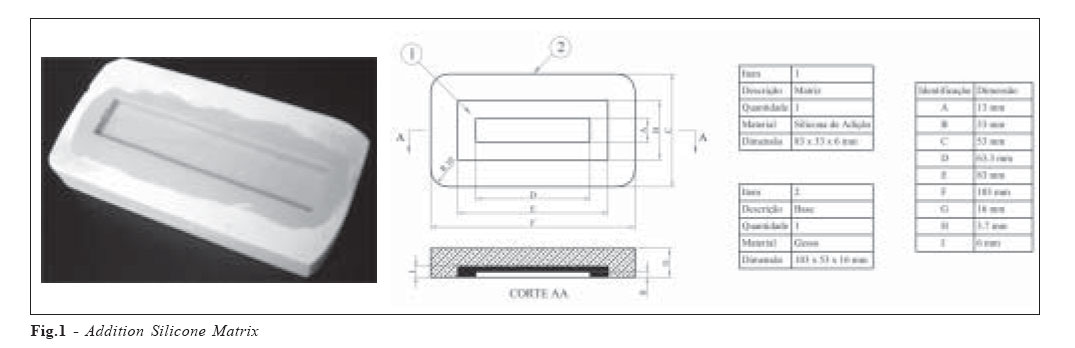
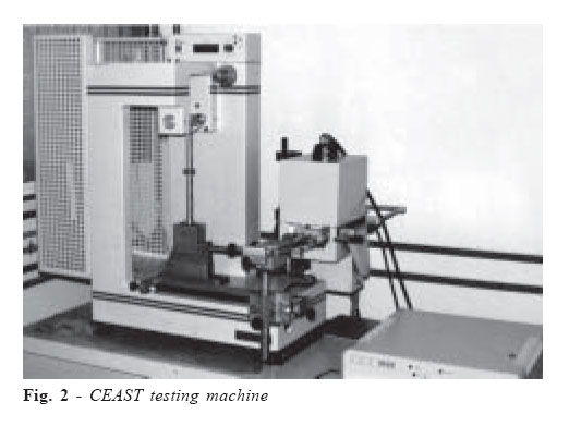
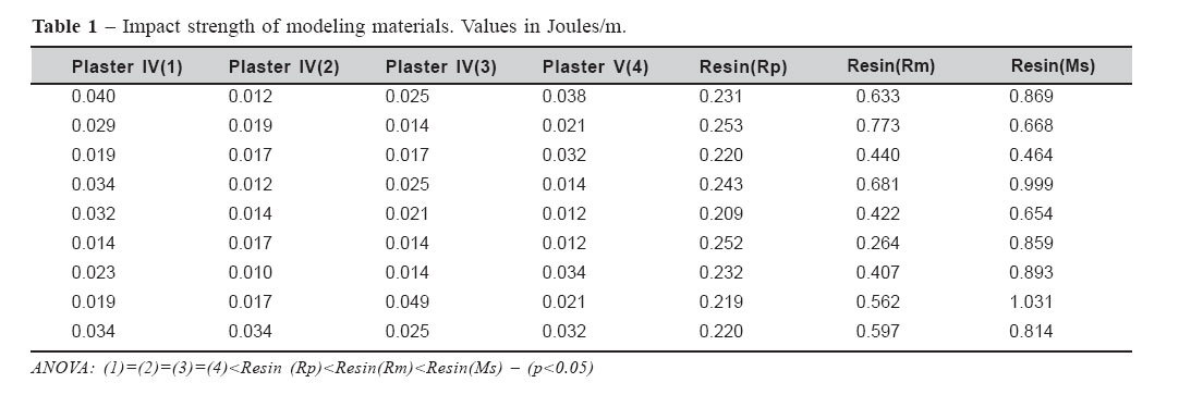
Brazilian Journal of Oral Sciences, Vol. 6, No. 21, April - June 2007, pp. 1349 - 1352 Impact fracture strength applied todental modeling materials Sérgio Candido Dias1, Marcos Ribeiro Moysés1, José Augusto M. Agnelli2, Gisseli Bertozzi Ávila3, José Carlos Rabelo Ribeiro1, Luciano José Pereira1 1 PhD, Professor Clinical Dentistry Post-Graduation Program, Vale do Rio VerdeUniversity – UNINCOR - Três Corações/MG –Brazil Correspondence to: Luciano José Pereira Universidade do Vale do Rio Verde, Três Corações, (MG), Brazil Av: Castelo Branco, 82 Bairro: Chácara das Rosas Três Corações, Minas Gerais, Brasil CEP: 37410000 Phone +55-35-3239-1276 Fax +55-35- 32352513 E-mail: lucianojosepereira@yahoo.com.br Received for publication: March 28, 2007 Code Number: os07019 Abstract The aim of the present study was to analyze the impact fracture strength using plaster and resin modeling materials. The analysis was made using nine dies having plaster III bases and internal portion of laboratory-type addition silicone. Nine test specimens were produced of each following material: type IV Durone plaster, type IV Fuji Rock plaster, type IV Rock Plus plaster, type V Durone plaster, Epoxiglass 1504 epoxy resin, epoxy resin modified with diatomite and epoxy resin modified with diatomite. The tests were performed in a Ceast testing machine. At the moment of fracture, the machine recorded a value (Energy), using a formula to obtain the impact strength value in joules per meter. Statistical and variance analyses and Student’s t test revealed that pure or silanized diatomite increased the impact fracture strength of models made of Epoxiglass 1504 epoxy resin and that silanizing the filler led to a further gain in impact fracture strength. The models made of Epoxiglass 1504 displayed greater impact fracture strength than those made of plaster types IV and V. No statistically significant differences were found among the analyzed plasters. Models prepared with epoxy resin displayed in general greater impact fracture strength than models made of plasters. Key Words: resin, plaster, dental impression materials IntroductionProsthetic rehabilitation is characterized by clinical and laboratorial phases, connected to each other by the moldmodel combination. Several steps need to be followed in order to make dental prosthesis. These involve direct and indirect treatment phases, which are potentially mistakable. Therefore, material and techniques were developed to minimize error possibility. Among all the stages concerned to indirect restorations, mold and modelling are extremely important to associate the dental office to the prosthesis laboratory1. According to Rudd et al.2, the model can be considered a real link between the oral cavity and the prosthetic professional, since it transfers to the technician all the relevant information needed to make indirect restorations. The mold techniques through history are considerably influenced by mold materials. It can be affirmed that consecutive changes are evident, despite several new materials are commercially available. The same can be stated to modelling techniques. An ideal mold material needs to reproduce dental shape and its relationship to adjacent structures. Additionally, it also needs to be sufficiently elastic in order to be removed from retentive areas and return to its original shape without distortion3. The most commonly used modeling material is dental plaster; however, due to its low resistance to abrasive wear, mechanical brittleness and dimensional instability, alternative systems for dental modeling have been proposed, among them die metallization4-5, low melt metal alloy sprays (atomization)6-7, and epoxy resin8-11 Dental models are subjected to constant handling, so they must be fracture resistant for safe laboratorial procedures. In response to this need, this study proposed to evaluate the impact fracture strength of modeling materials. Material and MethodsThe impact fracture strength was evaluated using nine dies made of laboratorial silicone with a type III plaster base with specific dimensions (Fig. 1). Nine test specimens were made of each of the modeling materials under analysis, i.e., pure Epoxiglass 1504 epoxy resin – hereinafter called (Rp); Epoxiglass 1504 epoxy resin doped with diatomite – dubbed (Rm); Epoxiglass 1504 epoxy resin doped with silanized diatomite – called (Ms); type IV Durone plaster (Dentsply) and herein identified as plaster (1); type IV Fuji Rock plaster (GC America Inc.-USA) and called plaster (2); type IV Rock Plus (Polidental) and herein identified as plaster (3); type V Durone plaster (Dentsply) and identified here as plaster (4). The Epoxiglass “1504” resin and Epoxiglass “1603” hardener (Epoxiglass), according to the manufacturer’s technical information, form a low viscosity, transparent epoxy resin system without filler material. This epoxy resin is used for finishing costume jewelry, in the production of gifts, in fusible systems, and in impregnation systems for the electrical industry in general. Once it has been handled, it has a work time of about 20 minutes, and a minimum hardening time of 6 hours. The resin and its hardener were loaded with diatomite in the proportion of 30%. Diatomite, also known as diatomaceous earth, is a very light powdery material formed through the accumulation of siliceous frustules of dead diatomaceous algae. The filler (diatomite) was superficially pretreated with silane (Silquest A187 –Crompton). The treatment consisted of the following: 50g of diatomite was placed in a glass beaker and the silane dripped onto it until the filler became completely moist, after which the mixture was homogenized, allowed to rest for 24 hours, and then oven dried. The modified resin was handled according to the manufacturer’s specifications, which call for a 35% weight ratio between the resin and the hardener, e.g., 20g of resin and 7g of hardener. The two materials were mixed manually in a slow circular movement for 5 minutes. The materials were weighed on a digital balance with a 3 Kg capacity and 0.5 g precision. The test specimens produced were arranged in a CEAST testing machine, following the aforementioned testing specifications (Fig. 2). A 4 joules pendulum was used. At the moment when fracture occurred, the device recorded a value (Energy). A formula was used to obtain the real value in joules per meter. The formula is: Impact fracture strength = (Value obtained – Standard calibration value) Thickness of the test specimen Results and DiscussionTable 1 shows the values of impact fracture strength, indicating that the plasters are less resistant to impact than the epoxy resins (p<0.001). An analysis of the values displayed by the resins indicates that the presence of diatomite (filler) increased the impact fracture strength and that silanization promoted a further gain of this strength (p<0.05). A statistical analysis of these values revealed the absence of significant statistical differences in the values shown by the plasters (1), (2), (3) and (4) (p=0.153). When analyzed separately, the resins (Rp), (Rm) and (Ms) proved to be statistically different (p<0.05). The Resin Ms group showed the best impact fracture strength, followed by the Rm and Rp respectively. The present research evaluated the impact fracture strength using plaster and resin modeling materials. Modeling materials are expected to be break resistant. The incorporation of rigid particulate filler in a polymeric matrix increases the composite’s modulus of elasticity. The filler’s effect on the impact fracture strength is complex12. The following characteristics of plasters and Epoxiglass 1504 epoxy resin employed in the preparation of dental models were analyzed previously in relation to compatibility with elastomeric molding materials, capacity to reproduce the details of the mold, superficial rugosity, and dimensional behavior. In general, the resin showed better behavior for dental modeling11. The presence of mineral filler in a polymer reduces the system’s impact resistance in comparison with virgin resin. This may be explained by the fact that inclusions in a polymeric matrix act as stress concentrators, reducing the energy required for the induction and propagation of cracks. Moreover, because inclusions differ considerably from the matrix in terms of their ductility, high modulus and little elongation, they embrittle the composite. However, rigid fillers could serve to deflect cracks and dissipate the energy associated with their growth, increasing the composite’s impact strength. This phenomenon may be enhanced by the presence of coupling agents, silanes and titanates, which possess an organic side of greater molecular weight, reinforcing the crack growth and propagation energy dissipation mechanisms13-14. It should also be noted that the filler packing fraction is a very important factor. Poorly packed fillers occupy larger volumes, contributing to larger numbers of stress concentrators or, analyzing the composite as a whole, they reduce the continuity of the matrix more effectively. Since it is the matrix that absorbs most of the impact shock, fillers with high packing fractions tend to reduce the impact strength to a much lesser extent with the same relative volume of filler. The present results showed that resins had increased impact fracture strength when compared to plaster. Moreover, when filler (diatomite) was added to resin, it presented the best results than all other materials tested. The fillers normally used are more rigid than plastics, and it is well known that they increase the hardness of the composite in relation to virgin resin13-14. Other factors that can influence the hardness of loaded systems are the degree of dispersion of the filler in the polymeric matrix; distribution of the filler; interfacial bonding, and probably many other factors. Dias11 conducted a study that revealed an increase in hardness in models prepared with epoxy resin loaded with zirconite. Analyzing the values expressed by the Epoxiglass 1504 epoxy resin loaded with diatomite, we found an increase in impact fracture strength when compared with virgin resin. This increased hardness was attributed to the fact that the diatomite’s pores were filled with resin, leading to the formation of a latticetype structure and increasing the composite’s strength. As proposed by Kats and Milewski13 and Nielsen14 silanization increases fracture resistance, a statement that we confirmed when the diatomite was treated with Silquest A187 silane used in the current study. The plasters displayed the expected brittleness due to their crystalline composition. Greater impact fracture strength was expected for the type V plaster, but the values displayed by the type IV and V plasters were very similar. Based on the results found of this research, concluded that: models prepared with Epoxiglass 1504 epoxy resin display greater impact fracture strength than models made of type IV and V plasters; the presence of pure or silanized diatomite in the Epoxiglass 1504 epoxy resin increased the impact fracture strength of the models tested here; silanization of the models made of Epoxiglass 1504 epoxy resin led to an additional gain in impact fracture strength; the plasters evaluated in this study showed similar impact fracture strength. AcknowledgementsThe authors gratefully acknowledge the financial support of CNPq (Brazil). References
© Copyright 2007 - Piracicaba Dental School - UNICAMP São Paulo - Brazil The following images related to this document are available:Photo images[os07019f1.jpg] [os07019f2.jpg] [os07019t1.jpg] |
| |||||||||
