 |
| About Bioline | All Journals | Testimonials | Membership | News |
|
||||||
|
||||||
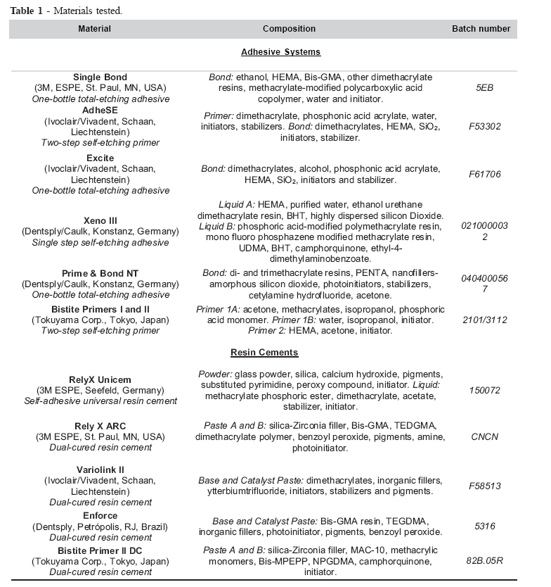
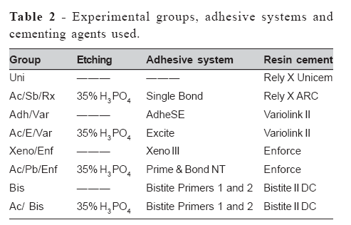
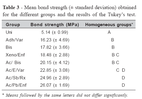
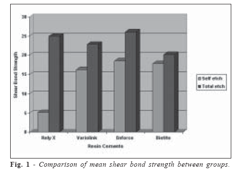
Brazilian Journal of Oral Sciences, Vol. 6, No. 22, 2007, pp. 1376-1382 Interaction between total-etch and self-etch adhesives and conventional and self-adhesive resin cements Carlos Rocha Gomes Torres2, Léia Quintanilha Pinto1, André Gilberto Leonel1, César Rogério Pucci2, Alessandra Bühler Borges2 1Academic Clinical Research Group (GAPEC),São José dos Campos School of Dentistry, SãoPaulo State University. Correspondence to: Carlos Rocha Gomes Torres Departamento de Dentística Restauradora – Faculdade de Odontologia de São José dos Campos Av. Engenheiro Francisco José Longo, 777, Jardim São Dimas, São José dos Campos, SP, Brazil, – CEP 12245-000 Phone: +55 12 3947 9048 Fax : +55 12 3947 9077 E-mail: carlosrgt@fosjc.unesp.br Received for publication: March 26, 2007 Accepted: September 20, 2007 Code Number: os07025 Abstract The aim of this study was to compare the bond strength to enamel between resin cements combined with total-etch and self-etch adhesive systems and a self-adhesive cement. Eighty bovine incisors had their buccal surface ground flat exposing a plane area in the enamel. Eighty Artglass resin cylinders measuring 3 mm in diameter and 4 mm in height were fabricated. The teeth were divided into eight groups of 10 teeth each and the resin cylinders were cemented with different adhesive systems and resin cements: G1: RelyX Unicem (self-adhesive cement); G2: H3PO4 + Single Bond + RelyX ARC; G3: AdheSE + Variolink II; G4: H3PO4 + Excite + Variolink II; G5: Xeno III + Enforce; G6: H3PO4 + Prime&Bond NT + Enforce; G7: Bistite Primers 1 and 2 + Bistite II DC; G8: H3PO4 + Bistite Primers 1 and 2 + Bistite II DC. After application of the adhesives, the cylinders were cemented according to manufacturer instructions. The specimens were submitted to 2000 thermal cycles at a temperature ranging from 5±5°C to 55±5°C, and shear bond strength was then tested at a velocity of 1 mm/min. The data were analyzed by ANOVA and the Tukey’s test (á=5%), obtaining a p value of 0.00. The following mean (±standard deviation) bond strength values were observed for each group: G1: 5.14(±0.99)a; G3: 16.23(±4.69)b; G7: 17.82(±3.66)b; G5: 18.48(±2.88)bc; G8: 20.15(±4.12)bc; G4: 22.85(±3.08)cd; G2: 24.96(±2.89)d; G6: 26.07(±1.69)d. Groups followed by the same letters did not differ significantly. For most of the resin cements tested, the application of adhesive systems using acid etching resulted in a higher bond strength when compared to the self-etch adhesive systems and to the selfadhesive cement. Key Words: cementation, shear strength, dental enamel Introduction Cementing agents are of vital importance during the process of indirect restoration of a tooth. In addition to providing a bond strength sufficient to maintain the restoration in position even at high masticatory loads, cements should present physicochemical properties similar to the tooth structure, biocompatibility, resistance to solubility, and a color similar to that of the substrate, among others1-2. In this context, the properties of adhesive cements most closely resemble these ideal characteristics2. However, the wide variety of types and commercial brands of resin cements makes the choice of material difficult for the dentist1. An extremely relevant factor for the success of cementation is the type of adhesive used together with the cement because the former is responsible for the bonding between the tooth structure and the cement itself3. The so-called totaletch adhesive systems require previous acid etching of the enamel and dentin surfaces, which leads to the formation of pores and permits mechanical imbrication to the enamel, and exposition of collagen fiber network of dentin, resulting in the formation of hybrid layer. However, this technique consists of several clinical steps that have to be rigorously followed, a fact that makes this method complex and time consuming4. In addition, the possibility of overetching the dentin and the difficulty in clinically determining the ideal moisture point of the etched substrate may result in the presence of dentinal collagen exposed by the acid but not impregnated by the adhesive, affecting bond durability5. Self-etch adhesives contain acid monomers that etch the substrate and simultaneously fill the pores formed. In addition to the advantage of requiring a smaller number of clinical steps, which reduces the working time and speeds up the cementation process of indirect restorations, these agents eliminate the need for the presence of unprotected dentinal collagen since etching and impregnation of the substrate occur at the same time6. Furthermore, the mild etching provided by these agents might be more favorable since it does not completely remove the smear plugs present in the tubules, thus preventing an increase in dentinal permeability and reducing the chance of postoperative sensitivity due to inadequate sealing of the tubules7. Since no washing or drying of the cavity is required, excessive dehydration of the dentin is also avoided, thus eliminating some factors that may cause pulp irritation8. However, some studies have reported a greater difficulty in enamel etching with this type of adhesive system which contains weak acids, producing less effective demineralization of the enamel prisms than that obtained with phosphoric acid employed together with total-etch adhesives9-11. Thus, the enamel is the most critical dental substrate when considering the use of these materials9, and doubts exist regarding the best bonding strategy to be employed in the cementation of indirect restorations in which a margin of enamel to be etched should be preferentially present. Furthermore, in certain situations such as cementation of veneers, the preparation might be completely in enamel and unsatisfactory bonding to this substrate may result in treatment failure. In order to simplify even further the cementation process, some brands of self-adhesive resin cements have appeared on the market, consisting of monomer able to etch and bond to dental surface without the need for separate application of an adhesive system. According to Piwowarczyc et al.2, the use of this materials simplifies the bonding between the tooth structure and the indirect restoration, also reducing cement line thickness and the clinical time spent. However, doubts remain regarding the bonding effectiveness of these agents to substrates with variable degrees of mineralization. Based on the above considerations, the objective of the present study was to evaluate the null hypothesis that the type of adhesive system used, i.e., total-etch or self-etch, does not influence the shear bond strength of different resin cements to enamel. Material and MethodsPreparation of the specimens Eighty healthy bovine incisors extracted immediately after slaughter, cleaned with a scalpel blade and polished with a mixture of pumice and water were used. The roots were sectioned in the cervical third with a carborundum disk and discarded. The dental pulp was extirpated and the pulp chambers were irrigated with distilled water to eliminate residues. After this procedure, the samples were stored in distilled water and stored in a freezer at -18oC until the time for use12. After drying the pulp chambers with suction cannulae, the opening of the root canals was sealed with wax to avoid penetration of the resin used for embedding. The teeth were placed inside silicone matrices and embedded in self-curing acrylic resin (Jet, Clássico, São Paulo, SP, Brazil). Next, the buccal surface of the specimens was ground flat with a plaster cutter (Kohlbach, Jaraguá do Sul, SC, Brazil) under water refrigeration for exposure of a plane enamel surface measuring 4 mm in diameter. The surface texture and smear layer were standardized using 600 grit water sandpapers coupled to a circular polisher (DP-10, Panambra, São Paulo, SP, Brazil) applied for 1 min13. An antiadherent adhesive tape with a 3mm hole was fixed above the exposed enamel region to delimit the cementation area. Preparation of the resin cylinders Using a Teflon matrix, resin cylinders measuring 3 mm in diameter and 4 mm in height were fabricated using Artglass composite resin for laboratory use (Heraeus Kulzer, Wehrheim, Germany), color A2, cured by the incremental technique according to manufacturer instructions in a UniXS curing unit (Heraeus Kulzer) equipped with a stroboscopic light source. After this procedure, the cylinder bases were sandblasted with aluminum oxide particles (average size of 50 µm) using a Microetcher (Danville, San Ramon, CA, USA) in order to facilitate micromechanical retention. These surfaces then received a layer of C&B Liquid bonding agent (Heraeus Kulzer) applied for 5s and dried with a mild air stream for 5s. Groups and cementation The specimens were divided into eight groups of 10 teeth each and the resin cylinders were cemented with a combination of different total-etch or self-etch adhesives and resin cements according to manufacturer instructions. The composition of the materials used in the present study is shown in Table 1. Table 2 lists the different experimental groups. Each cement tested was combined with a total-etch or self-etch adhesive, except for the RelyX Unicem cement which did not receive any type of adhesive. The adhesive systems were used as recommended by the manufacturers and the cements were applied in their dualcured form to the base of the resin cylinders which were then carefully placed above the delimited area on the tooth surface. All samples were submitted to a constant pressure of 150 g for standardization of the cement layer thickness using a flat tip attached to a parallelometer (BioArt, São Carlos, SP, Brazil). After removal of excess cement with a brush, the specimens were light-cured twice for 40 s in diametrally opposite positions with an Optilight 600 lightcuring unit (Gnatus, Ribeirão Preto, SP, Brazil) with a power density of 600 mW/cm2. Next, a layer of glycerin gel (Oxiguard, Kuraray, Osaka, Japan) was applied to the cementation interface to permit better chemical curing and was removed after 3 min. Thermocycling and shear bond strength evaluation After cementation, the specimens were stored in distilled water at 37oC for 24 hours. Next, the specimens were submitted to 2000 thermal cycles at a temperature ranging from 5 to 55oC, with immersion in each bath for 30 s, using a thermocycling apparatus (Ética Equipamentos Científicos, São Paulo, SP, Brazil). Shear bond strength was evaluated in a universal testing machine (DL-1000, EMIC, São José dos Pinhais, PR, Brazil) equipped with a load cell of 50 kg at a velocity of 1 mm/min. Statistical analysis The results of the shear bond test were analyzed statistically by one-way analysis of variance (ANOVA), followed by the Tukey’s test. The level of significance was set at 5% for all analyses. The calculations were performed with the Statistica for Windows software (StatSoft, Tulsa, OK, USA). ResultsANOVA yielded a value of p = 0.000 (F=41.96 for 7 degrees of freedom) which permitted rejection of the null hypothesis, indicating the existence of significant differences between groups. Table 3 shows the mean shear bond strengths (in MPa) obtained for the different groups, as well as the results of the Tukey’s test where groups showing a similar performance were grouped together. Fig. 1 shows the comparison of mean values between the different groups. Analysis of Table 3 and Fig. 1 shows that for the Enforce and Variolink cements the combination with the total-etch adhesive system resulted in significantly higher mean enamel bond strength values compared to the use of self-etch adhesives. Regarding the Bistite cement, although previous acid etching had numerically increased mean bond strength, this increase was not statistically significant. The selfadhesive RelyX Unicem cement showed a significantly lower mean bond strength than the RelyX ARC cement which used a total-etch adhesive system. In addition, no significant differences were observed between the Variolink, RelyX ARC and Enforce cements when acid etching was employed (Table 3). Using the self-etch adhesives, no significant differences were observed between the Adhese/Variolink, Bistite and Xeno/Enforce groups. DiscussionIn view of the difficulty in obtaining recently extracted human teeth in sufficient numbers, bovine teeth were chosen in the present study as substitutes. Some studies have suggested that bovine teeth can be used as adequate substitutes in adhesion testing14-15. However, morphological differences such as the higher porosity of bovine enamel need to be taken into account when extrapolating the results to a clinical situation16-17. Regarding the type of adhesive tested, comparison of the two groups cemented with Variolink II revealed superiority of the combination of this cement with the Excite total-etch adhesive system compared to the AdheSE self-etch adhesive from the same manufacturer (Table 3). The same situation was observed for the Enforce resin cement combined with the Prime & Bond NT total-etch adhesive when compared to the Xeno III self-etch adhesive also from the same manufacturer (Table 3). The results of the present study agree with other investigations that also reported higher enamel bond strengths when adhesive cements were combined with total-etch adhesives compared to the use of self-etch adhesives4,8,18. One of the hypotheses to explain these results is the lower etching capacity observed for the self-etch systems. It is known that the bond strength to enamel is directly related to the demineralization capacity of the etching agents19-20. This is due to the fact that adhesion to the substrate occurs by micromechanical imbrication of the cured adhesive in the pores formed in the etched enamel, which is directly related to the extent of the contact area between the substrate and adhesive21. Studies have shown that in enamel etched by 37% phosphoric acid the adhesive system penetrates to a depth of 1 µm, whereas in enamel etched by components of the self-etch adhesive the adhesive only reaches a depth of 0.6-0.7 µm9-10. Thus, a larger surface area for penetration of the adhesive is obtained after etching of the enamel with phosphoric acid compared to that obtained by the action of acidic components present in the self-etch systems22. To simulate the behavior of these materials in vivo, where they are subjected to hydrolytic degradation and contraction and expansion forces caused by the different temperatures of the ingested foods, all experimental groups were submitted to thermal cycling before the shear bond strength test. Differences in the coefficients of thermal expansion between dental tissues and the restorative material can induce mechanical stresses at the bonded interface, which result in crack propagation and amplification of interfacial defects23-24. According to De Munck et al.8, small penetration of the adhesive into the enamel leads to the formation of short resin tags which seem to be unable to withstand the variations in contraction and expansion of the material induced by thermal cycling, causing adhesive failures at the toothadhesive interface. Furthermore, acid etching prior to application of the selfetch adhesive system accompanying the Bistite cement yielded higher mean shear bond strengths, although this difference was not significant when compared to samples of the group cemented with the same material but using only the etching capacity of the primers provided by the manufacturer (Table 3 and Fig. 1). These results seem to support the hypothesis that the lower bond strength of cements combined with self-etch adhesive systems is due to their lower acid etching efficacy of dental enamel. De Munck et al.25 and Van Landuyt et al.26 compared the use of self-etch adhesive systems with or without prior selective phosphoric acid etching of enamel and observed an increased bond strength as a result of the greater penetration of the adhesive when combined with acid etching. Another factor that might explain the difference in shear bond strength observed in the present study when comparing the groups cemented with the same agent but using different types of adhesive systems is the discovery that self-etch systems may present a surface layer that contains uncured acid monomers after light-curing due to inhibition caused by oxygen27-28. These acid monomers seem to attack amines responsible for the initiation of composite curing, including resin cements, especially in cases of prolonged contact of the uncured composite with this layer, causing inadequate polymerization at this interface and creating an area susceptible to crack propagation. This unfavorable surface is called “acid inhibition layer”. Some studies confirm this observation and report that the application of a layer of hydrophobic monomers as a bond component of fourth generation adhesive systems to the self-etch adhesive layer would avoid direct contact of the acid monomers with the initiator components of the composite, thus preventing interference of the acids with chemical or dual (chemical and light) curing29-30. Tay et al.31 reported that one-step self-etch adhesives may act as permeable membranes after polymerization because of the high concentration of hydrophilic monomers and the lack of application of a layer of hydrophobic adhesive. This increased permeability permits the diffusion of water present in the dentinal tubules through the adhesive and the formation of water droplets at the adhesive-composite interface, creating a fragility zone in this area. However, in the present study this effect was probably not significant since no pulp pressure was observed in the teeth studied, with no significant difference being detected between onestep and two-step self-etch adhesives (Table 3). The self-adhesive RelyX Unicem cement was released on the dentistry market with the promise to reduce as much as possible the number of surgical steps necessary for cementation of an indirect restoration. The cement consists of a phosphorylated methacrylate monomer which, due to its acid nature, demineralizes the dental surface, thus permitting penetration of the cement. According to the manufacturer, similar to glass ionomer cements, an acid-base reaction occurs involving alkaline components of the material itself, which neutralizes the acid compounds after the beginning of the curing process and also leads to the release of fluorides into the tooth structure. The proposal of this cement is to combine the use of a simplified technique, which eliminates various surface pretreatment steps, and effective mechanical properties, in addition to the flexibility that dual curing offers to daily clinical practice 2,32. In the present study, however, the group cemented with RelyX Unicem presented a significantly lower bond strength compared to the other materials used (Table 3 and Fig. 1). These findings agree with those reported in other studies showing lower bond strengths of this material when compared to other resin cements available on the market, such as Variolink II and Panavia F (Kuraray, Osaka, Japan), irrespective of the adhesive system employed25,33-35. We believe that the low bond strength values observed for this self-adhesive cement might be due to insufficient etching of the enamel surface (Table 3). De Munck et al.25 compared the bonding effectiveness of this material to the tooth structure with and without prior application of phosphoric acid and observed a significant increase in bond strength with prior acid etching of enamel, in agreement with the above hypothesis. These inferior results obtained with the self-adhesive cement may also be attributed, in addition to its low etching effectiveness, to the high viscosity of the cement25. The latter causes poor penetration of the material into the enamel prisms, which results in low resistance to hydrolytic degradation and to the contraction and expansion forces caused by the different temperatures of the ingested foods, leading to long-term adhesive failures at the tooth-cement interface11. Abo-Hamar et al.33 compared the bonding performance of self-adhesive RelyX Unicem cement to enamel before and after thermal cycling and observed that the bond strength of the cement significantly decreased after cycling. The authors suggested that the self-adhesive cement should only be used for cementation of indirect restorations exclusively in dentin or when only a small amount of enamel is left. Comparison between the self-adhesive RelyX Unicem resin cement and the RelyX ARC resin cement of the same brand, combined with the Single Bond total-etch adhesive cement, showed a significantly lower bond strength to enamel for the group cemented with RelyX Unicem (Table 3). Therefore, although a longer clinical time is required, the use of cement combined with total-etch adhesive yielded a higher bonding effectiveness. In summary, despite the availability of a new set of materials on the market, resin cements combined with total-etch adhesives continue to offer better results in clinical practice because of their higher bonding effectiveness to the dental surface, although these materials require a longer clinical time and cause more aggressive etching of the dentin 4,8. The measurement of bond strength permitted the observation that the type of adhesive system used significantly influenced shear bond strength. The total-etch systems provided higher bond strengths than the self-etch adhesives when combined with dual-cured resin cements. The selfadhesive cement tested presented significantly lower bond strength than the other combinations of adhesive systems and resin cements studied. References
© Copyright 2007 - Piracicaba Dental School - UNICAMP São Paulo - Brazil The following images related to this document are available:Photo images[os07025t1.jpg] [os07025t2.jpg] [os07025f1.jpg] [os07025t3.jpg] |
| |||||||||
