 |
| About Bioline | All Journals | Testimonials | Membership | News |
|
||||||
|
||||||
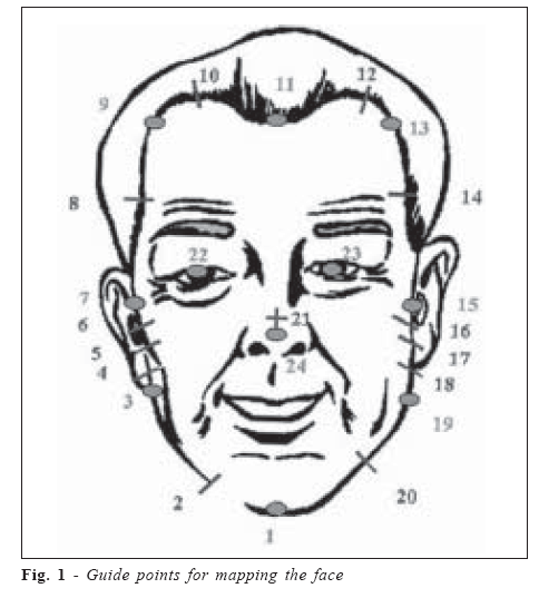
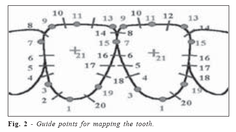
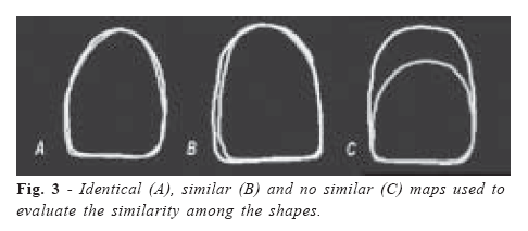
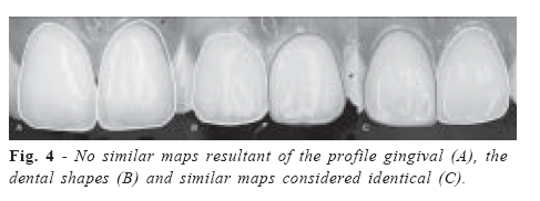
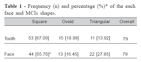
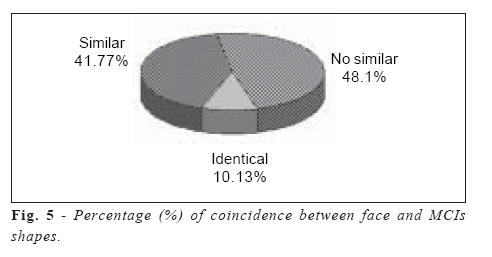
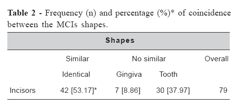
Brazilian Journal of Oral Sciences, Vol. 6, No. 22, 2007, pp. 1383-1386 Digitized study of the correlation between the face and tooth shapes in young adult individuals Frederico Augusto Peixoto Silva 1* Norma Lúcia Freitas de Almeida 2 Daniel Filgueiras Ferreira 3** Marcelo Ferraz Mesquita 4** Wagner Araújo de Negreiros 5** 1DDS, MS, PhD. Adjuntc Professor. Received for publication: June 20, 2007 Accepted: August 15, 2007 Correspondence to: Daniel Filgueiras Ferreira University of Campinas - School of Dentistry of Piracicaba Department of Prosthodontics and Periodontology Av. Limeira 901, Zip code: 13414903 - Piracicaba - Brazil Phone: 55 - 19 - 3433-7712 E-mail: danielfilgueiras@ig.com.br Code Number: os07026 Abstract This study evaluated the clinical validity of the “Williams’s law of harmony” to select artificial teeth in complete dentures, in which there is a similarity between the face and maxillary central incisor (MCIs) shapes. Two photographs were taken (face and MCIs) from 79 students aged 18-25 years, and digitized using a Genius HR-7 scanner. Face and teeth tracings were performed from the photographs using a Easy Digitizer 1.2 software. A non-parametric Chi Square test was applied in the statistical analysis (p<0.05). Results showed a predominance of the square shape (55.70%) in relation to the triangular (27.85%) (p<0.01), and the ovoid (16.45%) (p<0.001) ones. Considering the MCIs shape, the square (67.09%) predominated in relation to the ovoid (18.99%) (p=0), and the triangular (13.92%.) (p=0) ones. The analysis among the shapes showed a lower proportion of identical (10.13%) when compared with no similar (48.10%) (p=0), and similar (41,77%) (p<0.001) ones. Authors concluded that the “Williams’ law of harmony” was not confirmed in the majority of the individuals, and further studies are necessary to actually define which theory satisfies the clinical requirements. The clinical experience and critical sense seem to be more important for a satisfactory esthetic result. Key Words: denture, complete, teeth, artificial, esthetics, dental, criteria, selection, forms Introduction The replacement of missing teeth has always been a concern of the dentists, especially due to the esthetic features. Historical data revealed the use of animal and human teeth by Phoenicians and Egyptians in 2500 BC1. The art of selecting artificial teeth has high impact in dentistry because it restores the facial harmony, enhances the beauty, and brings psychological comfort to the patients. Many theories were developed regarding to objectively selecting artificial teeth, aiming to simplify the process of choice, and make it more suitable. Among these theories, the “Theory of Temperaments”2 is based on the relation between the teeth shape and the patient’s temperament; in the “Dentogenics” concept of Frush and Fisher3, the selection of artificial teeth was related to the patient’s sex, personality, and age; the nasal width as a guide for the selection of maxillary denture anterior teeth was also previously employed4-5; the “Williams’ law of harmony”6 says that there was harmony between the face and the maxillary central incisor shapes, classifying the teeth into square, triangular, and ovoid shapes. The Williams’ theory obtained great acceptance and has been used for manufacturing artificial teeth, being currently considered as a standard. In spite of the importance of selecting artificial teeth, this procedure has been neglected by dentists that do not correctly use the manufacturers’ folders, guides, and scales7. Therefore dentists usually delegate this responsibility to the laboratory technicians that have no information about the patients’ physical characteristics. Considering the importance of knowing the technical principles to select artificial teeth and the lack of a consensus in the literature about the efficacy of the Williams’ theory, this study has the aim to evaluate the predominant shapes of the face and maxillary central incisor, the similarity between the face and MCIs shapes, and the relation between the predominant facial shape and the shape of the MCIs. Material and MethodsThis research was approved by the ethical committee of the Tiradentes University of Aracaju (Sergipe, Brazil) and a written consent was obtained from all subjects. Seventy-nine students (26 men and 53 women, aged 18-25 years) from that University were selected according to the following criteria: for the MCIs, it was observed the absence of restorations, prosthetic crowns, accentuated cervical wear, wear caused by abrasion, attrition or erosion, gingival retraction and hyperplasia, and the use of orthodontic appliance; for the face, it was observed the absence of accentuated deviation of the middle line, accentuated facial asymmetry, excessive body weight, and negative history of trauma. The volunteers were seated on a dental chair with the trunk perpendicular and the Camper’s plane parallel to the ground. Two photographs were taken from each subject, one of the face and the other of the MCIs. The camera used was a Canon EOS Elan II-E (Canon Inc, Tokyo, Japan) with a 100 mm macro objective lens Canon (Canon Inc, Tokyo, Japan) and a circular flash Canon MS3 (Canon Inc, Tokyo, Japan). This set was supported on a Tron VPT-30 tripod (BMA Inc, São Paulo, Brazil). The objective camera lens was placed parallel to the long axis of the face to avoid image distortions8-9. The focal distance of all photographs was also standardized, being the focus fixed at the beginning of the procedure, and permitting that all photographs had a fixed distance between the Kodak Pró-image ASA 100 film (Eastman Kodak Company Kodak, Rochester, NY, USA) and the face. The photographs were digitized using the Digitization Software Easy Digitizer 1.2 (DSED) (Black Sea Technology Transfer Center, Trabzon, Turkey) and were scanned using a Genius HR7 table scanner (KYE Systems Corp, Taipei, Taiwan). The concept of apparent face8 used to trace the face followed a standard of points10, serving to digitize linear and angular measurements. However, the 21, 22, 23, and 24 points (Figure 1) were excluded of this analysis. The faces were classified into three basic forms, according to pre-defined standards10-12. Triangular face, with edges converging from the condylar point to the chin, taking the zygomatic arch and the angle of the jaw as references; square face, with the sides almost parallel each other; ovoid face, in which the surfaces are rounded, presenting a double curvature at the margin of the chin, and the most prominent point being below the zygomatic arch. The concept10 used to determine the points of the MCIs served to digitize the linear and angular measurements of the teeth, however, for this analysis, the 21 point was excluded (Figure 2). The teeth were classified into three basic shapes according to the adopted concept2. The classification13 had the following characteristics: square shape, with proximal walls parallel one another; triangular shape, with proximal walls converging from the incisal border to the root; and ovoid shape, with the greatest mesio-distal diameter being in center of the tooth. The similarity between the face and MCIs shapes was verified by superimposing the exported tracings from the DSED to the Power Point of Microsoft Office XP program (PPP) (Microsoft Corp, Washington, USA). The classification of the tracings8 determined: identical, when there was a perfect superposition or a negligible difference; similar, when there was a close approximation without an accurate superposition; no similar, when the shapes presented different tracings (Figure 3). The relation between the predominant facial shape and the shape of the MCIs was analyzed considering the compatibility, without attempting to verify a perfect symmetry between the shapes. The coincidence between the MCIs shapes was analyzed by exporting the tracings of the MCIs from DSED to PPP in the figure form. The shapes were classified into three groups: identical tracings; no similar tracings due to the gingival profile, and no similar tracings due to the tooth external form in height, width, or both (Figure 4). Significant differences (p<0.05) were calculated using the non-parametric Chi Square test. ResultsTable 1 shows the frequency and percentage of each face and MCIs shapes of the volunteers, presenting a predominance of square shape in relation to triangular (p<0.01) and ovoid (p<0.001) ones. For the MCIs, the square shape (67.09%) predominated in relation to the ovoid (18.99%) (p=0), and the triangular (13.92%) ones. The Figure 5 presents the percentage values resulting of the analysis between the face and MCIs shapes related to identical, similar, and no similar, showing a lower proportion of coincident shapes in relation to no similar (p=0) and similar (p<0.001) ones. The result of the assessment of coincidence between the shapes of the MCIs presented in Table 2 showed statistical differences only in the analysis of no similarity between the shape of the tooth and the gingival profile (p<0.001). The compatibility between the predominant facial shape and the shape of the MCIs was observed in 44 volunteers, corresponding to 55.70% of the sample, but with no statistical significance difference. DiscussionThe results demonstrated in Table 1 are not consistent with those reported by Wright14, in which the square shape was observed in 7% of the sample, ovoid in 11%, and triangular in 82%. DeSouza et al.15 also found different results: the triangular shape was the most frequent one (56.7%), followed by the square (35.1%), and the ovoid shape (8.1%). Considering the MCIs, this study confirmed the results of Wright 14 that observed square shape in 55% of the sample, followed by the triangular (31%), and the ovoid (8%). Kawachi16 also found similar result: the square shape predominated in 42% of the sample. Conversely, DeSouza et al.15 found a triangular facial shape in 45.9% of the individuals, followed by the square (40.5%), and the ovoid shape (13.5%). In the present study, the coincident values between the face and MCIs shapes were close to those recorded by Mavroskoufis and Ritchie8, that verified the aspect no similar in 68.7% of the sample, similar in 25.6%, and identical in 5.7%. Results are also consistent with those of Sellen et al17: no similarity in 44%, similarity in 34%, and coincidence in 22%. This study agrees with a previous one7 that related no relation between the shape of the face and the inverted shape of the maxillary central incisor. Conversely, the results disagree with Wright14 that found similar shapes in 64% and different in 36%, and with those published by DeSouza et al.15: a coincidence in 70.2% of the individuals. A previous study18 recorded that “no tooth selection system elaborated will automatically be perfect, as nothing can relieve the dentist of the responsibility for cultivating the esthetic sense of suitability that guarantees that he/she visualizes a harmonious combination, or detects a discordance in the relation of the face and the prosthesis”. Thus the theory advised by Williams6, in spite of the wide use, seems not to be completely reliable. Authors concluded that the “Williams’ law of harmony” to select artificial teeth had no efficiency to justify its acceptance. Further studies are necessary in this sense. Thus dentists should consider their clinical experience and critical sense in order to reaching a good esthetic result in complete dentures. AcknowledgementsFinancial support provided by FAPESB, Bahia State Agency for Research, Bahia, Brazil. References
© Copyright 2007 - Piracicaba Dental School - UNICAMP São Paulo - Brazil The following images related to this document are available:Photo images[os07026t2.jpg] [os07026f2.jpg] [os07026f4.jpg] [os07026t1.jpg] [os07026f5.jpg] [os07026f3.jpg] [os07026f1.jpg] |
| |||||||||
