 |
| About Bioline | All Journals | Testimonials | Membership | News |
|
||||||
|
||||||
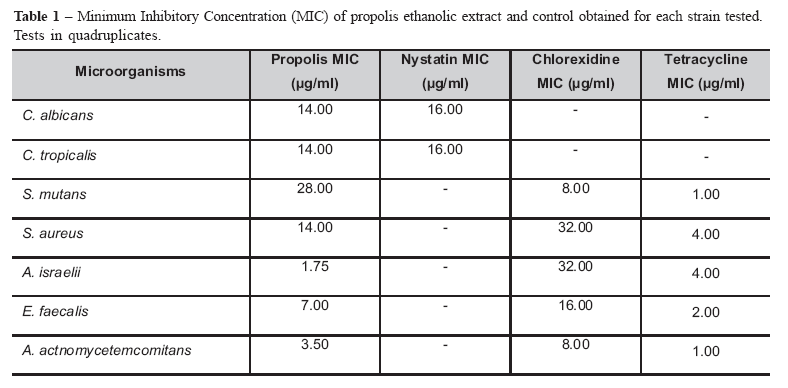
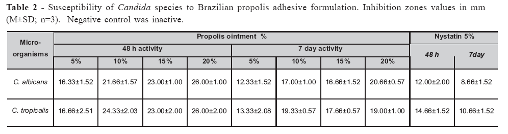
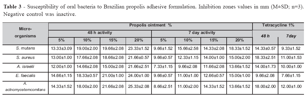
Brazilian Journal of Oral Sciences, Vol. 6, No. 22, 2007, pp. 1387-1391 Antimicrobial activity of a propolis adhesive formulation on different oral pathogens Rafael Tomaz Gomes1*Karina Imaculada Rosa Teixeira1*Maria Esperanza Cortés2*Vagner Rodrigues Santos3* 1DDS, Laboratory of Microbiology and Biomaterials 2DDS, PhD, Department of Restorative Dentistry 3DDS, PhD, Department of Oral Pathology and Surgery Correspondence to: Maria Esperanza Cortés Universidade Federal de Minas Gerais -Faculdade de Odontologia Avenida Presidente Antônio Carlos, 6627 -Pampulha - Belo Horizonte - MG CEP: 31.270-901 Phone: +55 31 3499-2470; Fax: +55 31 3499-2430 E-mail: mecortes@yahoo.com Received for publication: June 17, 2007 Accepted: September 20, 2007 Code Number: os07027 Abstract Recent studies have shown that propolis has appreciable antibacterial, antifungal and antiviral actions, as well as cytostatic and antitumoral activity. In light of these studies, the antimicrobial activity of a new adhesive formulation containing propolis was evaluated in this in vitro study. Susceptibility of the oral strains tested (Candida albicans, Candida tropicalis, Streptococcus mutans, Staphylococcus aureus, Enterococcus faecalis, Actinomyces israelli, Actinobacillus actinomycetemcomitans) was evaluated using the agar diffusion method with different concentrations of propolis (5, 10, 15 and 20%). All of the assayed bacteria and fungi species were susceptible to propolis, with minimum inhibitory concentrations (MIC) ranging from 1.75 to 14.0 μg/ml. The positive results suggest that propolis in this sustained release formulation should be further tested as an alternative therapy of infectious conditions of the oral cavity, such as denture stomatitis and periodontitis. However, in vivo studies of the effect of this new adhesive formulation of propolis are needed to determine its possible effects on the oral mucosa. Key Words: propolis, ointment, oral bacteria, oral fungi, susceptibility, MIC Introduction Propolis is a resinous material collected by bees from plant buds and exudates, which is employed for construction and repair of the honeycomb1. The name comes from the Greek ‘pro’, in front, and ‘polis’ meaning town or city, and bees use propolis to seal their hives against invasion by other insects and the weather2. The primary function of propolis in the hive is to act as a biocide, being active against invasive bacteria, fungi and even invading larvae1,3. There are a number of studies documenting the biocidal functions of propolis, its extracts and constituents. Several biological activities have been described for propolis, including antibacterial4-7, antifungal8-9, antiprotozoan10, antiviral11, antitumor12-13, immunomodulation14-15 and antiinflammatory16-17 activities, among others. The antimicrobial activity of propolis ethanolic extract has been studied by several authors; however few studies have investigated its activity towards oral pathogens5,18-20. The activities of propolis suggest its possible use in the local treatment of infectious conditions, especially in the stomatological field. For topical oral administration, the conventional formulations such as lozenges, troches, oral rinses, or mouthwashes would be the simplest dosage forms for delivery of active components through the mucosa of the oral cavity. However, these conventional dosage forms have the disadvantage of initial burst effect followed by a rapid decrease in concentration21. Successful topical treatment of oral diseases is difficult at best for reasons related to the constant flow of saliva and the mobility of the involved tissues. Buccal mucoadhesive formulations which control the drug release are expected to overcome these problems22. Despite increasing use of propolis worldwide3,23 only a few studies have been carried out to determine the inhibitory effect of propolis against some bacteria and fungi of relevance in dentistry19,24. Within this context, the aim of the present work was to evaluate the antimicrobial activity of a new adhesive formulation of propolis against seven oral pathogens. Material and MethodsMaterial Brazilian propolis was supplied by Pharmanectar (Belo Horizonte, Brazil), hydroxylpropyl methylcellulose by Aquapec (Japan), propylene glycol, ethanol and polysorbate 20 by Synth (Brazil), Brain Heart Infusion and Sabouraud dextrose broth by Biobrás (Brazil). All materials were used as received. Propolis sample The sample of propolis (reference #FO20569) from the state of Minas Gerais came from an area of mixed native and introduced vegetation near the city of Belo Horizonte, Brazil. Propolis extract solutions of 5, 10, 15, 20%, and 70% were used in this study. Propolis Adhesive Formulation To formulate the ointment, 10 ml of each solution of propolis (5, 10, 15, and 20%) was mixed with 100 ml of a solution of polysorbate 20 (0.15%) in 3.6 ml of propylene glycol (38%). The polymer hydroxymethylpropyl cellulose was dispersed in the resulting mixture (3% w/v) using mechanical stirring at 50ºC. Ethanol was evaporated in a hot plate (50ºC/24 h) under mechanical stirring. Ethanol evaporation was checked by measuring the loss in weight by mean of a laboratory scale balance. The air was removed under vacuum and the ointments were stored in an amber glass flask under refrigeration (10ºC) in order to avoid the substances decomposition. The resulting ointments were left in rest for 48 hours. Finally, the resulting product was obtained dropping 4.5 ml of sterile water at room temperature to formulate a sticky solution. Ointments containing Tetracycline 1% and Nystatin 5% were used as positive controls for bacteria and fungi, respectively. Pure ointment containing only the polymer was used as negative control. Susceptibility test Microorganisms provided from the Laboratory of Microbiology and Biomaterials at the Federal University of Minas Gerais included Candida albicans (ATCC 18814), Candida tropicalis (ATCC 20962), Streptococcus mutans (ATCC 25175), Staphylococcus aureus (ATCC 27664), Enterococcus faecalis (ATCC 4083), Actinomyces israelli (ATCC 12103), Actinobacillus actinomycetemcomitans (Y4FDC). Aliquots of frozen stocks in 20% glycerol of the different strains were inoculated in agar plates. C. albicans and C. tropicalis were grown for 2 days in Sabouraud dextrose agar. S. mutans, S. aureus and E. faecalis were cultured in Brain Heart Infusion agar for 1 day. A. israelli and A. actinomycetemcomitans were cultured in enriched Brain Heart Infusion agar for 5 days. The resultant cultures were diluted in PBS (phosphate buffer solution), to reach concentrations equivalent to Mac Farland scale nº 1. Determination of minimal inhibitory concentration (MIC) was performed by the agar dilution method and the susceptibility test to the resulting ointments containing propolis was performed by agar diffusion, both according to the National Committee of Clinical Laboratory Standard Guidelines25-26. MIC values were determined using the propolis extract (70%) in serial concentrations: 0.1, 0.2, 0.4, 0.8, 1.75, 3.5, 7.0, 14.0, 28.0 and 56.0 µl/ml. Control plates with serial concentrations of ethanolic alcohol solution were also tested. The strains were inoculated by a Steer apparatus. All tests were performed in quadruplicate. Nystatin was used as control for fungi. Chlorexidine and Tetracycline were used as controls for bacteria. C. albicans and C. tropicalis were grown in Sabouraud agar, and incubated at room temperature for 4 days. S. mutans, S. aureus and E. faecalis inoculated in Brain Heart Infusion agar were incubated aerobically at 37ºC for 2 days. Brain Heart Infusion agar enriched with hemin (0.1%) and menadione (0.01%) was used to grow strains of A. israelli and A. actinomycetemcomitans; the plates were incubated anaerobically (Gas-Pak-BBL) at 37ºC for 7 days. For the susceptibility test, sterile steel cylinders were put into the agar and filled with 150 µl of the experimental formulation. After 48 hours and 7 days of incubation at 37ºC, the diameters of the inhibition zones were measured and compared. The results were reported as Means ± Standard Deviation (M±SD). The data were submitted to analysis of variance using ANOVA test. The significance level chosen was p<0.05. Results and DiscussionMIC was determined as the lowest concentration of the propolis extract, which inhibited the growth of the tested microorganisms (Table 1). Tables 2 and 3 present the growth inhibition zones values (mm) obtained for each strain tested. All of the propolis concentrations succeeded in inhibiting the growth of the microorganisms. All of the strains grew on the control plates containing pure ointment, indicating that the formulation by itself did not exert an inhibitory effect on the microorganisms under these conditions. Among the strains tested, the most sensitive to propolis formulation was C. tropicalis, which showed the highest inhibition zones at 20% (26.00±2.00 mm). The most resistant strain was A. israelii with growth inhibition zones ranging from 12.00±1.00 mm (5%) to 21.66±2.51 mm (20%). The growth inhibition zones produced by propolis ointment (PO) 20% was statistically different of PO 5% for all microorganisms tested (p<0.05). It seems that the antimicrobial activity of propolis occurs in a dose-dependent manner. For all concentrations of propolis tested in this study, Candida strains were more susceptible to propolis than to the positive control. All the bacteria tested in this study were more susceptible to PO 20% comparing to the positive control (p<0.05). It was also observed a reduction in the growth inhibition zones after 7 days of culture. This observation suggests the growth of resistant microorganisms in the agar medium and a lasting antimicrobial effect of propolis. This fact can serve as base for studies of molecular inclusion of propolis constituents for slow release systems. Technically, ointments are semi-solid systems comprising small amounts of solid, dispersed in relatively large amounts of liquid, yet possessing more solid-like character27. Conventional ointments can also developed small levels of cross-links as a result of a gain in energy under the influence of shear forces, but this is reversible because of the involvement of weak physical forces28. Drug delivery has undergone a revolutionary advancement in the past few years. A lot of research is ongoing in various laboratories to explore stimuli-responsive ointments as drug delivery systems for better patient care29. The exploration of these polymeric networks for improved therapeutic efficacy will open newer arenas in drug delivery29-30. The development of new therapies for the treatment of the diseases of the oral cavity is of great relevance, since the systemic administration of antimicrobials has been reported to cause the development of multiresistant microorganisms, interbacterial transfer of resistance determinants, and side effects31. The propolis ointment used in this study showed good capacity of diffusion in agar; however it is important to remember that in vitro tests do not reflect the real conditions found in the oral cavity. In addition, determination of inhibition zones values depends on technical details that may vary between laboratories32. The size of the inhibition zones depends on molecular weight, negative charge, and composition, thickness and pH of the agar culture medium, which have an effect on the rate of diffusion of the antimicrobial agent in vitro33. Several studies have demonstrated the antimycotic and antibacterial effect of the ethanol extract of propolis19,24,34-37, but any of them has documented the development and the antimicrobial evaluation of a new adhesive ethanol free formulation of Brazilian propolis. In this study, all the concentrations of propolis used have been efficient in inhibiting the growth of all the microorganisms tested. The microorganisms studied in this work are of great relevance in dentistry and are involved with caries, periodontal diseases, endodontic injuries and oral mucosa lesions. Thus, the new formulation of propolis is expected to bring its therapeutic activity to the patient with greater comfort, since it not contains ethanol, which could has irritating actions on the buccal mucosa. The antimicrobial action observed in this new formulation suggests its use as an alternative adjuvant therapy for infectious conditions of the oral cavity, without causing major local or systemic adverse effects. A step further should be given to evaluate the citotoxicity of this new adhesive formulation of propolis and to verify if it will be able to be efficient against oral infections, such as denture stomatitis or periodontitis. Acknowledgements Partial support of this work by the National Foundation for Scientific Research (CNPq) is gratefully acknowledged. References
© Copyright 2007 - Piracicaba Dental School - UNICAMP São Paulo - Brazil The following images related to this document are available:Photo images[os07027t2.jpg] [os07027t1.jpg] [os07027t3.jpg] |
| |||||||||
