 |
| About Bioline | All Journals | Testimonials | Membership | News |
|
||||||
|
||||||
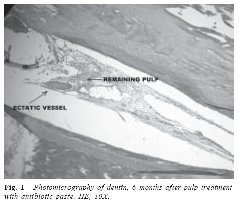
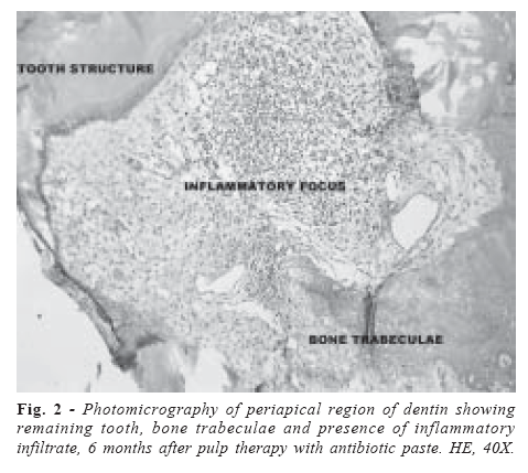
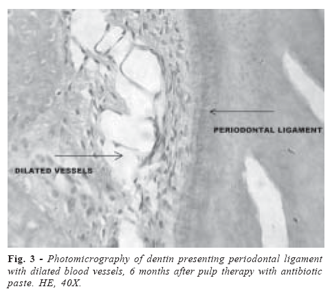
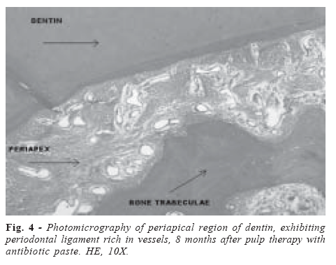
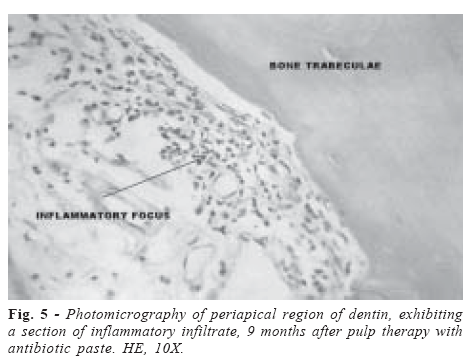
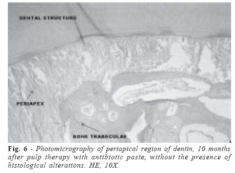
Brazilian Journal of Oral Sciences, Vol. 6, No. 22, 2007, pp. 1397-1401 Biocompatibility evaluation of an antibiotic paste after pulpotomy in dogs Glaucenira de Barros Bruno1 Ana Paula Nunes Alves1 Valdenice Aparecida Menezes2 Maria Cristina Germano Maia3 José Afonso Bruno4 Glauce Socorro de Barros Viana4 1Federal University of Ceará, School of Dentistry, Fortaleza, Brazil 2 Pernambuco State University, School of Dentistry, Recife, Brazil 3University of Fortaleza, School of Dentistry, Fortaleza, Brazil 4Faculty of Medicine of Juazeiro do Norte, Ceará, Brazil Correspondence to: Glaucenira de Barros Bruno Departamento de Clínica Odontológica FFOE/UFC Av. Senador Virgilio Távora, 233 / Apto. 702 – Meireles CEP: 60170-250 Fortaleza – Ceará Phone: (85) 3242- 2435 and (85) 99 55 9890 E-mail: glaucenirabruno@hotmail.com Received for publication: June 05, 2007 Accepted: July 30, 2007 Code Number: os07029 Abstract The purpose of this study was to evaluate by histological analysis, the biocompatibility of an antibiotic paste following treatment of pulpotomies in dogs. Four male adult dogs were used and their mandibular and maxilar premolars and molars (PM3; PM4; M1; PM4; M1) of both sides were submitted to pulpotomies. The antibiotic paste (CTZ) was prepared with chloramphenicol, tetracycline, zinc oxide and eugenol. After 6, 8, 9 and 10 months post-pulpotomy the dogs were sacrificed. Histological analysis of the teeth revealed an intense inflammatory process in the coronal pulp at 6 months after pulpotomy, with inflammatory cells dispersed throughout the entire apical region in this period. This process became partially reduced at 8 and 9 months after pulpotomy, and totally disappeared by the end of the experiment, at 10 months. In conclusion, the endodontic treatment with this antibiotic paste seemed to be biocompatible under the present experimental conditions. However, further clinical studies are necessary to assure that this paste is safe for use in pulpotomies in children. Key Words: pulp therapy, pulpotomy, antibiotic paste, biocompatibility tests Introduction The antibiotic paste (CTZ) composed of chloramphenicol, tetracycline, zinc oxide and eugenol was introduced in endodontic practice by Cappiello1, in 1964. At that time, operative proceedings were limited to cleaning the pulp chamber, followed by depositing the paste, and leaving the necrotic radicular pulp intact. The above-mentioned author used this technique as an alternative method for treating necrotic teeth with or without the presence of abscesses and/or fistulas. Although his studies showed that in treated deciduous teeth, the infectious process disappeared and masticatory functions returned to normality, few studies have been conducted since then to test the efficacy and biocompatibility of the CTZ paste. It is known that the biocompatibility of an endodontic material is determined by three methods: 1) evaluation of the material by means of a series of cytotoxicity trials in vitro; 2) subcutaneous or intra-bone implantation of the material, to observe local tissue reaction, and 3) evaluation of any reaction to the material in vivo, by pre-clinical and clinical trials in animals and humans, respectively2. The results of the in vitro cytotoxicity tests may not be completely related to the results obtained in vivo. Nevertheless, if the test material constantly induces a strong cytotoxic reaction in the tests with cell culture, one may rest assured that it will probably also exert some degree of toxicity in the living tissue3. CTZ paste has two antibiotics in its composition: chloramphenicol, which is hepatotoxic and potentially hematotoxic4-6 and tetracycline, which is hepatotoxic5-6. Biocompatibility is the main reference parameter for the study of biological properties, especially of materials that will be in contact with bone for a long period of time, since the use of an irritant material in direct contact with periradicular tissues may produce delay in the tissue repair process. In the literature, there are many studies on the biocompatibility of endodontic pastes used in the treatment of deciduous teeth. Among these, there are pastes that have calcium hydroxide7-10, iodoform10-12, a zinc-eugenol mixture8-9,12-13 and antibiotics in their composition10,14-15. It is known that the choice of an endodontic sealing material should mainly be guided by its biological properties, rather than by clinical characteristics, such as the absence of pain symptomatology or a reduction or stabilization of the periapical lesion, as shown by a radiographic image16. As very few studies have been performed to evaluate the biological properties of the antibiotic paste (CTZ) in pulp therapy10,14-15, it seems very important to evaluate it in laboratory animals through a pre-clinical trial, before it is indicated for routine use in primary teeth in children. Thus, the purpose of this experimental study was to evaluate, by histological analysis, the biocompatibility of antibiotic paste with CTZ following the treatment of pulpotomies in dogs. Material and Methods Four adult, male, mongreal dogs, weighing 15 to 20 kg, from the Zoonosis Institute in the city of Crato, State of Ceará, Northeast Brazil, were selected. All dogs were sent to the Faculty of Medicine of Juazeiro do Norte (FMJ) kennel, where the experiments took place. There, they were weighed, submitted to blood collection for hematological, biochemical and serological exams, immunized against rabies, medicated against parasites and verminosis, in order to acquire the necessary conditions for the study to be conducted. The following experimental protocol received the approval of the FMJ Research Ethics Committee. The animals were anesthetized with intravenous injection of sodium thiopental (30 mg/kg). During all surgical procedure, the animals had an isotonic saline solution with 0.9% glucose administered (iv), and when necessary, anesthesia was reinforced. After the animal was anesthetized and immobilized on a surgical table, pulp therapies were performed, beginning with isolation of the operatory field by means of rubber dams and clamps matching the teeth. The coronal opening was performed with spherical diamond tips as follows: the coronal pulp was amputated up to the level of the root canal entrance, with sharp endodontic curettes. Hemostasis was achieved by using sterile cotton pellets gently placed in the pulp chamber. The CTZ antibiotic paste was prepared according to Costa et al.14, by mixing the powder, consisting of 500 mg of tetracycline, 500 mg of chloramphenicol and 1000 mg of zinc oxide, with one drop of eugenol. After preparation, the paste was weighed and part of this material was gently placed over the pulp remainder, followed by zinc-eugenol cement filling the entire pulp chamber. Afterwards the cavity was restored with amalgam. The healthy teeth used in the present study were mandibular and maxilar premolars and molars (PM3; PM4; M1; PM4 and M1) of both sides. After the endodontic interventions, performed in 10 teeth of each animal, the dogs were sacrificed by intravenous perfusion with a 10% formol solution, at 6, 8, 9 and 10 postpulpotomy months, for posterior histopathological evaluation of the teeth. The specimens containing both teeth and adjacent bone tissue were sent to the Pathology laboratory for routine laboratorial processing. Initially, the teeth were decalcified in a solution of 10% nitric acid, with this solution being renewed every 12 hours. Decalcification time varied from 5 to 7 days, for pre-molars and molars, respectively. Afterwards, the teeth were included in paraffin, in order to obtain semi-serial cuts in the direction of the long root axis. The cuts were dyed with hematoxylin and eosin. Hematoxylin stained the cell nuclei and other acid structures blue or violet, and eosin stained the cytoplasm and the collagen pink. Sections were analyzed by optic microscopy. The following aspects were evaluated: pulp inflammatory response (present or absent) and periapical inflammatory response (present or absent). ResultsThe histopathological analysis was done in ten pulpotomized teeth of each dog, in the following periods: 6, 8, 9 and 10 months after pulpotomy. At the 6th month, the histopathological analysis revealed teeth without any alterations, but with a pulp remainder rich in congested blood vessels, hemorrhagic areas and presence of inflammatory lymph-plasmocytic cells, characterizing the development of an inflammatory process. In all the studied cases, the odontoblastic layer was preserved, and it was sometimes possible to see remainders of the applied sealing material. In the periapical region, fibrous areas with foci of mononuclear inflammatory (sometimes perivascular) cells and neo-vascularization were observed. The adjacent bone tissue consisted of normal bone trabeculae with occasional osteoclasts. The integrity of the periodontal ligament was present in all studied cases (Figures 1, 2, and 3). At the 8th month, the histopathological results showed that the inflammatory processes in the remaining dental pulp were intense, with the presence of dilated and congested blood vessels, hemorrhagic areas, and lymph-plasmocytic infiltrates in the pulp connective tissue. In some cases, a discontinuous odontoblastic layer was seen. In the periapex, fibrous areas intercalated with edema, dilated vessels, and dispersed or aggregated mononuclear cells were the major characteristics of the histopathological analyses. In some samples, a neutrophilic inflammatory infiltrate with an area of focal necrosis was found. The bone tissue consisted of preserved bone exhibiting rare osteoclasts (Figure 4). At the 9th month, the histopathological analysis revealed that the majority of dental structures presented no remaining pulp. Furthermore, the dental structures in which remaining pulp was still present, exhibited a layer of preserved odontoblasts, as well as the pulpal tissue conjunctivae with some ectatic vessels. In the periapical region, it was observed that the periodontal ligament and the alveolar bone, next to or distant from the tooth unit, presented histological signs of normality. In some cases, rare dilated blood vessels and chronic inflammatory foci were observed throughout the fibrous areas of bone trabeculae. Intense erythrocyte migration, osteoclasts and a small section of necrosis constituted occasional findings (Figure 5). At the 10th month, the remaining pulp was present as a small disorganized section in the middle of sealing material residues, however without inflammation. In the periapical region, preserved alveolar bone, large areas of fibrosis with some dilated vessels and foci of mononuclear inflammatory infiltrates, cementicles in the periodontal ligament and rare osteoclasts were observed (Figure 6). DiscussionThe main goal of biocompatibility tests is to stimulate a tissue reaction when a given endodontical material is in close contact with biological tissues. These tests are of fundamental importance in the development of new materials, reducing the incidence of adverse effects during the performance of pre-clinical, as well as the clinical assays. For instance, without the results of biocompatibility tests, pre-clinical trials would be more time consuming and costly17-19. Furthermore, it is recommended that only those materials previously submitted to biocompatibility tests in vitro and in vivo be considered for use in humans20. The endodontic treatment basically consists of the removal of septic contents from root canals, their neutralization and posterior filling with the most biocompatible material possible, due to the probability of its extravasation into the periapical region. In primary dentition, on account of the possibility of aggression to the permanent tooth germ, the technical procedure is more difficult, because of the continuous and irregular processes of root reabsorption that begins just after the end of the deciduous eruption. As a result of this lithic process, the materials applied for endodontic filling must be capable of reabsorption, and furthermore, the inflammatory reaction caused by them in the periapical region should be as light as possible, in order to respect the tissues biology. In human beings, the biological effects of pulp covering materials are evaluated primarily by the response of pulp and periapical tissues during the use of these materials. In clinical practice, the method of evaluating such response is represented by clinical signs and symptoms and by the radiographic evaluation. However, after pulp covering, the true response of pulp and periapical tissues could only be evaluated after avulsion of the tooth element in conjunction with periapical support tissues, by histopathological analyses21. This condition suggests the use of an animal model that allows the cell responses of teeth to be evaluated by means of microscopic examination, after the use of the material to be tested. Furthermore, the literature contains no preclinical screening tests accuracy for predicting human clinical responses. Data from Murray and Garcia-Godoy, 200722 demonstrated that the use of non-human primates for preclinical biocompatibility investigation provides an accurate method for evaluating clinical responses to dental materials. Studies7-13 have shown that, unlikely calcium hydroxidebased paste, the zinc oxide-based paste does not promote formation of a mineralized barrier, but induces acute and chronic inflammation and sometimes necrosis. However, its toxicity decreases between 3 and 6 months. In the present study, the histopathological analysis was initiated at the 6th month, considering that at this period all the teeth specimens presented some degree of inflammation. Since we worked with healthy teeth, the observation period was extended up to 10 months in order to evaluate the extension and duration of the lesion. In the present investigation, the histopathological analyses of dog teeth treated with the antibiotic paste CTZ showed an intense inflammatory process in the coronal pulp, at the 6th post-pulpotomy month. However, this process was partially reduced at the 8th and 9th months, and totally disappeared at the end of the experiment (10th month). These findings are in agreement with other studies23 that compared the biocompatibility of endodontic materials by subcutaneous implants in rats, finding that the materials initially induced an irritation that decreased with time. These authors also observed that the use of material whose reactions remain or increase in subsequent periods of experimental observation, is not acceptable. All these factors together suggest that the antibiotic paste CTZ has a slow but continuous action in the adjacent tissues. Similar data were observed in other biocompatibility test studies of this material in rats14 and dogs15. In the present study, the results of histopathological analyses done in dog teeth after pulpotomy with the antibiotic past CTZ, in different periods of time, demonstrated that this paste is biocompatible, thus being a promising material for use in pulp therapy. References
© Copyright 2007 - Piracicaba Dental School - UNICAMP São Paulo - Brazil The following images related to this document are available:Photo images[os07029f6.jpg] [os07029f1.jpg] [os07029f5.jpg] [os07029f4.jpg] [os07029f2.jpg] [os07029f3.jpg] |
| |||||||||
