 |
| About Bioline | All Journals | Testimonials | Membership | News |
|
||||||
|
||||||
Brazilian Journal of Oral Sciences, Vol. 6, No. 23, October-December, 2007, pp. 1462-1466 Bond strength evaluation of self-etch and total-etch adhesive systems on intact and ground human enamel Marina Di Francescantonio1 Marcelo Tavares de Oliveira2 Mirela Sanae Shinohara3 Gláucia Maria Bovi Ambrosano4 Marcelo Giannini5
1DDS, Graduate student (Master degree), Department of Restorative Dentistry 2DDS, MSc, Graduate student (Doctor degree), Department of Restorative Dentistry 3DDS, MSc, PhD, Assistant Professor, School of Dentistry, State University of Amazonas, Manaus, AM, Brazil 4 DDS, MSc, PhD, Professor, Department of Social Dentistry 5 DDS, MSc, PhD, Associate Professor, Department of Restorative Dentistry Piracicaba Dental School, State University of Campinas, Piracicaba, SP, Brazil
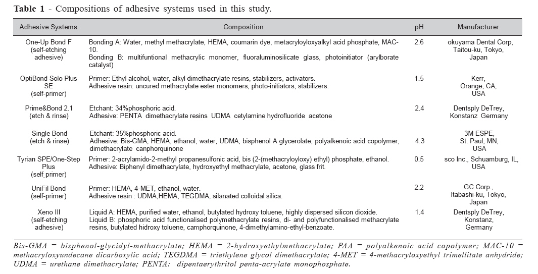
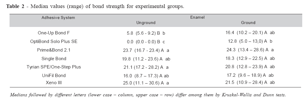
Received for publication: October 10, 2007 Accepted: December 20, 2007 Code NUmber: os07041 Abstract The aim of this study was to evaluate the bond strength of adhesive systems to intact and ground enamel. Enamel blocks from buccal and lingual surfaces of third molars were used for bonding procedures. Intact or ground (600-grit SiC paper) enamel surfaces were bonded using two “etch&rinse” adhesives (Prime&Bond 2.1 and Single Bond), three self-etching primer systems (Optibond Solo Plus SE; Tyrian SPE/One-Step Plus and UniFil Bond) or two self-etching adhesives (One-Up Bond F and Xeno III). A 6-mm composite crown was built on the bonded surfaces and samples were stored for 24 hs at 37°C. Samples were sectioned into 0.9-mm-thick slabs, each slab trimmed to a cross-sectional area of 0.8 mm2 and specimens loaded to failure at a crosshead speed of 0.5 mm/min, using a universal testing machine. Microtensile bond strength data (n=6) were analyzed using Kruskal- Wallis and Dunn tests. Differences in bond strength between intact and ground surfaces were not significant for both “etch&rinse” adhesives, Tyrian SPE/One-Step Plus, UniFil Bond and Xeno III systems. The enamel surface preparation resulted in higher bond strength for Optibond Solo Plus SE and One-Up Bond F systems. Enamel preparation using 600-grit SiC paper is unlikely to affect resin-enamel bond strengths. Keywords: dental enamel, dentin-bonding agents, tensile strength. Introduction Although phosphoric acid has been intensely used to etch the dental substrates (enamel and dentin) for bonding, selfetching adhesives are consider alternative methods to prepare the tooth for restorative procedures. Self-etching adhesive systems were developed in attempt to simplify the clinical use of dental adhesives, because they do not require separated phosphoric acid etching, water-rinsing or superficial moist controlling steps. The self-etching primers and adhesives are composed of aqueous solutions of acidic functional monomers and methacrylate components, with a pH relatively higher than that of phosphoric acid etchants1. While the adhesion to dentin produced by self-etching adhesives has been considered effective2, studies are in disagreement regarding the efficacy of conditioning and monomer infiltrations on enamel3-6. Morphological analyses of enamel surface treated with self-etching primers have showed not very demineralized surfaces and other areas that were predominantly unetched, which could impair the monomer infiltrations and hybridization process7-10. To improve the bonding of self-etching systems to enamel, it has been recommended, surface pre-treatments, increase in acidic monomer concentration, increase of etching time as well as different application methods 3,8,11-12. Moreover, the removal of superficial, aprismatic layer could facilitate the etching and the infiltration of adhesive monomers7,13-14, forming a strong and durable bonding. The purpose of this in vitro study was to evaluate the bond strength of self-etching adhesive systems and conventional systems (“etch & rinse” adhesives) to intact and ground enamel. The null hypothesis was that bond strength is not influenced by the characteristics of the enamel surface (intact and ground) regardless of the type of adhesive system used. Materials and Methods Forty-two extracted, caries-free erupted human third molars were used in this study according to protocols approved by the institutional review board of the Piracicaba School of Dentistry – University of Campinas (069/2003). Teeth were obtained by patients from 19 to 25 years old and stored in physiological saline solution with 0.1% thymol for no longer than 3 months. Tooth roots were severed and the crowns were longitudinally sectioned (mesio-distally direction) into two-halves, using a diamond blade (Isomet, Buehler Ltd., Lake Bluff, IL, USA) under water cooling. Eighty-four dental fragments were obtained (6 mm in length X 5 mm in width X 1.0 mm in thickness). A buccal or a lingual flat enamel surface of each fragment was chosen and selected for conditioning treatments and bonding procedures. All specimens were randomly assigned to fourteen groups (n = 6), according to surface treatment of enamel (grounded and intact) and type of adhesive system (selfetching and “etch & rinse” systems). The dental fragments from the grounded surface groups had their enamel surface abraded with a #600-grit SiC paper on a polishing machine (APL-4, Arotec S.A. Ind. Com., Cotia, SP, Brazil) under water cooling for 15 seconds. Seven adhesive systems: two 2-step “etch & rinse” single bottle adhesives (Single Bond and Prime&Bond 2.1), three 2step self-etching primer systems (OptiBond Solo Plus SE; Tyrian SPE/One-Step Plus and Unifil Bond) and two one-step self-etching systems (One-Up Bond F and Xeno III) were evaluated (Table 1). Adhesive systems were applied according to the manufacturer’s instructions. Resin composite build-ups were constructed incrementally on the adhesive-bonded surfaces of enamel in 3, 2-mm thick layers using a hybrid resin composite (Clearfil APX; Kuraray Medical Inc, Kurashiki, Japan). Each layer was light-cured for 40 s (670 mW/cm2) using a light-curing unit (XL 3000; 3M ESPE, St. Paul, MN, USA). After 24 hours of water storage, restored fragments were sectioned into 0.9 mm thick slabs with a diamond blade (Isomet 1000; Buehler Ltd, Lake Bluff, IL, USA). For each fragment sectioned, 2 slabs were selected and prepared for micro-tensile bond strength testing. Each slab was ground at the bonded interface and along both sides with a fine diamond rotary cutting instrument (#8835KR.314.012; Brasseler, Lemgo, Germany) under water irrigation to produce an “hour-glass” shaped specimen. This reduced the cross-sectional area of the specimen “necks” (0.7 × 0.7 mm) to approximately 0.8 mm2 for micro-tensile strength testing. Each specimen was fixed to the grips of a micro-tensile testing device with cyanoacrylate adhesive (Zapit; DVA, Corona, CA, USA) and tested in tension in a testing machine (4411, Instron Corp., Canton, MA, USA) at 0.5 mm/minute until failure. After testing, specimens were carefully removed from the device with a scalpel blade (BD, Franklin Lakes, NJ, USA). The cross-sectional area at the site of fracture was measured to the nearest 0.01 mm with a digital caliper (Starret 727-6/150; Starret, Itu, SP, Brazil) and used to calculate test results in units of stress (MPa). Means of the two specimens (“hour-glass” shaped) were calculated for each restored tooth fragment. Microtensile bond strength data were analyzed statistically by Kruskal-Wallis and Dunn tests. Results Table 2 shows the median values of tensile bond strength for the adhesive systems applied to intact and ground enamel. For Optibond Solo Plus SE and One-Up Bond F systems, the grounding of enamel surface before adhesive application improved the bond strength. When applied them on intact enamel surfaces, low bond strengths were found for One-Up Bond F and pre-testing debonding occurred for specimens restored with Optibond Solo Plus SE. For most adhesive systems (Prime&Bond 2.1, Single Bond, Tyrian SPE/One-Step Plus, UniFil Bond and Xeno III), the enamel surface preparation did not increase the bond strength. In the cases of bonding to grounded enamel, the adhesive systems produced similar bond strengths; however OptiBond Solo Plus SE had lower bond strength than Prime&Bond 2.1. Discussion Although some studies suggested to abraded the enamel surface before application of moderate or mild two-step selfetching primers or single-step, “all-in-one” adhesives7,10,13, the results of this study indicated that only two of them had theirs bond strengths increased by surface preparation with #600grit SiC paper. Thus, the null hypothesis was only partially confirmed because two self-etching adhesive systems tested were influenced by the characteristics of the enamel surface. The two etch & rinse, one-bottle adhesives (Prime&Bond 2.1 and Single Bond), which are used after conditioning with phosphoric acid, were not affect by abrading or not the enamel surface. The grounded or the unprepared enamel surfaces treated with phosphoric acids result in an etching pattern, which consist of the formation of a porous surface with exposed enamel crystallites and dissolution of both inter and intraprismatic areas. The aggressive etching effect of phosphoric acid on enamel surface overcomes the difficulty of conditioning of intact or unprepared surfaces. The formation of a deep etching pattern6,10,15 lead to similar bond strength when the adhesives are applied in both enamel surfaces. Self-etching systems contain acidic monomers based on esters from phosphoric acid, carboxylic acid or derivatives6-7,12-13. Their etching efficacy and bonding formation depends on type of acidic monomer, pH of adhesive solution, etching time and application method6,8,16. Acidic monomers are responsible for etching the dental substrates, whereas methacrylate components, such as HEMA, are available for monomer infiltration and polymerization of the bonding agent3-4,12. Selfetching systems can be classified as strong, moderate and mild, depending on their etching aggressiveness or acid dissociation constants (pKa values)2,6. In this study, it was selected a strong system (Tyrian SPE primer), two moderates (OptiBond Solo Plus SE and Xeno III) and two milds (Unifil Bond and One-Up Bond F). Studies have shown that most of the self-etching adhesives did not etch enamel as deeply as the phosphoric acid etchants did and the shallow etching pattern could compromise the bonding to enamel3,13,17. Pashley and Tay6 reported that the efficacy of self-etching primers in intact enamel does not depend solely upon their etching aggressiveness, but on monomeric composition of each material. It is also possible that the low enamel bond strengths might be caused by the high amount of unpolymerized acidic monomers remaining after curing4. Thus, no correlation among degree of primer aggressiveness, enamel etching pattern and bond strength to intact enamel has been established for self-etching adhesives18. The pH values of all self-etching systems tested were higher than phosphoric acid. In general, the demineralization effects of these systems are proportional to the acidity of the acidicprimers or self-etching adhesive solutions. The self-etching primers are less aggressive than phosphoric acid etchants, do not form a proper and defined acid etching pattern3,7,13-14 and the conditioning effects are also reduced in intact enamel surfaces, except for Tyrian SPE self-etching primer10. The Tyrian SPE primer is considered a strong self-etching adhesive with a very low pH (0.5). The etching pattern formed on both ground and intact enamel surfaces is similar to that promoted by phosphoric acid10. Moderate self-etch systems include Optibond Solo Plus SE and Xeno III adhesives with pH 1.4 and 1.5, respectively, while Unifil Bond (pH 2.2) and One-Up Bond F (pH 2.6) systems are considered mild. The bonding mechanism of these self-etching systems to enamel is based on nanoretentive interlocking between crystallites and adhesive resin18-19. These morphological features of the resin-enamel bonds are different from that formed with the etch&rinse adhesive systems18-20. This thin hybridized complex of resin formed in enamel and produced by self-etchings without the usual micrometer-size resin tags can be responsible for lower bond strengths presented by some self-etching systems and the questionable effectiveness of this type of dental adhesives on enamel surfaces9,21-22. Optibond Solo Plus SE and One-Up Bond F systems performed better on prepared enamel than on unprepared enamel. Although Optibond Solo Plus SE primer presents low pH (1.5), it did not bond to unprepared enamel, leading to pre-testing debonding in all specimens. Moreover, Shinohara et al.10 did not observed proper conditioning effects promoted by the acidic primer on intact enamel surfaces, which could compromise the adhesion to enamel, as observed in this study. However, Xeno III and Unifil Bond present similar and higher pH than Optibond Solo Plus SE, respectively, and were not affected by the mode of enamel surface preparation. Thus, other factors, such as, monomer infiltration, polymeric network formation and degree of conversion could also influence on the results of bond strength. For One-Up Bond F, SEM analysis of treated enamel surfaces with adhesive solution revealed an extremely mild etching pattern, regardless of the surface preparation10,13,15. This probably occurred due to the very mild pH (2.6) of the system, which would not provide adequate demineralization for infiltration of monomers. This pattern helps to explain the bond strength results, which were lower when compared to the ground surface. The higher bond strengths to the SiC-prepared surface can be attributed to the roughness of the surface that facilitates the activity of the self-etching adhesive to form a defined etching pattern for infiltration of adhesive resin. The removal of superficial, aprismatic layer by wet-grinding with 600-grit SiC paper improves the etching effects7,10,13. As the morphological structure and composition of the intact peripheral surface of the enamel is different from that of the middle enamel layer23, these differences can be favorable for etching effects in sub-enamel surface. Since in clinical situations the enamel is usually prepared with dental drills prior to application of the adhesive system, the concerns about effectiveness of self-etching adhesives can be reduced. In conclusion, most of the self-etching systems and the two “etch & rinse” adhesives showed the similar bond strength values for grounded or intact enamel surfaces. For Optibond Solo Plus SE and One-Up Bond F systems, the enamel surface preparation resulted in higher bond strength Enamel preparation using 600-grit SiC paper is unlikely to affect resinenamel bond strengths. Acknowledgement This study was supported by grants 01/13034-3 from FAPESP and by grants from Capes, Brazil. References
© Copyright 2007 - Piracicaba Dental School - UNICAMP São Paulo - Brazil The following images related to this document are available:Photo images[os07041t2.jpg] [os07041t1.jpg] |
| |||||||||
