 |
| About Bioline | All Journals | Testimonials | Membership | News |
|
||||||
|
||||||
Brazilian Journal Oral Sciences, Vol. 7, No. 24, Jan/Mar 2008, pp. 1493-1496 Effect of the flask contention method on the displacement of maxillary denture teeth Wagner Araújo de Negreiros1 Rafael Leonardo Xediek Consani2 Marcelo Ferraz Mesquita 2 Simonides Consani2 Thiago Assunção Valentino1 1DDS, MS. Graduate Student. 2DDS, MS, PhD. Professor Department of Prosthodontics and Periodontology, Piracicaba Dental School, State University of Campinas, Piracicaba, São Paulo, Brazil
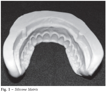
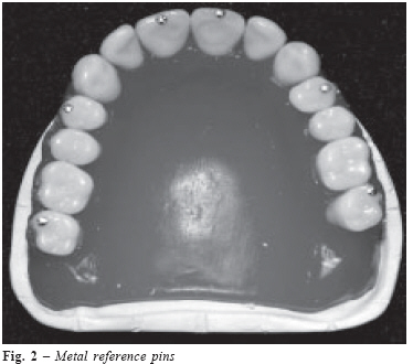
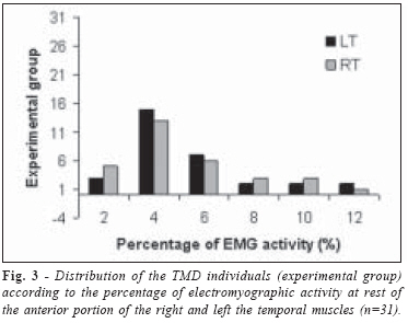
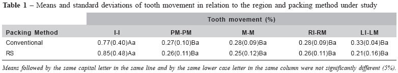
Received for publication: November 11, 2007 Accepted: February 12, 2008 Code Number: os08005 Abstract Keeping the flask closed may reduce the stress released by the resin dough after the pressing step, preserving the tooth relationship pattern. The aim of this study was to analyze the influence of the flask contention method on tooth displacement in acrylic resin complete dentures. Twenty identical maxillary complete dentures were made using the heat-cured acrylic resins Classico and QC-20, polymerized by long and fast polymerization cycles respectively, and randomly assigned to 4 test groups according to the conventional packing method and RS tension system. Transversal and anteroposterior distances between specific teeth were measured with a linear optical microscope before and after denture processing. Analysis of variance (ANOVA) and Tukey test were used to compare the groups (p<.05). Results showed that tooth movement was significantly greater for the distance RI-LI in comparison with the others, and the tooth movement was not significantly influenced by the packing method. Within the limitations of this study, the RS tension system presented a similar performance in reducing the tooth movement, when compared with the conventional packing method. The anterior region of the denture may present changes in the tooth position after processing, which need to be clinically adjusted. Key words: denture, complete; tooth, artificial; dental occlusion; polymerization; acrylic resin. Introduction A critical factor in the retention and stability of the complete denture is the dimensional change that occurs during the denture processing1-2. Previous studies have reported the importance of harmonious occlusal contacts to optimize the stability during mandibular movements and the consequent masticatory function3-5. The quality of a complete denture, however, depends on numerous processing variables that may generate base distortion and displacement of artificial teeth6-8. The combination of several factors, such as polymerization shrinkage, thermal contraction by flask cooling, and strain caused by stress release during deflasking causes undesirable dimensional changes9-10. Additional alterations may occur when the flask is transferred from the hydraulic press to the spring clamp after the final pressing step. This procedure may generate a great stress release because the resin forces the flask to open, as reported by Consani et al.11. Therefore the RS tension system (Precat, Piracicaba, SP, Brazil) has been proposed to avoid the flask opening and decrease the magnitude of the gap between base and cast. Considering the complexity of complete denture processing, the aim of this study was to compare the use of the RS tension system with the conventional packing method with regard to linear tooth displacement in acrylic resin complete dentures. Material and Methods A silicone mold (Elite Double; Zhermack, Rovigo, Italy) was formed from a metal master edentulous maxillary die without irregularities in the alveolar ridge walls. Twenty identical casts were poured from this mold with artificial type III dental stone (Herodent Soli-Hock; Vigodent, RJ, Brazil) with a ratio of 30 ml water to 100g powder. A uniform denture base was made with a 2mm thickness plate wax (Epoxiglass; Epoxiglass Chemical Products, Diadema, SP, Brazil) measured with a caliper. The height of the occlusion wax rim was 20mm in the buccal sulcus of the cast and 10mm in the second molar area. The maxillary stone cast was mounted on a Mondial 4000 semi-adjustable articulator (Bio-Art Dental Products, Sao Carlos, SP, Brazil) with the wax rim interocclusal relation according to the mandibular stone cast teeth, with the following references: intercondylar distance in M, Bennett angle at 15º, and condylar guide at 30º. The arrangement of the left anterior teeth began with the carved wax rim serving as a guide to the positions of the central and lateral incisors and canines. The same procedure was used on the right side. The posterior teeth were arranged starting with the first premolar until the second molar. The same procedure was used in the right arch. The tooth arrangement for the interocclusal relationship was anterior vertical overlap and posterior in Angle class I. After finishing the tooth arrangement of the first denture, a silicone matrix was made (Zetalabor, Zhermack, Rovigo, Italy) fitting in all buccal aspects of the denture, comprising the vestibular and incisal surfaces of anterior teeth and vestibular and occlusal surfaces of posterior teeth (Figure 1). The aim of this matrix was to guide a standardized tooth arrangement in all samples. Metal reference pins (Cadena, Coats Textil Ltda., Sao Paulo, SP, Brazil) were bonded with instantaneous adhesive (Super Bonder; Loctite, São Paulo, SP, Brazil) to the incisal border of the central incisors, buccal cusp of the first premolars, and mesiobuccal cusp of the second molars to serve as reference for quantifying the tooth movement (Figure 2). Therefore, the following linear distances were considered: I-I (incisor to incisor), PM-PM (premolar to premolar), M-M (molar to molar), RI-RM (right incisor to right molar), and LI-LM (left incisor to left molar). The distances were measured with a STM microscope (Olympus Optical Co., Tokyo, Japan), with an accuracy of 0.0005mm. The stone casts were randomly assigned to 4 test groups (n=5): Group 1: Classico acrylic resin flasking with conventional method and polymerization in water bath at 74°C for 9 hours; Group 2: Classico acrylic resin flasking with RS tension system and polymerization in water bath at 74°C for 9 hours; Group 3: QC-20 acrylic resin flasking with conventional method and polymerization in boiling water bath for 20 minutes; Group 4: QC-20 acrylic resin flasking with RS tension system and polymerization in boiling water bath for 20 minutes. The cast and wax patterns were flasked in the lower part of a traditional brass flask (Safrany; J Safrany Dental Metallurgy, Sao Paulo, SP, Brazil) with type II dental stone (Pasom; Dental Products, SP, Brazil). Petroleum jelly (Labsynth; Labsynth Chemical Products, Diadema, SP, Brazil) was used as a separating medium between the plaster in the lower part of the flask and the type III dental stone used in the upper portion. After 1 hour, the flasks were placed in boiling water to soften the baseplate wax. The flasks were separated, the wax removed, and the stone was cleaned with boiling water and liquid detergent (Ype; Chemical Amparo, Amparo, SP, Brazil). Two coats of sodium alginate (Isolak; Classico Dental Products, Sao Paulo, SP, Brazil) were used as a mold separator. Classico polymethyl methacrylate dough (Classico Dental Products, Sao Paulo, SP, Brazil) was used with a monomer:polymer ratio of 7:21 by volume. QC-20 polymethyl methacrylate dough (Dentsply, Dental Products, RJ, Brazil) was used with a monomer: polymer ratio of 10:21 by volume. Both resins were prepared in accordance with the manufacturer’s directions and each sample was processed according to its group assignment. Dentures from groups 1 and 2 were packed in conventional spring clamps after final pressing. In the RS tension system groups (3 and 4), the flasks were placed between the 2 plates of the RS tension system (modified from Getom polymerization plates). This assembly consisted of 2 iron plates, each measuring 150x40x8 mm. A 9 mm diameter screw was soldered onto each end of the lower plate; there were 2 corresponding holes with a cross-sectional diameter of 10 mm in the upper plate. During the definitive flask closure, the screws of the lower plate were fitted into the holes of the upper plate, and after applying hydraulic pressure, the screwnuts were tightly fastened onto the screws until just one stop (Figure 3). After polymerizing, the flasks were slowly cooled in a water bath and bench stored for 3 hours. After this period, the dentures were deflasked, polished, and the transverse and anteroposterior distances were measured again. Data collected in percent were transformed into arc sine of the root of x/100 and submitted to the analysis of variance (ANOVA) and Tukey test at 5% of significance. Results Table 1 shows that the tooth movement was significantly greater for the distance RI-LI in comparison with the others for the two packing methods used. It was also observed that the tooth movement was not influenced by the packing method in all regions studied. Discussion Previous studies investigated the linear magnitude of tooth displacement12-13, while others verified the direction of this tooth movement throughout the entire denture processing14-15. Understanding of this phenomenon may allow one to construct functional complete dentures that require less occlusal adjustment in the articulator and patient’s mouth16-17. Table 1 shows that tooth movement occurred in all distances evaluated, but most of them were not statistically significant (p<.05). In this study the bases were made 2mm thick, and according to previous studies18-19, this factor may reduce dimensional change in the base and the consequent alteration in tooth position. Two other aspects also need to be considered: 1) polymerization shrinkage of the resin may partly be compensated by the thermal expansion of the resin itself during the processing20; 2) the restrictive effect of investing plaster on keeping the tooth in position when the resin induces polymerization and cooling stresses21. The authors of previous studies reported that the greatest degree of base dimensional changes was observed in the denture posterior palatal seal2,22-23, and the greatest magnitude of tooth movement occurred in the posterior teeth, altering the occlusal relationship13-14. However, the present study only showed statistically significant tooth movement in the distance I-I (Table 1). This probably occurred due to technical reasons in processing: 1) the mesial space between the incisors was greater during the wax tooth arrangement, allowing their movement toward each other after pressing step; 2) the change occurred in the anteroposterior direction, increasing the transverse distance; 3) excessive polymerization shrinkage in the anterior region promoted rotation of anterior teeth, reducing the distance I-I. Therefore dentists should accurately observe the occlusal relationship of anterior teeth at the time of the clinical adjustments. A previous study11 reported that the RS tension system decreased the magnitude of the gap between the denture base and cast. In a clinical situation, this could contribute to improving the denture retention. The reduction of dimensional changes in denture bases processed by the RS tension system could occur because the flask halves remained in contact when the flask was removed from the hydraulic press. This condition may decrease the premature release of residual internal stresses from acrylic resin dough before polymerization. However, the results of the present study showed no statistically significant difference in the tooth movement between the packing methods used (p<.05). The RS tension system reduced the displacement of artificial teeth in most of distances analyzed (PM-PM, M-M, RI-RM, and LI-LM), but this difference was not significant (p<.05). It is possible that the positive action of the RS tension system in reducing the base inaccuracy was not able to preserve the tooth position in the denture14,24. The action of several other variables involved in denture construction could also explain the similar performance of the two packing methods studied. Within the limitations of this study, the RS tension system presented a similar performance in reducing the tooth movement when compared with the conventional packing method. The distance I-I (incisor to incisor) analyzed in this study presented the greatest dimensional change after processing. Acknowledgments The authors acknowledge with thanks, the financial support provided by FUNCAP (Ceara - Brazil). References
© Copyright 2008 - Piracicaba Dental School - UNICAMP São Paulo - Brazil The following images related to this document are available:Photo images[os08005f1.jpg] [os08005f2.jpg] [os08005t1.jpg] [os08005f3.jpg] |
| |||||||||
