 |
| About Bioline | All Journals | Testimonials | Membership | News |
|
||||||
|
||||||
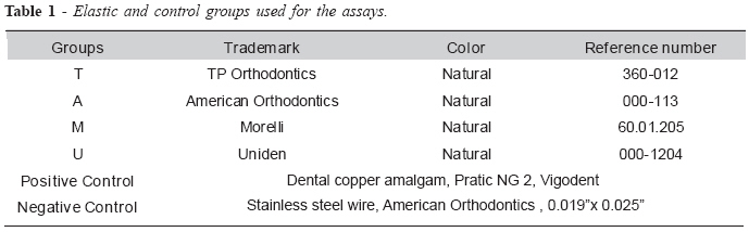
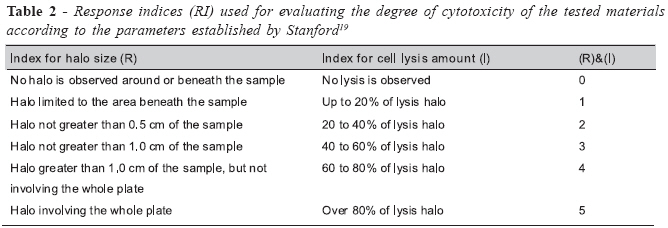
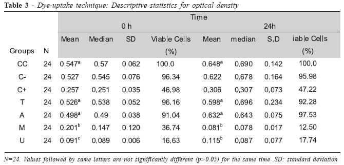
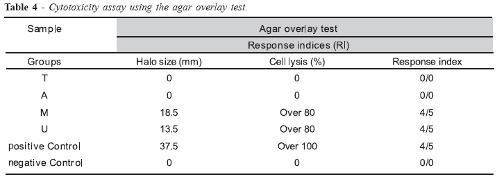
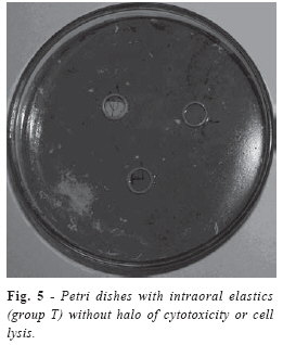
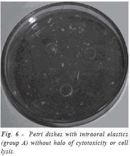
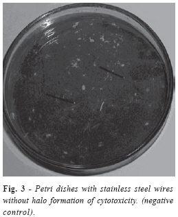
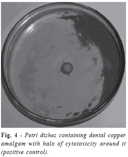
Brazilian Journal Oral Sciences, Vol. 7, No. 24, Jan/Mar 2008, pp. 1520-1526 Cytotoxicity of intraoral orthodontic elastics Rogério Lacerda Dos Santos1 Matheus Melo Pithon1 Márlio Vinícius de Oliveira2 Gabriella da Silva Mendes3 Maria Teresa Villela Romanos4 Antônio Carlos de Oliveira Ruellas5 1Student of the Master’s degree program in Orthodontics, Federal University of Rio de Janeiro-UFRJ, Brazil Received for publication: November 11, 2007 Accepted: April 15, 2008 Code Number: os08010 Abstract Aim: The present study compared in vitro four types of latex intraoral elastics (5/16 = 7.9 mm, mean load) regarding their possible cytotoxic effects. Key Words: cytotoxicity; elastics; biocompatibility; orthodontics Introduction The biocompatibility of dental materials has been theme of great speculation and uncertainty. There are, particularly in Orthodontics, several materials keeping direct contact with organic tissues for long periods of time. Recent studies have investigated the biocompatibility of different types of orthodontic materials1-2. Intraoral elastics are among these materials. Their contact with oral mucosa may be prolonged for months. Therefore, the possibility that elastomers release toxic substances has been addressed, as such substances could damage the cells1-2. Latex consists of cis-1,4-polyisoprepen and in liquid state it is conserved by adding a preserving agent (usually ammonia). When liquid latex is manufactured several chemicals are added so that the desired properties are achieved3. As variations exist in the composition of latex elastics, differences in their properties also occur. This may be one of the reasons why manufacturers produce elastics of different sizes, in order to compensate the variation of physical properties4. Depending on how the elastics are stored, alterations in their composition may occur3, since latex is highly sensitive to ozone and other free radical releasing systems, such as sunlight or ultra-violet light, which trigger the latex polymer chain. Under clinical conditions, orthodontic elastics are replaced with new ones before achieving this stage3. Prevulcanized latex consist of the pure natural latex having the highest molecular weigh3 and stabilisers, such as zinc oxide and chemically vulcanized products, then resulting in a mixture which is heated until 70o C5. Although zinc is known to be neurotoxic6, the amount released by orthodontic elastics can be ingested as research studies show no evidence of harm7. Anti-ozone and anti-oxidant agents are also added to latex during manufacturing of orthodontic elastics3. This process has the advantage of producing latex with higher mechanical properties, thus increasing its strength and elasticity5,7. However, natural latex is not into the category of materials known to be entirely inoffensive8-9. Allergy caused by latex proteins has been well documented10, including immediate hypersensitivity reactions11. Amongst the allergic reactions caused by orthodontic elastics, swelling and stomatitits, erythematous oral lesions, respiratory reactions, and even anaphylactic shock, the most severe form of allergy1213, can be cited. Latex allergy occurs in 3-17% of the cases14. It is necessary, therefore, to test the biocompatibility of dental materials by toxicity assays15. The use of cell culture medium for testing the toxicity of dental products is a valid way of understanding the biological behavior of such materials8. The objective of the present in vitro study was to test the hypothesis that there is no difference in the cytotoxicity between elastics of different manufacturers. Material and Methods Four different types of intraoral elastics (5/16" = 7.9 mm, mean load) made of natural latex were selected for studying their cytotoxicity on two in vitro methods (Table 1). The samples were allocated into four groups of 24 elastics each, according to their manufacturers: Group A (American Orthodontics, Sheboygan, Wisconsin, USA), Group M (Morelli, Sorocaba, São Paulo, Brazil), Group T (TP Orthodontics, Lodi, California, USA), and Group U (Uniden, Sorocaba, São Paulo, Brazil). The elastics used in this study came from the same production lot for each tested trademark. Dental copper amalgam (Vigodent, Rio de Janeiro, Brazil) with standard size and weight was used as positive control, whereas stainless steel wire (American Orthodontics, Sheboygan, Wisconsin) was used as negative control (Table 1). Cell culture containing Hep-2 line cells (human laryngeal carcinoma) was maintained in Eagles’ minimum essential medium (Cultilab, Campinas, São Paulo, Brazil) by adding 0.03 mg/mL of glutamine (Sigma, St. Louis, Missouri, USA), 50 µg/ml of garamicine (Schering Plough, Kenilworth, New Jersey, USA), 2.5 mg/ml of fungizone (Bristol-Myers-Squibb, New York, USA), 0.25% sodium bicarbonate solution (Merck, Darmstadt, Germany), 10 mM of HEPES (Sigma), and 10% fetal calf serum (Cultilab, Campinas, São Paulo, Brazil) for growth medium or no fetal calf serum for maintenance medium only. Next, the cell culture medium was incubated at 37oC for 48 h. The elastics had their both sides previously sterilised with ultraviolet light (Labconco, Kansas, Missouri, USA) during 30 min. Two methods were used for determining the cytotoxicity of orthodontic elastics: the first one, called “dye-uptake”16, is based on neutral red dye incorporated into live cells; the second one, called agar overlay, uses the agar diffusion technique17-18. It was used in this experiment only two times of evaluation 0 and 24 h, a time that, usually these elastics are changed to each 24 h for the patient. Time zero represents the contact and immediate removal of the elastic of cell culture medium. The timing of 24 h represents the maintenance of elastic in the cell culture medium for 24 h after removal. Dye-uptake Volumes of 100 µL of Hep-2 cells were distributed into 96well microplates. After 48 h, the growth medium was replaced with 100 µL of Eagles’ minimum essential medium (MEM) obtained following incubation in the different types of elastics at 0 and 24 h. Positive and control groups consisted of culture medium put in contact with amalgam and stainless steel wire, respectively. After 24-h incubation, 100 µL of 0.01% neutral red dye (Sigma, St. Louis, Missouri, USA) were added to the culture medium in the 96-well microplates, which were incubated again for 3 h at 37oC so that the red dye could penetrate the live cells. Following this period, 100 µL of 4% formaldehyde solution (Vetec, Rio de Janeiro, Brazil) in PBS (130 mM of NaCl; 2 mM of KCl; 6 mM of Na2HPO4 2H2O; 1 mM of K2HPO4 1 mM; pH 7.2) were added in order to promote cell attachment to the plate. After 5 min, 100 µL of 1% acetic acid (Vetec, Rio de Janeiro, Brazil) and 50% methanol (Vetec, Rio de Janeiro, Brazil) were added in order to remove the dye. After 20 min, a spectrophotometer (BioTek, Winooski, Vermont, USA) at 492 nm wavelength ((l = 492 nm) was used for reading the data. Agar Overlay Technique For this test, Hep-2 cells were grown in 10-cm-diameter Petri dishes of. After reaching a confluence with cell density of approximately 106 cells/mL, the cell culture was replaced with two-fold concentrated MEM mixture and 3% agar, and 3 samples of each elastic were carefully placed on the agar layer. Next, the Petri dishes were incubated for 24 h at 37oC in 5% CO2 atmosphere. Thereafter, the cells were attached by using 4% formaldehyde in PBS and stained with 1% crystal violet dye (Vetec, Rio de Janeiro, Brazil) so that the lysis halos could be measured by using a millimeter rule. The results were recorded as response indices (RI) according to parameters established by Stanford19, being represented by two numbers separated by a slash in which the first number means the halo size (diffusion of toxic substance) and the second one means the amount of cell lysis (Table 2). Such indices were calculated in relation to the halos measured following use of crystal violet dye. Data were compared by analysis of variance (ANOVA). Tukey multiple-comparisons test was used for identifying differences between the groups. Significance level was set at p<0.05. Results Dye-uptake test There were statistically significant differences between groups T (TP Orthodontics) and M (Morelli), T (TP Orthodontics) and U (Uniden), A (American Orthodontics) and M (Morelli), A (American Orthodontics) and U (Uniden), and M (Morelli) and U (Uniden) (p=0.00). No statistically significant difference was found between Groups T (TP Orthodontics) and A (American Orthodontics) (p=0.754) at 0 h for dye-uptake method (Table 3). After immersing the elastics, the 24-h cytotoxicity assay using dye-uptake method showed results similar to those of 0-h cytotoxicity and agar overlay assays. There were statistically significant differences (p=0.00) between groups T and M, T and U, A and M, and A and U. On the other hand, Groups T and A as well as M and U had no statistically significant differences between them (p=0.975 and p=0.972, respectively) after 24 h for the dye-uptake assay (Table 3). Agar Overlay Technique The positive control had RI = 4/5 (Table 4), which means a diffusion of toxic substance greater than 1.0 cm in length and more than 80% of lysed cells, but not involving the whole Petri dish. The negative control had RI = 0/0, which means absence of cell lysis (Figures 1 and 2). Morelli and Uniden elastics were cytotoxic, as their RI was 4/5 (Table 4). This means that the diffusion of toxic substance was greater than 1.0 cm and 80% of the cells within the halo had been lysed (Figures 5 and 6). On the other hand, TP Orthodontics and American Orthodontics elastics showed biocompatibility with respect to cytotoxicity as their RI was 0/0 (Figures 3 and 4). Discussion In the present study, dental copper amalgam and stainless steel wire were used as positive and negative controls (Table 1), respectively, because they have proved to be adequate for this type of assay18,20-21. Dental amalgam is potentially cytotoxic due the presence of mercury, but there are also other neurotoxic substances depending on their composition and manufacturer6. Toxic materials show zones corresponding to cell death22. The positive control had RI = 4/5 (Table 4), which means a diffusion of toxic substance greater than 1.0 cm in length and more than 80% of lysed cells, but not involving the whole Petri dish. The negative control had RI = 0/0, which means absence of cell lysis (Figures 1 and 2). These results were similar to those obtained by Moreira Pacheco18,21. Sterilization is a prerequisite for cytotoxicity assay and it can be done by autoclavation23. However, the elastics become hardened after this procedure because of the heat17, resulting in degradation and subsequent release of toxic substances. In this study, ultraviolet light was used in the present study for sterilizing both sides of the elastics24 during 30 min. All elastics were found to have the same color and malleability following UV light sterilization. The results showed that Morelli and Uniden elastics were cytotoxic, as their RI was 4/5 (Table 4). This means that the diffusion of toxic substance was greater than 1.0 cm and 80% of the cells within the halo had been lysed (Figures 5 and 6). On the other hand, TP Orthodontics and American Orthodontics elastics showed biocompatibility regarding cytotoxicity as their RI was 0/0 (Figures 3 and 4). However, variations occur in the composition of latex elastics4, which might explain the difference in the results regarding both trademarks. By recognizing the importance of the biological characteristics of dental materials, the American Dental Association (ADA) have developed a series of tests in order to develop a reliable method for assessing both toxicity and irritation such materials may cause to oral tissues22. The cytotoxic potential can be assessed by means of in vitro and in vivo (animals) experiments22, which represents one of the ways of analyzing the safety and efficacy regarding dental materials25. Regarding in vitro experiments, three different methods can be employed: evaluation of chromium release, Millipore membrane filtration and agar overlay test19. The dye-uptake technique16, in which neutral red dye is incorporated by live cells, allowing that the initial cytotoxic effect of dental materials on cells can be evaluated by a spectrophotometer was used for reading the data through optical density. The percentage of viable cells was obtained by comparing the mean optical density (OD) of control cells (no contact with the materials) with that of cell cultures put in contact with different elastics, resulting in 50% toxicity for the cell cultures (CC50) (Table 3). According to Schmalz8, the major risk factor for using potentially cytotoxic intraoral elastics would be that substances released by them could be ingested by the patient over time, which might result in diseases caused by accumulation of the toxic substance. It is known that latex is not entirely biocompatible as it may cause allergic reactions3,14 and interact with food14,26 and medications27. Nevertheless, the potential for hypersensitivity may not be related to the potential for cytotoxicity. In other words, the material may be allergenic but not cytotoxic, although the contrary may not be true. As dental latex elastics are widely used in clinical orthodontics, the cytotoxic effects they may produce should be taken into account, particularly intraoral elastics because of their continuous and prolonged contact with the mucosa. Therefore, materials proved biocompatible should be selected if such a concern occurs. Previous studies on the toxicity of orthodontic latex elastics have shown that they were toxic to gingival fibroblasts9. The cytotoxic effect was demonstrated after exposing the elastics to the culture medium as the Morelli and Uniden elastics were found to cause more cell death in comparison to TP Orthodontics and American Orthodontics elastics, thus suggesting that the former are less biocompatible than the latter. However, as in vitro experiments cannot reproduce the oral environment in all its aspects, those elastics should not be considered clinically inert. Variations occur in the composition of the latex elastics and this could explain the difference of results between the trademarks in this study. Although orthodontic elastics are cytotoxic under in vitro conditions, this effect is not always reflected in the clinical practice7,9. It is important for the orthodontist to know how to manage patients with a latex allergy and how to deal with this problem28. An alternative for patients with allergy to latex, is to use latex-free elastics, which can be used in orthodontics without jeopardizing the orthodontic treatment29. In conclusion, TP Orthodontics and American Orthodontics intraoral elastics were found to produce less cell lysis, whereas Moreover, Morelli and Uniden intraoral elastics were found to cause significantly more cell lysis, suggesting a less biocompatibility of these elastics and greater possibility of stimulation to the allergic reactions. References
© Copyright 2008 - Piracicaba Dental School - UNICAMP São Paulo - Brazil The following images related to this document are available:Photo images[os08010t2.jpg] [os08010f8.jpg] [os08010f3.jpg] [os08010t4.jpg] [os08010t3.jpg] [os08010f4.jpg] [os08010f5.jpg] [os08010f6.jpg] [os08010f7.jpg] [os08010t1.jpg] |
| |||||||||
