 |
| About Bioline | All Journals | Testimonials | Membership | News |
|
||||||
|
||||||
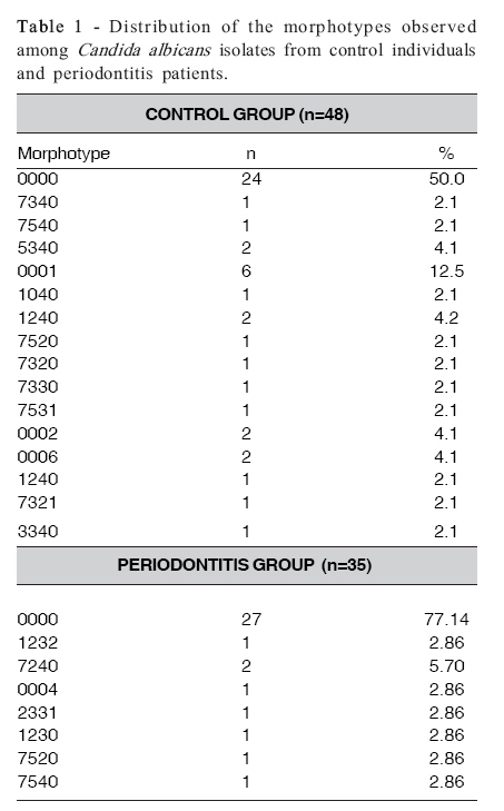
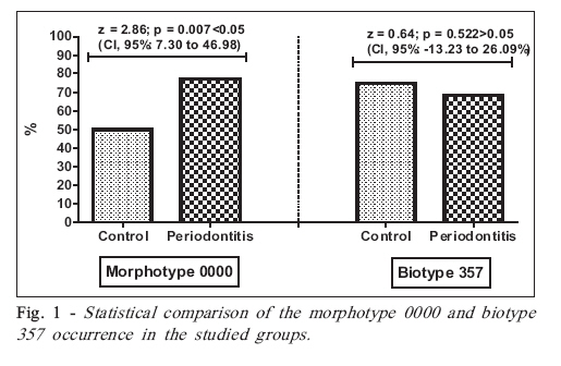
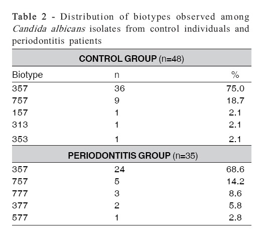
Brazilian Journal of Oral Sciences, Vol. 7, No. 25, Apr-Jun, 2007, pp. 1531-1534 Phenotypic characterization of Candida spp. isolates from chronic periodontitis patients Cristiane Yumi Koga-Ito1; Edson Yukio Komiyama2; Clélia Aparecida de Paiva Martins2; Silvana Soléo Ferreira dos Santos3; Ivan Balducci4; Antonio Olavo Cardoso Jorge1 1PhD in Microbiology and Immunology, Department of Oral Biosciences and Diagnosis, Dental School of São José dos Campos, São Paulo State University, Brazil; Received for publication: September 05, 2007 Accepted: May 30, 2008 Code Number: os08012 Abstract Aim: Several typing methods for Candida spp. have been suggested in the literature in order to distinguish isolates for studies about the virulence or infection routes of these microorganisms and, in particular, for epidemiological purposes. The aim of this study was to establish a comparison between the phenotypic profile of oral Candida isolates from periodontitis patients and control individuals. Key words: periodontitis, Candida albicans, biotyping, morphotyping Introduction Candida spp. have been correlated to cases of severe and refractory periodontal infections, particularly in immunocompromised patients or individuals under antimicrobial therapy for long periods1-5. Several Candida spp. typing methods have been suggested in the literature in order to distinguish isolates for studies about the virulence or infection routes of these microorganisms and, in particular, for epidemiological purposes6-10. Among the methods based on phenotypic characteristics, serotyping, biotyping, morphotyping and sensitivity to killer toxins are included7. There are also methods based on genotypic characteristics, such as immunobloting11 and DNA fingerprinting techniques12. Morphotyping is considered an efficient, reproducible and low cost method for C. albicans characterization7,13. Moreover this method reveals a good discriminatory power. The variation in the morphology of Candida colonies was firstly observed by Negroni14. Later, based on Brown- Thomsen’s16 observations about the morphological variations of colonies due to alterations in the incubation temperature or medium composition, Phongpaichit et al.15 proposed a typing method with codes. This coding system was based on the characteristics of the colonies as well as on their surfaces. Hunter et al.17, studying the morphotype distribution among 446 C. albicans strains, suggested a typing method based in the characteristics of colony surfaces, represented by 4 digits. The phenotypic switching and Candida morphotypes have been associated to their virulence. Different phenotypic expressions in relation to colony growth are related to variations that are considered responsible for the several degrees of virulence7,18. Phenotypic switching occurs frequently and causes changes in colony morphology and cell surface properties, such as alteration in the adherence to epithelial cells19. The biotyping technique proposed by Odds and Abbott20 has been reported as a method with an adequate discriminatory power mainly when associated with the morphotyping method21. Considering the adequate discriminatory power of the combination of morphotyping and biotyping methods, the purpose of this study was to establish a comparison between the phenotypic profile of oral Candida isolates from periodontitis patients and control individuals, aiming to correlate specific phenotypic features to the occurrence of periodontal disease. Material and Methods This research project has been independently reviewed andapproved by the Bioethics Research Committee of the Dental School of São José dos Campos, São Paulo State University, Brazil (Protocol #72/99-PH/CEP). Candida albicans isolates from chronic periodontitis (n=35) and control individuals (n=48) were included in this study. These strains were previously isolated and belonged to the strain collection of the Microbiology Laboratory at the Dental School of São José dos Campos, São Paulo State University. Isolates from chronic periodontitis were obtained from 88 individuals aged 25 and 62 years (mean age of 41.33 ± 5.54 years), with at least two 5-mm deep periodontal sites and diagnosed clinically as chronic periodontitis patients. Control group isolates were obtained from 68 individuals aged 25 to 55 years (mean age of 34.45 ± 7.93 years) diagnosed as periodontally healthy patients. Candida isolates were morphotyped according to the methodology proposed by Hunter et al. 7 and Pongpaichit et al 15. Briefly, the isolates were plated on Sabouraud dextrose agar and incubated at 25ºC for 48 h. Then, yeast suspensions were prepared in sterile distilled water adjusted to the turbidity of tube #3 of McFarland scale. Using sterile swabs, the suspensions were plated on the surface of malt extract agar (Difco, Detroit, MI, USA). Plates were maintained at room temperature in a light-proof environment for 10 days. After this period, the macromorphological aspects of the fringes and surface of the colonies were evaluated. The results of morphotyping were recorded using 4-digit codes. Biotyping methods were performed according to Odds and Abbot22, with a combination of tests of tolerance (to pH 1.4 and NaCl), resistance (5-fluorocytosine, safranine and boric acid), enzymatic activity (proteinase) and growth in presence of urea, sorbose and citrate. The isolates were plated on Sabouraud dextrose agar and incubated at 37ºC for 24 h. Then, saline suspensions adjusted to the turbidity of tube #3 of McFarland scale were obtained. Aliquots of 100 µL of these saline suspensions were deposited in the wells of a Steers’ inoculator and plated in the test culture media and the positive control (Sabouraud agar). All tests were performed in triplicate. Plates were incubated at 37ºC for different periods of time (3 to 4 days for control, safranine, urea, citrate, boric acid and sodium chloride; 6 to 7 days to resistance to pH 1.4 and tests of sorbose, proteinase and 5fluorocytosine). Positive tests were considered when the strains grew at pH 1.4, 5-fluorocytosine, sodium chloride, boric acid, urea, sorbose and sodium citrate, as well as for the strains that formed colonies with diameter grater than 2 mm in presence of safranine. The results of biotyping were recorded using 3-digit codes, according to Odds and Abbott22. Values of 1, 2, 3 and 4 were attributed to the positive tests and 0 to the negative tests. Statistical Analysis The data regarding the occurrence of the morphotypes and biotypes were compared between the periodontitis and control groups by two proportion Z test at 5% significance level. Results The morphotype 0000 was the most frequently observed (50%) among the oral isolates from control individuals. The morphotype 0001 was observed in 12.5% of the isolates. The morphotypes 1232, 7240, 0004, 2331, 1230 and 7520 were also observed (Table 1). Among the isolates from chronic periodontitis patients, the morphotype 0000 was also the most frequently found (77.14%) differing significantly from the control group (p=0.007) (Figure 1). Other 7 morphotypes were observed among the isolates of this group (Table 1). Biotypes 357 and 757 were the most frequently observed in both groups without statistically significant differences (p=0.522) (Table 2). Discussion Candida genus yeasts are considered opportunistic microorganisms23,24 and may lead to severe periodontal infections in immunocompromised patients or individuals under antimicrobial therapy for long periods1. Under specific situations, such as in immunodepressed patients25 , superinfection by Candida can be refractory to conventional periodontal treatments. In fact, C. albicans presents virulence factors that can have an important role in the pathogenesis of periodontal disease such as the ability of penetrating the epithelium, inhibiting polymorphonuclear cells and causing lysis of monocytes8,26. Also, Lu et al. 27 have described the hyphal invasion capacity of C. albicans to inhibit the production of antimicrobial peptides by oral epithelium. Candida species have been isolated from the subgingival microbiota of the gingival tissues of patients with periodontal abscesses1, periodontitis28,29, and patients with chronic periodontitis under therapy with antibiotics17 . Morphotyping has been employed in epidemiological and virulence studies. Ribeiro et al. 30 studying children with Down’s syndrome showed that the C. albicans isolates from these patients induced more formation of fringes compared to the isolates obtained from control individuals, and the authors suggested a correlation with increased virulence. In fact, Hunter et al. 17 had previously suggested a correlation between invasive infections and isolates with discontinued fringes. The increased virulence of isolates with fringes was also related by Spadari et al.31. These authors observed a higher degree of adherence in these isolates compared to strains without fringes. In the present study, this tendency was not observed, as similar proportion of isolates in the groups showed discontinued fringes. Four isolates from the control group (8.33%) and 3 from the periodontitis group (8.57%) presented discontinued fringes. These results suggest that no correlation between colonies with discontinued fringes and periodontitis occurrence could be done. The morphotype 0000 (complete absence of fringes) was the most frequently isolated from the oral cavity of control and periodontitis patients, and this result corroborates the absence of correlation between the presence of discontinuous fringes, which is associated to filamentation, and periodontitis occurrence. Previous studies6,17,32 also observed the higher incidence of this morphotype among isolates from the oral cavity. This result may suggest that the mycelial form formation might not be an essential feature of Candida in the periodontal milieu, and other factors (i.e. proteolytic enzymes and toxins production) might be more important contributors to the pathogenesis of this disease. For more detailed data, further studies on these features could be useful. Also, studies on genotyping could provide important results. In fact, several authors have pointed out the importance of combining phenotyping and genotyping methods6,9,33,34 . Hunter et al. 7 have demonstrated the occurrence of the morphotypes 7540 and 7340 among isolates from several sites of the body. These morphotypes were also observed among the isolates of the present study. The great variability of morphotypes in the control group (16 models) has been reported elsewhere17. Ribeiro et al.29 did not observe the occurrence of morphotype 0000 and found a high prevalence of morphotype 5530 in Down’s syndrome patients. The biotyping method proposed by Odds and Abbott20 has been considered as a method with adequate discriminatory power. However, in the present study, it was not able to distinguish between periodontitis and control isolates. Biotyping permitted the identification of 5 different biotypes among the isolates from the oral cavity of control individuals. The biotypes 357 and 757 were the most frequently observed (75% e 18.8%, respectively). Among the isolates from periodontitis patients, the biotypes 357, 757, 777, 377 and 577, and the model 357 (68.6%) were the most frequently observed. In the present study, most isolates assimilated urea and citrate and did not assimilate sorbose. Also, these isolates were tolerant to salt and developed in presence of boric acid and safranine. Neely et al.35 also verified a high prevalence of isolates with these features among Candida isolates from children. In conclusion, the results obtained by biotyping of the isolates did not allow distinguishing a characteristic model related to periodontal disease, whilst the morphotype 0000 was most frequently isolated from periodontitis patients. References
© Copyright 2008 - Piracicaba Dental School - UNICAMP São Paulo - Brazil The following images related to this document are available:Photo images[os08012t2.jpg] [os08012f1.jpg] [os08012t1.jpg] |
| |||||||||
