 |
| About Bioline | All Journals | Testimonials | Membership | News |
|
||||||
|
||||||
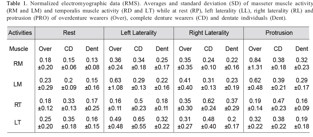
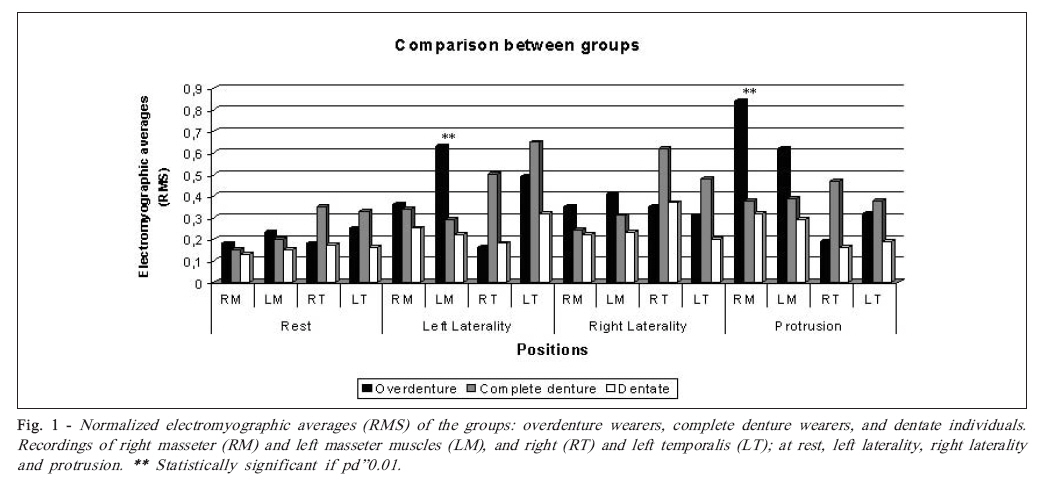
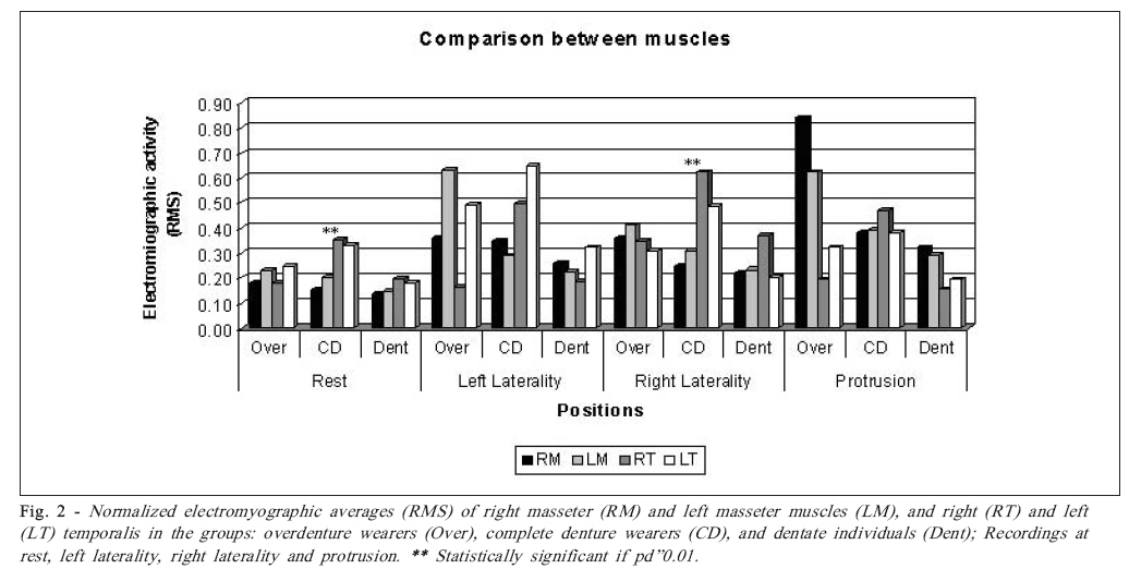
Brazilian Journal of Oral Sciences, Vol. 7, No. 25, Apr-Jun, 2007, pp. 1550-1554 Using overdenture on implants and complete dentures: effects on postural maintenance of masticatory musculature Carla Moreto Santos1; Mathias Vitti2; Wilson Matsumoto3; Renato José Berro4; Marisa Semprini2; Jaime Eduardo Cecílio Hallak5; Rodrigo Galo6; Simone Cecílio Hallak Regalo2 1DDS, PhD student, Department of Morphology, Stomatology and Physiology, Dental School of Ribeirão Preto, University of São Paulo, Brazil Received for publication: March 06, 2008-08-18 Accepted: June 12, 2008 Code Number: os08016 Abstract Aims: Thanks to advances in osseointegration, oral rehabilitation specialists have had the option of using implants to improve retention and stability in treatments with complete dentures. This study compared the masticatory muscle electromyographic activity in implant-supported overdenture wearers, complete denture wearers and dentate individuals. The electromyographic activity of the right and left masseter muscles, and the right and left anterior temporalis muscles was analyzed in 10 implant-supported overdenture wearers (Group 1), 10 conventional complete denture wearers (Group 2), and 10 dentate individuals (Group 3), with mean age of 65 years, at rest and during postural position maintenance. The analysis was performed using the MyoSystem-Br1 electromyographer with differential active electrodes. Analysis of variance tests were carried out to compare the groups and muscles and revealed different electromyographic values that were statistically significant at 1% significance level. Duncan’s pos-hoc test showed that Group 3 presented the smallest values (pd”0.01). The electromyographic contraction pattern was similar between Groups 1 and 3 (p>0.01), and hyperactivity of anterior temporalis muscles was observed in Group 2 (pd”0.01). Key Words: Masticatory muscle, overdenture, complete denture, electromyography. Introduction Geriatric dentistry developed as a consequence of world population aging and has contributed to improve the quality of life of elderly individuals by both preventive and curative measures. Elderly people who are edentulous or use dentures that are worn out or inappropriately adjusted may have functional limitations, such as chewing difficulties. Due to such limitations, these individuals may limit their food choices1. In addition to reducing their eating pleasure, this change in feeding may compromise their overall health, since they often choose foods that have less fiber and are of a low nutritional value. Aging has different effects on the organs and systems of each human being. In the oral cavity, it appears to have a great influence and may reduce chewing efficiency due to either tooth loss and/or bad conditions of remaining teeth, which results in difficulties in eating appropriately1-2. The aging process produces a typical structural deterioration of the stomatognathic system as well as muscles and nerves throughout the entire body. In case of tooth loss, parts of the mandibular bone are reabsorbed and the oral mucosa loses its morphological characteristics, the muscular fibers become atrophic, a great part of motor neurons and their receptors are lost, and there is also a reduction of neurotransmitters3. As age advances, sensory functions, including taste, smell, and touch (for texture and temperature) become less precise. Studies of mandibular masticatory movements have shown there is a reduction in vertical development during the chewing cycle in elderly individuals, complete denture wearers, when compared to dentate young individuals4-6. Karlsson and Carlsson7attributed this difference to several factors such as volume reduction in masticatory muscles, poor neuromotor coordination, and energy reduction in muscle cells. Oral rehabilitation treatment may have a direct influence on various structures, including muscle activity. This may occur because rehabilitating intervention is performed in the oral cavity, which is part of the stomatognathic system, in which all structures function harmoniously8-9. Good denture fitting is essential for complete and partial denture wearers to achieve proper speech articulation and to preserve chewing and swallowing functions. Thus, it is of utmost importance to study muscular activity of the masticatory system in cases of oral rehabilitation. Only with complete understanding of such functions one can obtain results with the highest level of efficiency. Many authors consider elderly edentulous individuals as oral invalids and, like complete denture wearers, they have a reduced capacity in many masticatory system functions compared to individuals with natural dentition10-11. Complete dentures supported by osseointegrated implants in the edentulous jaw have provided occlusion stabilization that shows considerable improvement in muscular activity and mandible movements. This type of treatment has been increasingly used by oral rehabilitation specialists to improve the satisfaction and functional comfort of edentulous individuals. This study compared the masticatory muscle electromyographic activity in implant-supported overdenture wearers, complete denture wearers and dentate individuals (control group). The results may provide valuable data to be considered when choosing one of these total oral rehabilitation treatments for elderly individuals and allow for detailed diagnosis and prognosis, which will improve their quality of life. Material and Methods Volunteers This study was approved by the Research Ethics Committee of the Dental School of Ribeirão Preto, University of São Paulo (FORP-USP) in compliance with the Resolution 196/ 96 of the Brazilian National Health Council. The volunteers were informed about the experiment and agreed to take part in the study by signing an informed consent form (Process number 2003.1.752.58.9). Thirty subjects with mean age of 65 years were enrolled in this study. As inclusion criteria, the volunteers should have overall good health conditions, present similar clinical buccofacial conditions, no complaints of orofacial pain, no history of previous orthodontic treatment and no evidence for other pathologies. The selected subjects were allocated into 3 groups, as follows. Group 1: 10 individuals (6 women and 4 men), aged between 46-75 years (mean age = 64.4 years), wearers of upper complete dentures and lower implant-supported overdentures for at 6 six months. The prostheses were evaluated considering the main factors that promote acceptable support, stability, and retention for appropriate functioning of the conventional upper complete dentures. The lower overdentures were supported by 4 implants located in the foramen mental region. A bar was attached to these implants, connected to the prostheses by a plastic clip located in the midline. One ball was placed on either end of the bar and connected to capsules using an O-ring system. The disocclusion guides were grouped, bilaterally balanced. Factors such as occlusal vertical dimension, resting vertical dimension and centric relation were evaluated by methods proposed by Misch12 and were considered as appropriate. Group 2: 10 volunteers (5 men and 5 women), aged between 51-82 years (mean age = 68 years), wearers of upper and lower conventional complete dentures for at least 6 months. These participants were clinically evaluated in the same way as in Group 1 and were selected according to the satisfactory conditions of their dentures. The disocclusion guides were grouped, bilaterally balanced. Factors such as occlusal vertical dimension, resting vertical dimension and centric relation were evaluated in the same way as in Group 1 and were considered as appropriate. Group 3: 10 dentate volunteers with natural dentition (4 men and 6 women), aged between 60-75 years (mean age = 67.9 years). The inclusion criteria for this group were to present complete permanent dentition, Angle class I occlusion and no symptoms of temporomandibular joint dysfunction. An examination of the oral cavity was performed, including a periodontal evaluation. Electromyography This analysis was performed using a MyoSystem-Br1 electromyographer with differential active electrodes (silver bars 10 mm apart, 10 mm long, 2 mm wide, x20 gain, input impedance 10 G&! and 130 dB CMRR). Surface differential active electrodes were placed on the skin over the belly of the left and right masseter, and on the skin over the anterior portion of the left and right temporalis muscles. Electrode positions were determined by palpation and the electrodes were fixed using adhesive tape, with the silver bars perpendicular to muscle fibers. A circular stainless-steel electrode (3 cm in diameter) was also used as a reference electrode (ground electrode), fixed on the skin over the sternum. The skin region where electrodes were to be positioned was previously cleaned with alcohol and shaved, if needed. The electromyographic (EMG) signals were analogically amplified with a gain of 1000·, filtered by a pass-band of 0.01-1.5 kHz and sampled by a 12-bit A/D converter with a 2 kHz sampling rate. The signals were digitally filtered by a pass-band filter of 10–500 Hz for data processing. EMG signals were captured with volunteers comfortably seated in an office-type chair with their arms next to their body and hands on their thighs. Muscle activity was recorded while subjects maintained the following postures for ten seconds: rest position (RP), left laterality (LL), right laterality (RL), and protrusion (PR). At the end of the exam, activity during dental clenching at maximum habitual intercuspation (MHI) was recorded and these values were used to normalize the subjects’ activities of posture maintenance. The data comprised the gross values’ root mean square (RMS) of the EMG signal collected during the exam. Statistical Analyses Analysis of variance (ANOVA) was used to compare the EMG activity of the masseter and temporalis muscles in overdenture wearers, complete denture wearers and dentate individuals (controls) at rest and during different postural positions. Two ANOVA tests were used: one to compare the groups (overdenture wearers, complete denture wearers and dentate individuals), and another to compare the muscles (right and left masseter, and right and left anterior temporalis muscles). Statistical analysis was performed using the SPSS software (SPSS Inc., Chicago, IL, USA) with significance level set at p d”0.01. Results Complete denture and overdenture wearers presented higher EMG activity than dentate individuals (Table 1) during all positions tested in this study, including rest. No statistically significant difference (p>0.01) was observed for mandibular rest when the groups (overdenture wearers, complete denture wearers and dentate individuals) were compared. However, comparing the masticatory muscles, statistically significant difference (pd”0.01) was observed in the group of complete denture wearers. Duncan’s posthoc test revealed higher EMG activity for the right temporalis. Dentate individuals were the only group that showed a balance of muscle activity for the right and left masseter, and for the right and left temporalis (Figures 1 and 2). There was a statistically significant difference (pd”0.01) for the right temporalis muscle while maintaining the posture of left laterality (Figure 1). The group of complete denture wearers had the highest EMG values. For right laterality, the right temporalis muscle presented significantly higher EMG activity in the group of complete denture wearers (Figure 2) by analysis of the Duncan’s complementary test results. In protrusion, the right and left masseter muscles showed slightly higher EMG activity than the right and left temporalis muscles for overdenture wearers and dentate individuals. On the other hand, complete denture wearers did not show this correlation. When the three groups were compared, a statistical significance (pd”0.01) was found for the right temporalis muscle activities in all subjects (Figure 1). Duncan’s post-hoc test showed higher EMG activity for the group of complete denture wearers when the right temporalis was analyzed. Discussion It is known that complete denture wearers have lower masticatory efficacy in comparison to that of individuals with natural dentition13, 14. Allen et al.15 performed a study in which the satisfaction of complete denture wearers with the treatment was assessed by validated questionnaires. The results revealed that the individuals who received new complete dentures had a lower level of satisfaction than those who received overdentures. However, those who received implant-supported overdentures showed the highest level of satisfaction. The best way to ensure masticatory efficacy, and consequently health on postural muscular function, with aging is to maintain the highest possible number of healthy teeth7-8. In cases of tooth loss, patients turn to rehabilitation seeking for a function similar to that of the natural teeth. However, it cannot be provided by dentures supported by edentulous ridges. This is why the use of implant techniques in dentistry has increased continuously15 . The main topics concerning the treatment of edentulous patients researched by rehabilitation specialists include the relations of masticatory muscle functions, dental occlusion, craniofacial relations, and temporomandibular joint dysfunctions. This highlights the importance of the evolution of EMG methods in order to make possible performing electromyographic exams routinely in a near future. Analysis of masseter and temporalis muscles permits determining muscle activity during fucntion. Ferrario et al.16 analyzed EMG activity of the masseter and temporalis muscles of edentulous overdenture wearers during unilateral gum chewing and during maximum tooth clenching. These authors found that when compared to complete denture wearers, dentate individuals and overdenture wearers showed higher EMG contraction. Furthermore, only the temporalis muscle showed different values between groups during maximum tooth clenching. Tallgren et al.17 observed the EMG activity of the masseter and temporalis muscles of 21 complete denture wearers and observed that EMG activity of the temporalis increased soon after fixing the complete dentures, whereas activity of the masseter remained constant throughout the tests. It was concluded that the temporalis muscle was sensitive to changes in intermaxillary relation and to the stability of complete dentures during deglutition. This may explain the hyperactivity found in the present study for the temporalis muscle compared to the activity of the masseter muscle in complete denture wearers during all postural positions. It is likely that such a factor did not occur in overdenture wearers due to the greater stability provided by this type of prosthesis during function. Therefore, in this case, the temporalis muscle is not hyperactivated, permitting the musculature to remain balanced and maintain posture and rest positions. Chen et al.18 performed EMG tests on 40 denture wearers, of which 14 were complete denture wearers, 12 wore overdentures supported by natural teeth, and 14 wore overdentures supported by osseointegrated implants, during fixed masticatory sequence with standardized portions of 2 food staples. The authors observed that temporalis muscle activity prevailed when compared to the activity of the masseter muscles in all groups. Thirty-two of the 40 subjects showed predominant activity of the temporalis muscles over the masseter muscles. Masticatory efficiency was also tested and the results revealed that the group of subjects with implant-supported overdentures had the highest masticatory efficiency, followed by subjects with overdentures supported by natural teeth. The group of complete denture wearers showed the lowest masticatory efficiency. In the present study, there was also a prevalence of activity for the temporalis muscles in the studied groups to stay at rest and all studied postural positions. This means that because complete denture wearers have reduced masticatory efficiency, the temporalis muscle becomes hyperactive during mastication in an attempt to overcome this deficit. Rather than producing strong movements, the temporalis are muscles specialized in producing fine movements, and therefore become fatigued and hyperactive even at rest and during posture maintenance. The elevator musculature may become hyperactive due to fatigue and stress, which may also cause parafunction of the musculature during posture maintenance19,20. Advances of oral implantology brought better conditions to support complete dentures in edentulous individuals with limitations to receive fixed treatments. This research showed that overdentures wearers have masticatory conditions more similar to that of dentate individuals than complete dentures wearers. This advantage is so important to provide ideal nutritional conditions and better quality of life to the increasing elderly population. In conclusion, the instability inherent to the biomechanical function of complete dentures sensitizes and unbalances the EMG activity of temporalis muscles. The temporalis musculature is fatigued by complete dentures due to factors such as instability and masticatory ineffectiveness inherent to this type of prosthesis. Hence, the musculature becomes hyperactive at rest and during posture maintenance. The use of overdentures supported by 4 implants with a barclip and O’ring retention system cause muscular activity, at rest and during posture maintenance, that is more similar to that of dentate individuals than complete denture wearers. Acknowledgements This work received financial support from FAPESP (Process number 04/05324-0). References
© Copyright 2008 - Piracicaba Dental School - UNICAMP São Paulo - Brazil The following images related to this document are available:Photo images[os08016f1.jpg] [os08016f2.jpg] [os08016t1.jpg] |
| |||||||||
