 |
| About Bioline | All Journals | Testimonials | Membership | News |
|
||||||
|
||||||
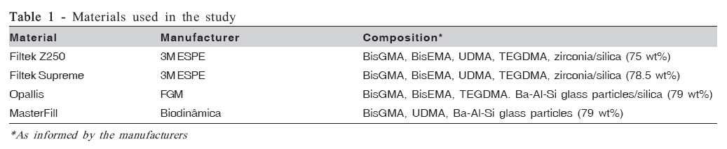
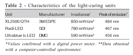
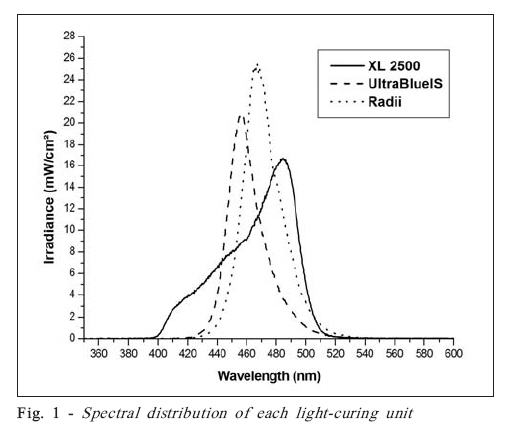
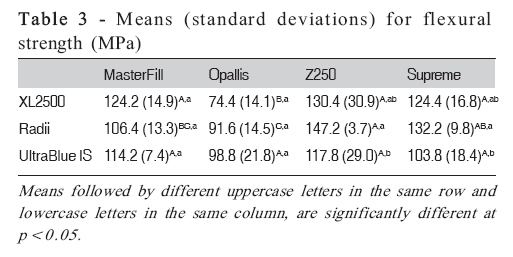
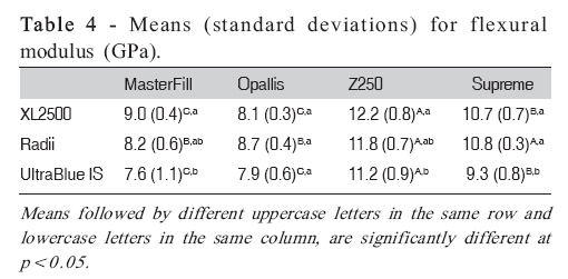
Brazilian Journal of Oral Sciences, Vol. 7, No. 25, Apr-Jun, 2007, pp. 1555-1558 Influence of light-curing units on the flexural strength and flexural modulus of different resin composites William Cunha Brandt1; Leandro Cardoso2; Rafael Ratto de Moraes1; Lourenço Correr-Sobrinho3; Mário Alexandre Coelho Sinhoreti3 1 DDS, MS, Graduate Student Department of Restorative Dentistry, Dental Materials Division, Piracicaba Dental School, University of Campinas, Brazil Received for publication: June 25, 2008 Accepted: July 18, 2008 Code Number: os08017 Abstract Aim: This study aimed to evaluate the flexural strength and flexural modulus of different resin composites (MasterFill, Opallis, Z250, Supreme) after photoactivation with quartz-tungsten-halogen (QTH - XL2500) or light-emitting diode (LED - Radii and Ultrablue Is) light-curing units (LCUs). Key words: Composite resin, light, physical properties, polymers, photoactivation. Introduction The ultimate goal of dental restorative materials is to replace the biological and functional properties of healthy tooth structures, with physical and esthetic properties matching those of natural teeth. Since their introduction about 40 years ago, restorative resin composites have proven to be successful1, and it is expected that their use for restoring both anterior and posterior teeth will continue to increase. Although considerable improvements have been made in the properties of these materials over the last decade2, Brazilian formulations are increasingly available, but little is know about their performance. For this reason, studies evaluating their properties are warranted. Likewise composites, several light-curing units (LCUs) are available, each one having specific characteristics of light emission, such as irradiance level and spectral emission range and peak. Although quartz-tungsten-halogen (QTH) LCUs are the most traditional ones, blue light-emitting diodes (LEDs) are increasingly popular among clinicians. These LCUs emit a narrow spectrum of wavelengths that is better correlated with the spectral absorbance of camphorquinone (CQ), the most commonly used photoinitiator in dental composites3. In comparison to LEDs, QTH LCUs also present some drawbacks that may interfere with their long-term exposure efficiency, such as high heat generation within the quartz bulb, degradation of filters over the course of time, and relatively overall short efficient working life span4-5. However, although some studies describe similar curing efficiency for QTH and LED units, the actual efficiency of LEDs still needs further evaluation. Therefore, the aims of this study were to evaluate the light characteristics (irradiance level and spectral emission) of LED and QTH LCUs, and to investigate their influence on flexural strength and flexural modulus of different resin composites. The tested null-hypothesis was that no significant differences in strength and modulus would be observed regardless of the LCU or resin composite. Material and Methods Four commercial hybrid resin composites, shade A3, were evaluated: MasterFill (Biodinâmica, Ibiporã, PR, Brazil), Opallis (FGM, Joinville, SC, Brazil), Filtek Z250 and Filtek Supreme (3M ESPE, St. Paul, MN, USA). Materials’ compositions are shown in Table 1. Light characteristics Three LCUs were investigated: XL2500 QTH (XL - 3M ESPE), UltraBlue Is LED (UB - DMC, São Carlos, SP, Brazil) and Radii LED (RD - SDI, Victoria, Australia). The LCUs were connected to a voltage stabilizer and the output power (mW) of each unit was measured with a digital power meter (Ophir Optronics Inc., Danvers, MA, USA). The diameter of each light guide tip (cm) was measured with a digital caliper (Mitutoyo, Tokyo, Japan) accurate to 0.01 mm. Irradiance (mW/cm2) was computed as the ratio of the output power by the area of the light guide. Additionally, the spectral distribution of each LCU was obtained using a computer-controlled spectrometer (USB2000; Ocean Optics, Dunedin, FL, USA). Flexural strength Flexural strength test was conducted in accordance with the ISO4049 standard specifications6. Five rectangular barshaped specimens (25 x 2 x 2 mm) were prepared for each material-unit combination by placing the composites into a stainless steel mold held between two glass microscopic slides. After light-activation procedures, the specimens were removed from the mold and stored in distilled water at 37°C in the dark. After 24 h, the height and width of the specimens were measured with a digital caliper (Mitutoyo) and the samples were subjected to a three-point bending test in a mechanical testing machine (DL500; EMIC, São José dos Pinhais, PR, Brazil), at a crosshead speed of 0.5 mm/min until failure. Flexural strength (FS) was determined as follows: FS = 3Pf L / 2WH2 where Pf is the measured maximum load (N) at the time of specimen fracture, L is the distance between the supports on the tension surface (20 mm), W is the mean specimen width, and H is the mean height of the specimen between the tension and compression surfaces. Data were submitted to two-way ANOVA and Tukey’s test (p<0.05). Flexural modulus A chart plotter recorded the stress-deformation profile during the flexural test. Flexural modulus (FM) was calculated from the linear-elastic range, between bending force and specimen displacement before fracture, as follows: FM = (DF / Dy) x (L3 / 4WH3) where DF / Dy is the change in force (DF) per unit change in deflection of the center of the specimen (Dy). Data were submitted to two-way ANOVA and Tukey’s test (p<0.05). Results Light characteristics Table 2 presents the light characteristics for each LCU. Figure 1 shows a comparison for the spectrum of wavelengths. For XL, a spectrum concentrated in the 400-520 nm wavelength range was observed. For RD and UB, the spectrum was concentrated in the 430-530 and 420520 nm range, respectively. Flexural strength Results for flexural strength are shown in Table 3. The factor ‘LCU’ was not significant (p=0.179), while the factor ‘material’ (p<0.001) and the interaction unitmaterial (p=0.018) were significant. For XL, Opallis showed significantly lower flexural strength than the other composites (p<0.001), which were similar to each other (pe”0.947). Correspondingly, for RD, Z250 and Supreme were similar between them (p=0.554), while MasterFill was similar only to Supreme (p=0.119); Opallis showed significantly lower strength than Z250 and Supreme (pd”0.004), but similar to MasterFill (p=0.565). On the other hand, for UB, all composites presented similar results (pe”0.349). When comparing the different LCUs for each material, no significant differences were observed for MasterFill (pe”0.269) and Opallis (pe”0.091). For Z250 and Supreme, RD showed the highest flexural strength values, which were similar to XL (pe”0.31), but significantly higher than UB (pd”0.041); XL and UB presented similar results (pe”0.176). Flexural modulus Results for flexural modulus are shown in Table 4. The factors ‘material’ and ‘LCU’ were significant (p<0.001), but not their interaction (p=0.165). For XL and UB, Z250 and Supreme showed significantly higher values than both MasterFill and Opallis (pd”0.012), which performed similar (pe”0.147). For RD, Z250 and Supreme presented similar results (p=0.072), and both presented significantly higher modulus than MasterFill and Opallis (p<0.001), which were similar (p=0.677). When comparing the different LCUs for each material, no significant differences were detected for Opallis (pe”0.194). For MasterFill and Z250, XL and RD were similar (pe”0.155), and so were RD and UB (pe”0.295), but XL yielded significantly higher modulus than UB (pd”0.04). For Supreme, XL and RD were similar (p=0.992), generating significantly higher modulus than UB (pd”0.004). Discussion Flexural strength is a measure of composite strength: the higher the value, the stronger the material. The ISO4049 standard6 classifies dental polymer-based restorative materials into 2 types: Type I materials are those claimed by the manufacturers to be suitable for restorations involving occlusal surfaces, while Type II are all other polymer-based restorative materials. The minimum flexural strength requirement for Type I materials is 80 MPa6-7 . The results of this study showed that all composites presented flexural strength higher than 80 MPa, except for Opallis photoactivated with QTH. Indeed, Opallis showed the lowest strength values among all tested composites, regardless of the LCU. Therefore, the nullhypothesis is rejected. The probable explanation for this result may rely on the fact that the composites tested in this study present distinct compositions, and differences in filler load and type, organic matrix components, and even surface treatments of the particles, may affect the mechanical behavior of the materials. On the other hand, flexural modulus describes stiffness, a measure of material’s resistance to deformation under load. There are debates on how much modulus resin composites should possess8. Class V cervical cavities, for example, demand a low modulus restorative material to flex with the tooth. A relatively high modulus, on the other hand, is expected for posterior restorations to withstand the occlusal forces and preserve the adhesive interface. Theoretically, the ideal value would be similar to that of dental structures, so the restorative would have similar deformation to the surrounding tooth under loading. However, when compared to the modulus of human enamel (84 GPa)9, resin composites have much lower values; in comparison to human dentin (14 GPa)9-10, some composites may present similar values. In the present study, Z250 and Supreme generally presented higher modulus than the other tested composites. This result might be related to differences in resin formulation or inorganic load among the materials, which strongly affect the properties of the dental composites. Opallis and MasterFill possess irregular glass particles, while the other composites possess spherical ceramic particles that present higher strength and improve the packing of the inorganic fillers, enhancing the properties of the composite. Composites made with irregular particles could have lower flexural strength and modulus than those made with spherical fillers due to the fact that the stress concentration around the fillers would be expected to be greater for materials loaded with irregular-shaped particles11 . The LCUs produced different results among the resin composites. In general, RD and XL generated higher values for strength and modulus. This is probably explained by the fact that these LCUs presented higher irradiance than UB. The amount of light energy delivered to the specimens might affect the conversion of double bonds which, in turn, might interfere with the development of mechanical properties. Moreover, a photo-polymerization initiated at low light level is generally associated with relatively few centers of polymer growth, possibly resulting in a more linear final polymer structure, with lower network strength. On the other hand, irradiance at high levels produces a multitude of growth centers and leads to the formation of densely cross-linked polymers12-13, which might also explain the results observed for RD and XL. In addition to irradiance, the polymerization potential by photoactivation is also dependent on the correlation between the light spectrum emitted by the LCU and spectrum of absorption of the photoinitiator14-15. Therefore, one could expect better results for RD, since the peak of emission for this LCU was centered on the absorption peak of CQ, as shown in Figure 1. Nonetheless, similar results for flexural strength and modulus were detected for both RD and XL, irrespective of the resin composite. This result might be attributed to the fact that both LCUs presented high and similar irradiance values, which might have accounted for their similar results16 . The present results have clinical implications, since both flexural strength and flexural modulus were found to be dependent on the material and LCU tested. The Brazilian resin composites generally presented poorer properties as compared with the other composites tested, while highintensity LCUs generated polymers with enhanced strength and modulus. Notwithstanding, the results of the present study should be restricted to the conditions tested here. Further studies evaluating different resin composites and curing devices are necessary. Acknowledgements The authors are grateful to the Dental School of the Federal University of Pelotas, RS, Brazil, for the access to its facilities, and to Biodinâmica and FGM for donating the materials used in the study. References
© Copyright 2008 - Piracicaba Dental School - UNICAMP São Paulo - Brazil The following images related to this document are available:Photo images[os08017t4.jpg] [os08017t3.jpg] [os08017f1.jpg] [os08017t1.jpg] [os08017t2.jpg] |
| |||||||||
