 |
| About Bioline | All Journals | Testimonials | Membership | News |
|
||||||
|
||||||
Brazilian Journal of Oral Sciences, Vol. 7, No. 25, Apr-Jun, 2007, pp. 1559-1562 Cytogenetic damage in khaini users of Tamilnadu, Southern India Vellingiri Balachandar1; Balasubramaniam Lakshmankumar2; Kuppanan Suresh3; Pappusamy Manikantan3; Raman Sangeetha2; Subramaniam Mohanadevi3; Keshavarao Sasikala4 1M.Sc., M.Phil., PhD., Research Scholar 2M.Sc., Ph.D, Research Scholar 3M.Sc., M.Phil, Research Scholar 4M.Sc., Ph.D., Professor and Head Human Genetics Laboratory, School of Life Sciences, Bharathiar University, Coimbatore. Tamilnadu, India
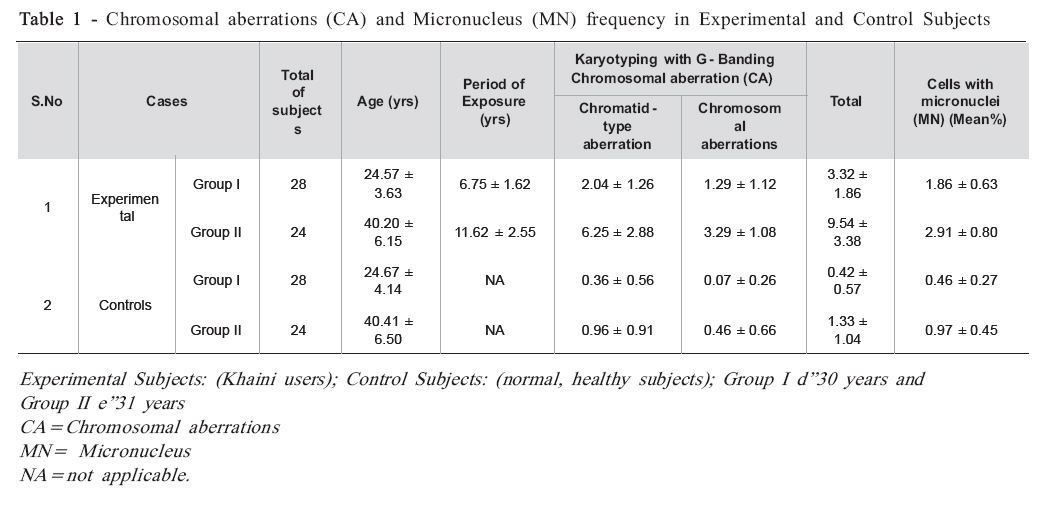
Received for publication: December 06, 2007 Accepted: June 10, 2008 Code Number: os08018 Abstract Aim: The smokeless tobacco (ST) has a strong association with the risk of oral leukoplakia (OL), oral submucous fibrosis (OSF) and oral cancer (OC). ST components exhibit genotoxicity and may alter the structure of DNA, proteins and lipids, resulting in the production of antigenicity. In this study, an attempt was made to estimate the cytogenetic damage [chromosomal aberrations (CA) and micronucleus (MN)] in people habituated to consume khaini (ST), which is one of the major forms of tobacco consumption in Tamilnadu, India, and believed to be a major risk factor for OC. Key Words: Oral cancer, smokeless tobacco, Khaini, chromosomal aberration, micronucleus. Introduction Cancer is currently one of the most dreaded diseases of which head and neck squamous cell carcinoma is the fifth most common. Oral cancer affects as many as 274,000 people worldwide annually, and the frequency is often indicative of the patterns of tobacco use1,2, which is a major contributor to deaths from chronic diseases. Smokeless tobacco (ST) contains considerable nicotine much more than is contained in cigarette tobacco3. ST perceived as a safer alternative to smoking, also contains 28 carcinogenic agents, including nitrites and alkylating agents4-5. Numerous different forms of ST have been used worldwide. Khaini, an interesting kind of ST, is commonly used instead of cigarettes in the Tamilnadu region, southern India, especially in Ooty and Coimbatore. Tobacco, slaked lime paste and areca nut are the major components in Khaini, and a small amount of this mixture (approximately 1 g) is applied to the mucosa of the lower lip for 10-15 min and then it is spit out. This procedure is repeated many times during the day. Pharmacological studies have shown that this kind of ST is a form of buccal nicotine use6,7. The present study underscores the concepts of a molecular epidemiology to detect humans with a high cancer risk with the aid of valid biomarkers, such as biomonitoring. Specific biomarkers on cytogenetic endpoints may help in establishing preventive measures to reduce cancer risks8. Cytogenetic analysis has also been applied to the biological monitoring of human populations exposed to mutagenic and carcinogenic agents. The mutagenic and carcinogenic effects of ST have been previously demonstrated in habitual users9,10. The frequency of chromosomal aberrations (CA) in peripheral blood lymphocytes is a relevant biomarker for cancer risk in humans, reflecting either early biological effects of genotoxic carcinogens or individual cancer susceptibility. Micronucleus (MN) in buccal cells is a sensitive method for monitoring genetic damage in human populations11-13. In the present study, the assessment of cytogenetic damage might help understanding the mode of action of these obnoxious agents, which provide an inexpensive alternative to the smoking types, and the cancer risk involved. Despite several earlier reports regarding the use of ST from various regions around the world, Tamilnadu being home to a more diverse number of such khaini products might aid in the assessment of chromosomal damage leading to tumorigenesis. Therefore, this study investigated whether individuals with khaini habit present more CA with the increase in the exposure period and assessed whether prolonged habitual ST leads to an increase in the number of buccal epithelial cells, by analysis using the MN assay. Although ST has been extensively investigated, to the best of our knowledge, this work is the first to perform a cytogenetic analysis in khaini users in this region. Material and Methods Subject Recruitment and Sample Collection The subjects for this study comprised 104 randomly selected male subjects with no immediate history of viral disease (i.e., during the past 3-4 months). The study groups comprised 52 individuals habituated to ST consumption but without any other habits like smoking, pan chewing or alcohol use. An equal number of healthy subjects who did not use any form of tobacco or alcohol and had not been exposed to any kind of chemicals or radiation were selected as controls. Cases and controls were omnivores, with no apparent nutritional deficiencies. A questionnaire arguing on lifestyle factors, smoking, alcohol consumption, personal information, age, health status, use of medication and exposure parameters, was used to interview the subjects. Age matched controls (±2 years) were recruited to the respective subjects. All cases were exclusively khaini users at the time of the study, consumers for a period of 5 years or more. The cutoff of at least 4 cans/ pouches per week was established to ensure subject safety considering that the use of nicotine patch doses up to 63 g/ day in ST users had only been previously reported in a case series14. The subjects were grouped as follows on the basis of age: Group I d”30 years and Group II e”31 years. Venous blood samples (5 mL) were drawn in heparinized syringes from each subject for the chromosomal analysis. The work was carried out in accordance with the ethical standards laid down in the 1964 Declaration of Helsinki. Chromosomal Aberration Assay All chemical reagents were purchased from Sigma Chemicals, except for colcemid, which was obtained from Gibco Laboratory. Blood samples were set up to establish leukocyte cultures in our laboratory following standard procedures15 . 0.5 mL blood was added to 4.5 mL RPMI 1640 medium supplemented with 10% calf fetal serum, 2 mM l-glutamine, 1% streptomycin-penicillin, 0.2 mL reagent grade phytohemagglutinin, and was incubated at 37 ºC. After 50 h, cultures were treated with 0.1 g/mL colcemid to arrest the cells at metaphase in mitosis. Lymphocytes were harvested after 52 h by centrifuging cell suspension to remove culture medium (800-1000 rpm), addition of hypotonic solution (KCl 0.075 M) at 37 ºC for 20 min to swell the cells, and treated twice with Carnoy’s fixative (3:1 ratio of methanol: acetic acid). Slides were carefully dried on a hot plate (56ºC, 2 min). Three days later, the slides were stained using the Giemsa technique. The observations were made by a researcher for CA on a routine basis. All authors in the research team made critical observations and recorded the results. For the CA analysis, 100 well spread complete metaphase cells in first cell cycle were evaluated per subject under a microscope at ×100 magnification to identify numerical and structural CA. Chromatid-type CAs: (chromatid gaps; chromatid breaks) Chromosome-type CAs: (break; gap; exchange) were observed. The collected data were registered on master tables and later transferred to a computer file. Collection of Cells and Micronucleus Assay Subjects were asked to rinse their mouths with water and a premoistened wooden spatula was used to sample cells from the buccal mucosa. The spatula was applied to a pre-cleaned microscope slide and, after drying, it was fixed with methanol-acetic acid solution (3:1). To visualize the DNA, specimens were subjected to Giemsa (2%) staining for 10 min, when the slides had dried. Finally, the slides were cleaned under running water and air dried. Slides were evaluated microscopically (×400) by an observer not in possession of the results of the questionnaire. MN was reported per 1,000 counted cells. The results were controlled by a second experienced independent observer to ensure the quality of the examination. Criteria used for identification of micronuclei were according to those described by Countryman and Heddle16. Results Individual data of the 104 subjects selected for the present study are presented in Table 1. In groups I and II, 28 (53.84%) and 24 (46.15%) subjects were enrolled in each category (experimental and control subjects). The mean percentage of MN cells in the experimental group I was 1.86 ± 0.63 in users whereas in group II was 2.91 ± 0.80. In the controls, the mean percentages of MN cells were 0.46 ± 0.27 for group I and 0.97 ± 0.45 for group II. Statistically significant results were obtained in experimental subjects compared to controls (p < 0.01), confirmed by chi-square test. The mean ± SD of CA in experimental and control subjects in group I was 3.32 ± 1.86 and 0.42 ± 0.57, respectively; in group II it was 9.54 ± 3.38 and 1.33 ± 1.04, respectively. Chisquare tests with CA data showed significant values with 0.730 for group I and 0.267for group II. Discussion In group I (d”30 years), 28 subjects were selected as this age group has been reported to present concentrate ST consumers, comprising students, athletes and young workers. Adolescent use of all tobacco products has increased (US-DHHS)17. The National Collegiate Athletic Association (NCHA)18 has found that use of smokeless tobacco among college athletes increased by 40% from 1985 to 1989. The majority of college users had begun “dipping” snuff and chewing tobacco before going to college (NCHA)18 . There is sufficient evidence that oral use of ST is carcinogenic to humans. Khaini addiction is frequent in some places of Tamilnadu region, India. In order to elicit the above issues, the present study has been carried out to determine the chromosomal damage in khaini users in Tamilnadu State. Assessment of normal levels of cytogenetic damage in the general population is essential for proper interpretation of data obtained by monitoring of populations that are occupationally or accidentally exposed to known or potentially genotoxic agents. CA have for many years been applied as biomarkers of genotoxic exposure and early effects of genotoxic carcinogens 19,20.The micronucleus test has received increasing attention as a simple and sensitive short-term assay for the detection of environmental genotoxicants21 . Hence, the assessment of cytogenetic damage performed in the present study shall aid understanding the mode of action of these obnoxious agents, which are a cheap alternative to the available smoking types, as well as the cancer risk involved in using them. The data presented in Table 1 show a larger number of CA in the experimental subjects compared to controls. Chromosomal instability has been described in many human dysplastic lesions and is considered a primary event in neoplastic transformation as well as a marker of progression to cancer22,23.A significant increase in the mortality ratio for all types of cancer in subjects with increased levels of CA in their lymphocytes has been found8,24-25 . The control subjects of groups I and II showed minimum number of CA when compared to the experimental subjects (Table 1). The CA in controls might have been due to factors like their age and lifestyle. Several studies have reported the relationship between aging and structural CA. Some studies have found a significant influence of age on CA frequency, whereas others have found no association at all. Recently, age and lifestyle factors have been found to be strongly associated with the frequency of CA measured by the chromosome painting technique26,27. The present study also confirmed that age along with duration of exposure plays a major role in genetic damage as presented in group I and group II experimental subjects, the latter presenting a larger number of CA and MN. As seen in the Results section, the binucleated MN cells are significantly higher in khaini users than controls (Table 1). Similarly, the incidence of micronuclei increased in the buccal mucosa cells of smokeless tobacco users28 . Carcinogenic and mutagenic compounds, including tobacco-specific nitrosamines present in ST forms29 are believed to be responsible for the induction of micronuclei. Moreover, the carcinogenic and mutagenic effects of ST forms have been attributed to tobacco-specific nitrosamines30,31. The ash that is mixed with tobacco leaf powder permits the absorption of alkaloids into the buccal mucosa by transforming them into a basic form6-7. These compounds are produced from nicotine by bacterial or enzymatic activity. The same formation occurs in the mouth under the influence of saliva32 . Controls showed a minimal number of MN when compared to experimental subjects because, in the present study, controls were healthy normal and omnivores, with no apparent nutritional deficiencies. Finally, the present study showed that the MN assay is a cost-effective procedure, accurate and easy to carry out for population-based studies. Furthermore, in vivo evaluations allow for considering the influence of the individual susceptibility in screened humans. Previous reports have established that buccal cells are useful not only for characterizing the molecular mechanisms underlying tobacco-associated oral cancers, but also as exfoliative cells that express diverse changes that appear promising as candidate biomarkers for the early detection of oral cancer. In conclusion, the experimental group II presented a larger number of CA and MN when compared to the experimental group I. On the other hand, the control groups I and II presented negligible numbers of CA and MN. The remarkably greater presence of CA and MN in the experimental subjects was exclusive from khaini users who are highly predisposed to oral carcinoma and head and neck squamous cell carcinoma. It is evident that cytogenetic assays can be used as a primary screening test for ones susceptibility to oral carcinoma. The consumption of khaini as a form of ST poses serious risks as a genotoxic and carcinogenic substance. The accompanying analyses of trends and modalities of induction of cancers associated with the consumption of tobacco in all forms point firmly to the understanding that the only way to eliminate completely the cancer risk linked to tobacco use is not changing from one type of consumption to another, but rather stop using tobacco in any form or, even better, never to start. References
© Copyright 2008 - Piracicaba Dental School - UNICAMP São Paulo - Brazil The following images related to this document are available:Photo images[os08018t1.jpg] |
| |||||||||
