 |
| About Bioline | All Journals | Testimonials | Membership | News |
|
||||||
|
||||||
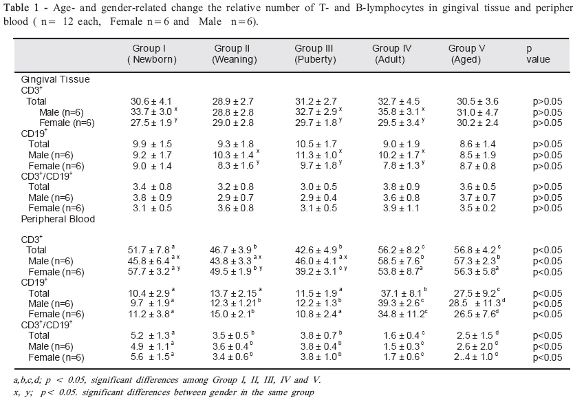
Brazilian Journal Oral Sciences, Vol. 7, No. 26, Jul/Sept, 2008, pp. 1609-1613 The effects of age and gender on CD3+ and CD19+ lymphocyte in gingival tissue and peripheral blood: an animal study Turgut Demir1; Varol Canakci1; Cankat Kara1; Cenk Fatih Canakci1; Fuat Erdem2; Mustafa Atasever3 1Department of Periodontology, Atatürk University, Faculty of Dentistry, Erzurum, Turkey Received for publication: May 26, 2008 Accepted: September 10, 2008 Code Number: os08028 Abstract Aim: The aim of the present study was to determine age- and gender-related values for healthy mice of CD3+ T and CD19+ B lymphocytes and CD3+/ CD19+ (T/B) ratios in peripheral blood and gingival tissue by the flow cytometry technique. Key words: lymphocytes, CD3+, CD19+, age, gender, gingiva, peripheral blood, mice. Introduction Although periodontal bacteria are the causative agents in periodontal diseases, subsequent progression and disease severity are thought to be determined by the host immune response1 . Social and behaviour modulations, and genetic or epigenetic traits of the host, each of which influenced and/or modulated by the host’s immune and inflammatory responses. The clinical entity of the periodontal disease results from the interaction of periodontopathic plaque bacteria and host immune response mechanisms2-3 . The infiltrate in periodontal disease contains mononuclear cells, which are mainly transmigrated mononuclear phagocytes and lymphocytes. Whereas T lymphocytes predominate in the established chronic lesion, the proportion of B cells and plasma cells increases with the progression of the disease4-7 . The immune response is promoted by secretion of bacterial products and involves T and B lymphocytes and macrophages. In fact, clear differences in the degree of inflammation and secreted cyto-kines were observed when periodontitis patients were compared with normal healthy individuals8-10 . Some studies have suggested that B cells occur in larger numbers than T cells in chronic periodontitis11-12 whereas in a study on progressive periodontitis, T cells and B cells were found to occupy similar proportions13 . Recently study show that the CD3+ / CD19+ (T/B) ratio in gingival tissue was about 4.2 times higher in the Adult Periodontitis than in the Localized Prepubertal Periodontitis units. The corresponding ratio for peripheral blood was 1.614 . It is suggested that certain differences may existed in the local defense mechanisms in different periodontal diseases. In order to understand the pathogenesis of periodontal diseases, the systemic factors and conditions that will affect the prevalence, progression and severity of periodontal diseases should be well observed and considered15 . Age, sex, stress levels, nutrition, systemic diseases, exercise, smoking and/or alcohol consumption habits play an important role that can affect the pathogenesis of different periodontal diseases15-21 . However, the systemic immune response is affected by age, sex and other factors19-22 . If confounding factors such as stress levels, nutrition, systemic diseases, exercise, smoking and/or alcohol consumption habits are eliminated by means of periodontally healthy animal model, the study would standardize and we are able to clarify effects of age and gender on relative numbers of T and B lymphocyte, thanks to the short lifetime of the these animals. To our knowledge, there is no published study about effects of age and gender on relative number of CD3+ T CD19+ B lymphocytes in both gingival tissue and peripheral blood of periodontally healthy animals. The aim of the present study was to determine age- and gender-related values for healthy mice of CD3+ T and CD19+ B lymphocytes and CD3+ / CD19+ (T/B) ratios in peripheral blood and gingival tissue by the flow cytometry technique. Material and Methods Animals Healthy 60 BALB/c mice were obtained from Medical Experimental Practice and Research Centre, Atatürk University. The animals were maintained under a 12/12 h dark/light cycle at constant temperature 21±2 ºC. Each mouse was placed to a separate cage. The mice’s diet was stored at 4.5 ± 0.5°C in plastic containers and handled with plastic gloves and appropriated with utensils to avoid contamination. The diet was placed in shallow glass food cups with stainless steel follow-through disks to reduce food spills. The average of life time of the mice were 2-3 years (19).They were separated into five groups according to the life expectancy23: Group I (newborn, 1-10 days old), Group II (age at weaning, 21-28 days old), Group III (age of sexual maturity, puberty ,7-8 weeks old), Group IV (adult , 8 months old), and Group V (the-aged, 14 and over). Males and females were equally represented in each group. Ethics: The study protocol was reviewed and approved by Atatürk University Medical Experimental Practice and Research Centre Ethical Committee. Clinical evaluation The clinical evaluation consisted of plaque index24 , gingival index25 and probing pocket depths to determine gingival or periodontal health. The measurements were made in the Medical Experimental Practice and Research Centre, Atatürk University, by the same periodontist. The numerical scores of the plaque index and gingival index were obtained according to the formula Per mice = sum of individual scores /number of anterior teeth present for each mouse, and subsequently group score was calculated by adding together the individual scores and dividing the total into the number of mice included. The pocket depths were recorded by measuring the distance from the free gingival margin to the base of the pocket with a thin wire exerting a constant force of 1 gr. Examiner variability in using the dental examination criteria were tested by performing duplicate examinations on 12 randomly selected mice on consecutive days. Corresponding percentages of agreement were 97 % for plaque index and gingival index and 99% for probing depth. Thus, these clinical measurements were used to determine periodontal health of mice. Collection of Blood and Tissue samples All of the mice were killed by cardiac puncture after anaesthesia with thiopental. Blood was removed directly from the heart by injection. Blood (minimal 0.5 ml) collected into EDTA-containing hemogram tubes. They were sent to haematology laboratory at once. Gingiva was peeled from around the mandibular and maxillary incisors in upper and lower jaw and separated from tongue with scalpel and obtained gingiva cut into very small pieces with a scalpel and was resuspended in phosphate buffered solution (PBS). Lymphocyte subset identification T cells were defined as those cells express-ing the CD3+ antigen, and B cells were defined as those cells expressing CD19+ . Lymphocytes Levels Blood (100 μl) was incubated with antibody (10μl) for 15 min at room temperature. After incubation the samples lysed and fixed with 500 μl Optilyse® C. Finally, the cells were washed and resuspended in 500 ml of phosphate buffered solution (PBS) for flow cytometry analysis. Gingiva was peeled from upper and lower jaw with scalpel and obtained gingiva cut into very small pieces with a scalpel. Tissues were transformed into suspension through filtration. For lymphocyte subset analysis a flow cytometer (Coulter EPICS®XL-MCL) was used. The computer-assisted evaluation was made with a commercially available software program (Coulter System II Software version 2.0). With the use of volume and side scatter, lymphocytes were gated and specific fluorescence dot blot. The absolute and the relative (proportional) count of each lymphocytes subpopulation were calculated. Monoclonal Antibodies The following monoclonal antibodies used: Rat Anti-Mouse CD19-FITC (PN IM3233), Rat Anti-Mouse CD3-FITC (PN IM2768). IgG2a (rat) Isotypic Control (IM1271) was used as control. (Data Sheet Immunotech, Beckman Coulter). Statistical Analysis The significance of differences in number of T and B lymphocytes in groups was determined by two factor analysis. Duncan’s multiple range test was used to determine differences among groups. Student’s t-test was used for determine difference between male and female in each group. A value of p<0.05 was considered to be significant. All values are expressed as mean ± standard deviation. For these procedures, SPSS for Windows (version 11.0) was used. Results The mean relative number of CD3+ T lymphocytes and CD19+ B lymphocytes and CD3+/ CD19+ (T/B) ratios in gingival tissue and peripheral blood of periodontally healthy BALB/c mice according to age group and gender are shown in Table 1 There were no significant differences in the total number of CD3+ and CD19+ in gingival tissue for all age groups (p>0.05). In addition, The CD3+/ CD19+ T/B lymphocytes ratios in gingival tissue did not differ in all age groups (p>0.05).The mean relative number of CD3+ T lymphocytes in gingival tissue for group I (Newborn) (p<0.05), group III (Puberty) (p<0.05), and group IV (Adult) (p<0.05) in male was higher than in female. The relative number of CD19+ B lymphocytes in gingival tissue for group II (Weaning) (p<0.05), group III (Puberty) (p<0.05), and group IV (Adult) (p<0.05) in male was higher than in female. But, our data show that CD3+/ CD19+ ratios in gingival tissue were similar for all age group when considering the gender differences (p>0.05). Mean relative number of peripheral blood CD3+ T lymphocyte indicated a decrease in Group II (Weaning) and Group III (puberty) compared to the other groups (p<0.05), while relative number of peripheral blood CD19+ B lymphocyte increased in Group IV (adult) and Group V (aged). The peripheral blood CD3+/ CD19+ T/B lymphocytes ratios decreased in Group IV (adult) and Group V (aged) mice. Significant distinction occurring with regard to statistically and decrease at this rate is stemmed from the fact that the increase in CD19+ B lymphocytes is larger than that of CD3+ T lymphocytes. The mean relative number of CD3+ T lymphocytes in peripheral blood for group I (Newborn) (p<0.05), and group II (Weaning) (p<0.05) in male was lower than in females, while for group III (Puberty) (p<0.05), Group IV (adult) (p>0.05), and Group V (aged) (p>0.05) in male higher than in female. The mean relative number of CD19+ B lymphocytes and CD3+/ CD19+ (T/B) ratios in peripheral blood were similar for all age group when considering the gender differences (p>0.05). Discussion It is known that during the transition from gingivitis to periodontitis, a characteristic change in the predominant lymphocyte subpopulation occurs. In cases of mild gingivitis there is a slight preponderance of T lymphocytes, where B lymphocytes and plasma cells predominate in the sulcus tissue in cases of periodontitis. A study carried out by Gemmel et al demostrates that the mean percent CD3 lymophocytes did not vary significantly between the tissue from healthy/gingivitis and periodontitis subjects, while the percent B lymphocytes was increased in group with periodontitis tissue comparison with healthy/gingivitis tissue26. While most studies have focused T lymphocytes subgroups from the periodontally diseased tissue and peripheral blood, potential age- and gender-related changes of T and B lymphocytes from periodontally healthy tissues are not still characterized. In this study, we analysed the effects of age and sex on relative number of CD3+ T and CD19+ B lymphocyte and CD3+/ CD19+ ratios in gingival tissue and peripheral blood of periodontally healthy BALB/c mice with flowcytometric analysis. Lymphocyte can be analyzed at the level of one cell through flow cytometry, and physical and biological features of the cells can be accurately and quantitatively measured27. Because of all these qualities, the flow cytometry method in present study is preferred. Our data revealed no difference in the relative number of CD3+ T and CD19+ B lymphocytes in gingival tissue for all age groups. However, the relative number of CD3+ T lymphocyte in gingival tissue was higher in males than in females in newborn group, puberty group and adult group. The relative number of CD19+ B lymphocyte in gingival tissue was significantly differences between male and female in weaning group, puberty group and adult group. The CD3+/ CD19+ ratios in gingival tissue were same in all age group and gender. A study carried out by Lazuardi et al.28 revealed no differences in number and size or percentage of B lymphocytes between young and old individuals. In another study, Fransson et al.29 used the experimental model to evaluate the host response to plaque in periodontally healthy young (20-25 years) and old subjects (65-80 years), and gingival biopsies were obtained at days 0, 7 and 21 of plaque formation. Authors concluded that although the ratio between T-cell and Bcell markers (CD3+/CD19+) was found to be higher in the young (14.8) than in the old subjects (5.9) on day 0 (healthly gingiva), the ratio decreased during the course of the experiment in both groups. They also reported that during the 3-week period of plaque accumulation, older subjects developed a larger sized inflammatory cell infiltrate (ICT) with a greater proportion of B cells than younger subjects. Their study suggested that differences exist between young and old periodontally-healthy individuals in the inflammatory response during the course of experimental gingivitis In another study, Zitzmann et al.30 reported sites treated with non-resective methods (Open Flap Debridement; ‘‘old gingiva’’) resulted in larger ICT and higher B-cell proportions than at sites exposed to resective procedures (Gingivectomy; ‘‘young gingiva’’). The findings of our study are not consistent with findings reported by Fransson et al.29 and Zitzmann et al.30 but in agreement with Lazuardi et al.28 . On the other hand, the data of the present study revealed difference in the relative number of CD3+ T and CD19+ B lymphocytes in peripheral blood for all age groups. While the relative number of CD3+ T lymphocyte in peripheral blood was higher in newborn group than in weaning and in puberty group, it was lower in newborn, in weaning and in puberty group than in adult and in the aged group significiantly. Although the relative number of CD3+ T lymphocyte in peripheral blood in females was higher than males in newborn and weaning group, it was significantly lower in females than males in puberty group. The relative number of CD19+ B lymphocyte in peripheral blood significantly elevated in adult group, it was decrease in aged group. Previously, Weksler31 revived a decrease in B lymphocytes with aging. Our finding comfirms the data of Weksler ME. The CD3+/ CD19+ ratios in peripheral blood decreased gradually from newborn group to the aged group. The results of the present study suggest that significant differences observed with periods of life and gender in periferal blood of peridontally healthy mice, but significant differences exist with only gender in gingival tissue of periodontally healthy mice. These age- and sex-related differences may have a bearing on the natural pro-gression of periodontal disease with aging in humans. However, further studies are required to establish whether these mechanisms are important during the normal course of periodontal disease. This kind of information could represent a useful tool both in clinical practice and in the understanding the physiological process of immunescence. Acknowledgements This study was supported by a grant (PN-2003 / 274) from Ataturk University,Turkey. References
© Copyright 2008 - Piracicaba Dental School - UNICAMP São Paulo - Brazil The following images related to this document are available:Photo images[os08028t1.jpg] |
| |||||||||
