 |
| About Bioline | All Journals | Testimonials | Membership | News |
|
||||||
|
||||||
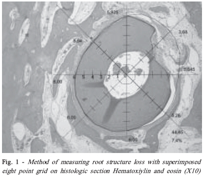
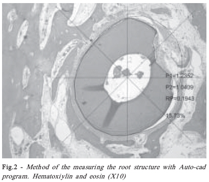
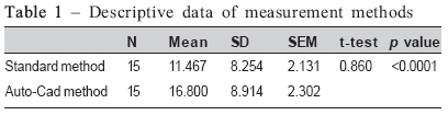
Brazilian Journal Oral Sciences, Vol. 7, No. 26, Jul/Sept, 2008, pp. 1620-1623 Evaluation of root structure loss using AutoCAD assisted histomorphometry Prohic Samir1; Nakas Enita2 1DDS,Msc, PhD, Assistant professor, Department of Oral Surgery and Implantology Received for publication: September 11, 2008 Accepeted: October 14, 2008 Code Number: os08030 Abstract Aims: The quantitative measurement and characterization of microscopical images using a computer is histomorphometry. The purpose of this experimental research is to evaluate the validity of two methods of histomorphometry of root structure loss measuring antiresorptive efficacy of topical application of alendronate in delayed tooth replantation on a canine model. Key words: dental trauma; AutoCAD; alendronate Introduction The most frequent therapy used on patients with avulsed teeth is the method of replantation. After replantation, there may be ankylosis and root resorption, which frequently lead to tooth loss in 4–6 years.1 Clinical and histological studies have shown that the main factors that have impact on replanted teeth are: the length of the extra-alveolar period, storage condition and maturation of the root. Many studies have tried to identify a medicament that would act in the manner of inhibiting root resorption of replanted teeth. These treatments have focused on the use of various forms of fluoride and antibiotics.2- 5 Aforementioned studies do not take into account the active role of osteoclast in root resorption. The possibility of osteoclast inhibition could increase the likelihood of PDL cell proliferation, differentiation, and maintenance of the cemental layer.6 Because of the similar morphology, enzymatic properties and function of the cells leading to resorption of dentin, cement and bone, the processes of root and bone resorption may be considered similar7 . Thus, drugs that may inhibit bone resorption may also be effective for the treatment of tooth resorption. Alendronate (ALN) is a third-generation bisphosphonate, which demonstrates the osteoclast inhibitory activity that can slow down the resorptive process. Solutions containing different concentrations of alendronate may inhibit the bone resorption; moreover, the higher the concentration, the higher the effect8,9 . Measurement of root structure loss as indicator of antiresorptive efficacy is important factor in estimation of drug effectiveness. In order to get precise data we measure root structure loss by standard method6,10 . Nowadays AutoCAD as well as other computer programs are used in medicine for image analysis and they can be used for precise measurements.11-17 The purpose of this experimental research is to evaluate the validity of two methods of histomorphometry of root structure loss measuring antiresorptive efficacy of topical application of alendronate in different concentrations of 3 mMol and 1 mMol in delayed tooth replantation on a canine model. Materials and Methods We used five mongrel dogs skeletally mature with a mean weight of 13.9 ±1.5 kg. The animals were handled according to international standards of animal welfare that are accepted by The Research and Bioethics Committee of the Dentistry School, of the University of Sarajevo (Issue number 09/133-3/04). All experimental procedures were performed with the animals under general anaesthesia. This was accomplished by preaneasthetic sedation with acepromazin maleate (Vetranquil, Sanofi, France) at a dose of 0.05 mg/kg IM, and anaesthetic induction with Propofol (Propofol Abbott,Pakistan) at the dose of 4 mg/kg IV, followed by intubations and maintenance with 2,5% halothane (Fluothane, Zeneca, UK) with the oxygen flow of 2 liters/ minute. Immediately after intubations, the animals were given a one-time dose of tramadol hydrochloride (Lumidol, Belupo, Croatia ) at a amount of 7 mg/kg SC. Forty-eight mandibular premolar mature roots of five dogs were used in the study, each dog served as its own control. It was used two rooted mandibular premolar teeth (P2, P3, and P4). P1 premolar was excluded due to size of the root and impossibility of immobilization. Also 12 roots (6 teeth) were excluded due to root or bone damage. The two-rooted premolars were hemisected and accessed, instrumented with stainless steel K-type files to the apical delta, and irrigated with sterile saline. Canals were dried with paper points, obturated with laterally condensed gutta-percha (VDW, GmbH, Germany) and Roth’s 801 sealers. Teeth were extracted as atraumatically as possible, and dried for 45 minutes at room temperature in sterile petri dishes. Furthermore, roots were soaked in: 1 mMol of ALN, 3 mMol of ALN, physiological saline, for 5 minutes and replanted. Alendronat solutions were prepared by dissolving Alendronat (Fosamax® 70mg alendronat sodium oral solution, Merck &Co Inc. Whitehouse station NJ, 08889, USA) in distilled water. Due to the stability of the replanted teeth, splinting of teeth was determined to be unnecessary. Because of divergence of the roots of mandibular premolar teeth in canine model only sutures for fixation were performed (Mersilk 3-0, 26 mm ½ c, round bodied, Ethicon Inc.USA). The dogs were maintained on a soft diet for 2 days. They tolerated the treatment procedures well, their food intake and behaviour did not change following the treatment. All procedures were done based on double blind investigation principle and were performed by experienced specialist. After four months, the animals were sacrificed by an overdose of a 6 % solution of sodium pentobarbital administered IV, and the bone blocks were prepared for analysis. Block specimens containing the teeth and surrounding alveolar bone were dissected using diamond separators, and fixed in 10% neutral formalin. Following this, the bone blocks were placed in 5% nitric acid HNO3 for seven days and then in 5% sodium sulphate Na2SO4 for 24 hours. Bone blocks were placed into a tissue processor (Microm STP 120, MICROM International GmbH, Germany) for the process of dehydration through various percentages of alcohol, from 70% to an absolute alcohol, after in organic solvent Xylol (changing the solution every 60 minutes for two times I, II). For tissue impregnation the blocks were immersed in liquid paraffin at the temperature of 56°C. After the process of impregnation was completed, the specimens were embedded in paraffin blocks sized 2x2x2 cm and sectioned on rotary microtome (Leica RM 2145, Leica Microsystems, Swiss). The bone blocks were sectioned horizontally in reference to the longitudinal axis of the teeth to the thickness of 5 μm, at 140 μm intervals. The process of staining was than completed by haematoxylin-eosin method in digester. Histological slides were photographed with digital camera (Olympus C 5060 5.1 megapixel) which is directly attached to microscope (Carl Zeiss, Axiolab,Jena, Germany) connected to a PC. The images obtained were stored as figures (TIFF) for further interpretation. Root structure loss due to resorption was measured. Histomorphometry methods To evaluate the extent of root structure loss due to resorption, the original circumference of the root was determined. The radius of the remaining root structure was ranked on a linear, integer 0–6 scale at each of the eight points of the superimposed grid, with the value of 6 given to an unaffected radius and the value of 0 given to a point without any remaining root structure.6 (Fig. 1) Our original method of measuring the root structure was used, with further assistance from the computer-assisted program, Auto-cad 2002 for Windows OP. The program imported the images and, by means of multiple points (Polyline tool), it outlined the image of the original circumference of the root (P1), later we outlined the image of the remaining root structure (P2). Then, by calculating differences between area P1 and area P2, we obtained the value of the loss of the root structure (RP). Measurements were made in a manner as to evaluate the circular area. (Fig. 2) Root structure loss was assessed by the difference between the P1 and P2 areas. All measurement was performed by a blinded investigator in three different time periods. For statistical analysis we used mean value. Statistical analysis The root mass loss data were subjected to Scheffe F multiple comparison tests to identify differences between groups. The t-test for two independent samples was used to evaluate root mass loss calculation between different means of measurement. Statistical calculations were carried out with statistic software programme SPSS® for Windows®. Results In order to evaluate root structure loss we compared previously published method of determining root structure loss and our method 15 slides were used and the histomorphometry was made using both methods. There was evident more root structure loss when measured with AutoCAD (p<0.05). Average root structure loss in different groups In the group of 1 mMol, the root structure loss was registered at a value of 17.21%. In the group of physiological saline, root structure loss was 34.28%. The result difference satisfies statistical significance (p<0.005). On comparing the group of 1 mMol and the group of 3 mMol, it was observed 17.21% of root structure loss in the first group and 12.74% in the second group, with statistical significance (p<0.005). Discussion To date research indicates that the main mode of action of bisphosphonates in the prevention of bone structure loss is inhibition of osteoclast function through the break-down of enzyme passages, which are very important for osteoclast activity and survival7,11,18. Results of root structure loss in our study are 17.21% root structure loss in the 1mmol group, while a loss of 34.28% was found in the group with the physiology solution (p<0.005). Furthermore, in support of a more potent anti-resorption action with regards to the concentration, when we statistically compared 1 mMol and 3 mMol groups in our research, we found out that the former registered a 17.21% mass loss, and the latter a loss of 12.74%, (p<0.005). This certainly affirms the fact that locally applied alendronate is a strong inhibitor of resorption processes, regardless of the concentration. It proves unable to stop, but can only potently slow down resorptive processes in the root of the replanted tooth. The doses of systemic application of alendronate in the work of El-Shinnawi et al.18, which proved a positive effect of bisphosphonate on bone density in patients affected by periodontal disorder, were undoubtedly selected on the basis of abundant clinical experiences in the per oral ordination of Fosamax®; however, local doses have no such kind of support and they are not sufficiently examined, which is an additional reason and justification for our study. Digital Image Quantification (Morphometry and Image Analysis) involves the computer-assisted quantification of various measurement parameters performed on digital photographs of histologic sections. Typical measurement parameters include object counts, linear measurements, area measurements and relative color intensities. Digital Image Quantification has proven valuable in studies that concern the identification and documentation of subtle induced changes in tissues.19 In this study it was used AutoCAD software program for histomorphometry. The accuracy of the results obtained in calculations of root mass loss was tested both through mathematical calculations of percentages expressing the root mass loss, and using measuring methods published in earlier works. It can easily be noticed that, from a geometric aspect, computer method displays a considerably higher precision. The obtained results showed additional accuracy and precision by computer method that enables future researchers in the area of traumatic injuries of dento-alveolar system, for applying in patho-histology scientific approach, and for measuring the resorption changes on the roots of replanted teeth. References
© Copyright 2008 - Piracicaba Dental School - UNICAMP São Paulo - Brazil The following images related to this document are available:Photo images[os08030f1.jpg] [os08030t1.jpg] [os08030f2.jpg] |
| |||||||||
