 |
| About Bioline | All Journals | Testimonials | Membership | News |
|
||||||
|
||||||
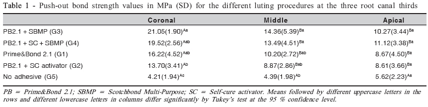
Brazilian Journal Oral Sciences, Vol. 7, No. 27, Oct/Dec, 2008, pp. 1653-1656 The effect of luting techniques on the push-out bond strength of fiber posts André Luis Faria-e-Silva1;André Figueiredo Reis2; Luis Roberto Marcondes Martins3 1DDS, MS, Graduate Student, Department of Restorative Dentistry, Piracicaba Dental School, University of Campinas, Piracicaba, SP, Brazil 2DDS, MS, PhD, Professor, Department of Restorative Dentistry, Guarulhos University, Guarulhos, SP, Brazil 3DDS, MS, PhD. Professor, Department of Restorative Dentistry, Piracicaba Dental School, University of Campinas, Piracicaba, SP, Brazil Received for publication: May 27, 2008 Accepted: September 22, 2008 Code Number: os08038 Abstract Aim: This study aimed to evaluate the effect of using a chemical co-initiator and/or an additional coat of a hydrophobic resin on the bond strength of fiber posts luted with a dual-cured resin cement. In addition, the use of the resin cement only was also evaluated. Introduction Some single-bottle etch-and-rinse adhesives have been demonstrated to be incompatible with chemically cured composites due to an adverse chemical interaction between unpolymerized acidic adhesive resin monomers and the basic tertiary amine catalyst in the composite1. This fact prevents composite polymerization and can create an area susceptible to fracture propagation. Thus, different types of chemical co-initiators have been introduced to overcome this shortcoming2. However, this adverse interaction is only partially responsible for such an incompatibility. The other factor responsible for impairing bonding is the fact that some adhesives can behave as permeable membranes that permit the passage of fluids after polymerization3-5. In the presence of a slow-setting composite, water diffusion from dentin tends to be exacerbated. The water that migrates to the composite-adhesive interface can be trapped as water blisters, which might act as stress raisers that may result in debonding of the resin-dentin interface6 . Clinically, this incompatibility can occur during luting of endodontic posts. The use of dual-cure resin cements is recommended in order to provide a controlled working time, which cannot be controlled in self-cure cements. However, Pfeifer et al.7 demonstrated that incompatibility between single-bottle adhesives and dual-cured cements can occur when the resin-based luting cements are not photoactivated. The most apical areas of the root canal rely mostly on the chemical cure rather than the physical cure8 . During fiber post cementation, the low compliance of the canal space renders it impossible to accommodate resin cement polymerization shrinkage. Thus, in addition to the low bond strength that can occur due to the high complexity of the adhesive procedures in the root canal, debonding can occur even in the absence of incompatibility. Goracci et al.9 observed no difference in post retention with or without the application of a dentin bonding agent. For the authors, the main factor contributing to displacement resistance of the bonded and unbonded fiber posts was the sliding friction. This study examined the effect of using a chemical coinitiator and/or an additional coat of a more hydrophobic bonding resin used with a single-bottle etch-and-rinse adhesive on the push-out bond strength of a fiber post luted with a dual-cure resin cement. In addition, the importance of application of a dentin adhesive on post retention was evaluated. The tested null hypotheses were: (1) the different luting procedures do not affect the pushout bond strength and (2) the push-out bond strength is not different at the different regions tested. Material and Methods Twenty-five bovine incisors with mature apices and roots with no curves were selected for this study. The crowns were removed in order to obtain a remaining 17-mm long root segment. For the endodontic treatment, a step-back preparation technique was used with stainless-steel K-files and Gates-Glidden drills #2 to #4. All enlargement procedures were followed by irrigation with 2.5% sodium hypochlorite. The prepared root canals were obturated with gutta-percha cones using the lateral condensation technique and Sealer-26 resin sealer (Dentsply Indústria e Comércio Ltda., Petrópolis, RJ, Brazil). The filled roots were stored in 100% relative humidity for at least 72 h to allow the resin sealer to set. Post-holes were prepared by standardization of the length at 9 mm and the preparation was performed with a size 5 largo drill. A 1.5-mm diameter glass fiber-reinforced composite post system Reforpost® (Angelus, Londrina, PR, Brazil) was used in this study. Roots were randomly divided into three groups (n=5) according to the adhesive procedure to be used. The bonding procedures were carried out as follows: Group 1: The canal walls were etched with 35% phosphoric acid (Dentsply Indústria e Comércio Ltda.) for 15 s, rinsed for 15 s and gently air-dried. Excess water was removed from the post space with absorbent paper points. Two coats of Prime & Bond 2.1 (Dentsply Indústria e Comércio Ltda.) adhesive system were applied, gently air dried and lightcured for 40 s. Group 2: The bonding procedure was performed as described for Group 1. However, before adhesive application, Prime & Bond 2.1 was mixed with the selfcure activator (Dentsply Indústria e Comércio Ltda.) at a 1:1 ratio. Group 3: The bonding procedure was performed as described for Group 1. Afterwards, one coat of a hydrophobic adhesive resin (Bonding Agent, Scotchbond Multi-Purpose: 3M/ESPE, St. Paul, MN, USA) was applied, the excess was removed with paper points and the adhesive was light-cured for 40 s. Group 4: The bonding procedure was performed as describe for Group 2. After adhesive light-curing, the bonding agent of SBMP was applied and light-cured for 40s Group 5: The resin cement was used with no adhesive. In all groups, the fiber posts were treated with a silane coupling agent and then the respective bonding agent of each group was applied onto the posts (the hydrophobic resin was used for Group 3 and 4, while no adhesive was applied onto the posts of Group 5) and light cured for 20 s. Afterwards, the dual-cured resin cement Enforce (Dentsply Indústria e Comércio Ltda.) was inserted into the root canal with a #40 lentulo spiral. The post was cemented into the root canal with light pressure, and excess luting material was removed. Light activation was performed at the coronal portion of the root for 60 s. The specimens were stored in distilled water for 1 week at 37º C. After the storage period, the specimens were sectioned transversally. Three 1.5-mm-thick slabs were obtained per root and identified as coronal, middle and apical specimens. Each slab was positioned on the push-out jig, which was placed in a universal testing machine (Model 4411, Instron Corp., Canton, MA, USA) with a cell load of 500 N. Load was applied at a crosshead speed of 0.5 mm/min until post dislodgement occurred. Statistical analysis was performed using split-plot two-way ANOVA followed by Tukey’s post-hoc test at 95% confidence level. Results Split-Plot ANOVA showed that there were statistically significant differences for the factors “bonding procedure” (p<0.0001), “root region” (p<0.0001) and for interaction between factors (p = 0.0059). Tukey’s test was used for the interactions between factors and the results are shown in Table 1. In the coronal third, G3 presented higher mean push-out bond strengths than G2. The no-adhesive group (G5) presented the lowest values in this third. When adhesive was used, there was no statistically significant difference between the other groups. In the middle third, G3 and G4 (hydrophobic resin) presented higher values than G5 (no adhesive). However, there were no differences between adhesive procedures in this third. In the apical third, all groups presented similar push-out bond strengths. In all groups, except for G5, the coronal third presented higher bond strength values than the middle and apical thirds, which were not significantly different from each other. In G5, there were no significant differences between the bond strengths recorded in the different regions evaluated. Discussion The first null hypothesis was rejected based on the results of the push-out bond strength test. It was demonstrated that the push-out bond strengths differed depending on the luting procedure used. The only difference between adhesive methods occurred in the coronal third, where hydrophobic resin adhesive application produced higher values of fiber post retention than the use of self-cure activator only. In this third, the resin cement benefits largely from both light and self-curing as they are readily accessible to the curing light10. Thus, incompatibility is not expected to occur. One possible reason for this is a difference in adhesive degree of conversion and in adhesive layer quality. When the adhesive is applied, it can flow to the apical region and reduce the adhesive layer thickness in the coronal region. The solvent content in Prime & Bond 2.1 is approximately 80 wt%11. Solvent evaporation might result in additional reduction of the adhesive layer thickness. The self-cure activator contains only 2% of chemical co-initiator and the remainder is solvent (ethanol and acetone). The use of self-cure activator increases the solvent content and can result in a thinner adhesive layer12 As the adhesive layer is thin, blisters can be formed even when the resin cement is light-cured due to rapid water movement across the adhesive13, reducing the bond strength. Even though positive pulpal pressure is not present in endodontically treated teeth, dentin is still hydrated, which might compromise adhesion. Rinsing the etchant with water during bonding procedures probably result in the retention of substantial amounts of water within the widened tubular entrances created by acid-etching14 This water is not completely removed with the use of paper points and may be responsible for the occurrence of fluid droplets in the adhesive layer. These droplets may act as stress raisers and contribute to crack propagation during the push-out testing, reducing post retention15 . In addition, Zheng et al.16 reported that thin adhesive layers are not adequately polymerized due to the inhibition caused by oxygen. Thus, the additional application of a more hydrophobic bonding resin might have improved the polymerization degree of the adhesive layer and eliminated or reduced the permeability17-18. However, this effect did not occur when the hydrophobic resin and selfcure activator were used in the same procedure. This might be attributed to the low degree of conversion of the first adhesive layer formed by Prime&Bond 2.1 and the selfcure activator (unpublished data). The increased solvent content by use of self-cure activator hinders its evaporation from the adhesive layer19. Residual solvents might impair monomer conversion, forming pores that can act as flaw initiating sites during push-out testing, and increase adhesive permeability. An interesting observation in the present investigation was that the highest values of fiber post retention occurred in the coronal third for all adhesives used. The apical and middle thirds presented the lowest values and no difference was observed among them. Thus, the second null hypothesis was also rejected. The direct light curing of both adhesive and resin cement promoted higher bond strengths in the coronal third, independently of the adhesive method. Moisture control and adhesive light activation are even more critical procedures in the apical region, which can contribute to the lower bond strengths to dentin. In addition, resin cement photoactivation is also compromised, and the self-cure component of the polymerization system is responsible for most or all polymerization reaction. In the absence of photoactivation, a chemical incompatibility between single-bottle adhesives and dual-cured cements may occur7. The slower curing rate of resin cement in these thirds can result in water flow to the cement/adhesive interface. The morphology of root dentin at different areas (coronal, middle and apical) and the hybridization ability of these areas may also help to explain the results. Ferrari et al.20 demonstrated a reduction of bonding ability toward apical third of root canal dentin. This is related mainly to the density of dentin tubules in. the coronal and middle thirds compared to the apical third. All these factors can be responsible for the reduction in fiber post retention. It is important to emphasize that incompatibility is not expected to occur when a hydrophobic adhesive resin is used, but low bond strength is still likely to occur in the more apical regions. In the coronal third, all adhesive procedures produced higher fiber post retention compared to the use of resin cement alone. However, in the middle third, adhesive application produced higher bond strengths than the nonadhesive treatment only when a hydrophobic resin was used. Lack of differences among the groups was found only in the apical third. The highest push-out bond strength obtained with the use of hydrophobic adhesive in the middle third probably occurred because this last adhesive coat eliminated the incompatibility between the singlebottle adhesives and the dual-cured resin cement. However, the same effect was not observed in the apical third. In the groups that used the hydrophobic resin adhesive, the first adhesive layer was formed by Prime & Bond 2.1 or by its combination with the self-cure activator. Due to adhesive flow, the adhesive layer is thicker in the apical third. As vapor pressure is reduced in this area, an increase in residual solvent is also expected to occur5. This fact can result in a reduced degree of conversion and in the presence of flaws in the adhesive layer. These two events probably reduced the bond strength to dentin. Both bonding and sliding friction contribute to the resistance to dislocation of fiber posts during the push-out testing. As the bond strength is very low in the apical region, it is likely that the sliding friction has the main contribution to fiber post dislocation resistance9. This friction results from the contact between the resin cement and the root canal walls. However, it is important to observe that the degree of conversion of the resin cement did not influence the sliding friction, since there was no difference between the three regions when only the resin cement was used. Sigemori et al.8 demonstrated that the degree of conversion of Enforce resin cement decreases remarkably as cavity depth increases. A high degree of conversion often results in improved physical and mechanical properties of resin cements. Despite the improvement in the mechanical properties, higher degree of conversion increases the polymerization shrinkage. Since friction occurs by contact, it is reasonable to assume that closer contact between resin cement and root dentin improves fiber post retention. The highest bond strengths were observed in the coronal third of the root. In this region, both sliding friction and adhesive bonding opposes fiber post dislocation. A decrease in bond strength was observed towards the apical regions and the post retention did not benefit from the adhesive application in the apical area. The clinical implications of the reduced bond strengths observed in the middle and apical regions have not yet been reported, but the development of materials with improved adhesive properties to the root canals would certainly improve the quality and reliability of restorative procedures in endodontically treated teeth. In conclusion, the highest bond strengths were observed in the coronal third. The application of a hydrophobic resin produced increased fiber post retention, except in the apical third. On the other hand, application of the self-cure activator did not provide any additional benefit to the interface. References
© Copyright 2008 - Piracicaba Dental School - UNICAMP São Paulo - Brazil The following images related to this document are available:Photo images[os08038t1.jpg] |
| |||||||||
