 |
| About Bioline | All Journals | Testimonials | Membership | News |
|
||||||
|
||||||
Brazilian Journal Oral Sciences, Vol. 7, No. 27, Oct/Dec, 2008, pp. 1662-1665 Quantitative role of mast cells in odontogenic cystic enlargement Shailja Chatterjee1; Sumita Mahajan2; Karen Boaz3; Thomas George4 1MDS, Senior lecturer, Received for publication: June 06, 2008 Accepted: September 16, 2008 Code Number: os08040 Abstract Aim: Mast cells have been hypothesized to play a significant role in pathogenesis of odontogenic cysts. The aim of this study was to evaluate mast cell distribution in cystic lining and the capsule to formulate a mechanism of cystic expansion. Key words: mast cells, degranulation, toluidine blue, odontogenic cysts Introduction Odontogenic cysts are possibly the most common benign destructive lesions in the human maxillofacial skeleton. Three most common jaw cysts- Radicular cysts, Odontogenic Keratocysts and Dentigerous cysts (of developmental odontogenic origin) are characterized by an expansile non – infiltrative growth, resulting in a smooth and usually unilocular cavity containing fluid or semi fluid material, lined by an epithelium and supported by a fibrous connective tissue capsule1 . The expansion of the jaw cyst involves destruction of the extra-cellular matrix due to proteolysis of collagen fibers, osteoid – derived gelatin and protein components of basement membrane1. Mast cells contain numerous cytoplasmic granules, which are degranulated into the extra-cellular space upon activation. In addition to preformed granule contents, activated mast cells can synthesize de novo vasoactive mediators, for example, platelet – activating factor, chemotactic mediators, and several proinflammatory cytokines such as IL-1α, IL-3, IL-6 and TNF – α. Furthermore, mast cells are a rich source of heparin and proteolytic enzymes, such as tryptase, chymase and hyaluronic acid, which participate in connective tissue breakdown in the capsule during normal metabolic turnover, as well as in inflammation1 . Products released by mast cell activation and subsequent breakdown products of connective tissue elements are released into the cyst lumen increasing the hydrostatic pressure with subsequent enlargement. The aim of this study was to evaluate mast cell distribution in cystic lining and the capsule to formulate a mechanism of cystic expansion using morphometric analysis. Material and Methods Paraffin – embedded formalin fixed tissue blocks of Odontogenic Keratocysts, Dentigerous cysts and Radicular cysts (10 each) were retrieved from the archives of the Department of Oral Pathology, Manipal College of Dental Sciences, Mangalore. Sections of 5μm were cut and stained with freshly prepared Toluidine blue solution (1% tolonium blue in 1% sodium chloride), mounted with DPX, and followed by mast cells counting under 40X magnification. Counting of mast cells: Mast cells were counted in ten areas under 40X magnification using an ocular grid with a total area of 0.30625mm2, divided in four zones:
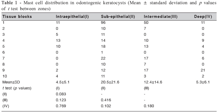
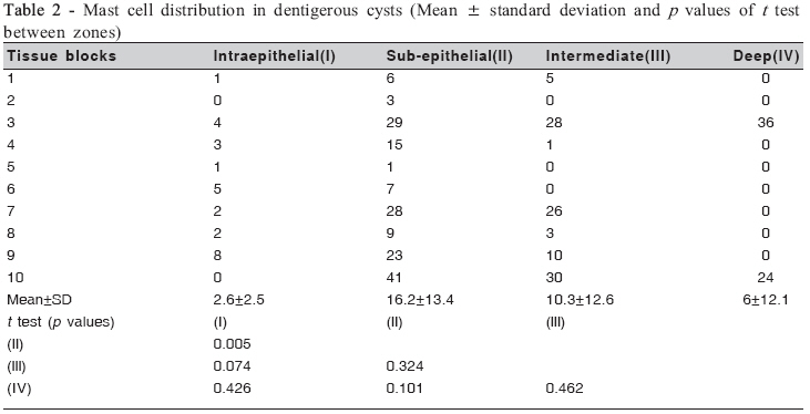
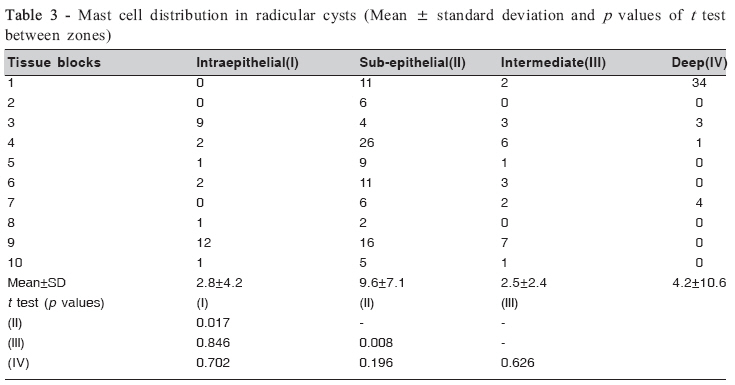
For intraepithelial counting, the graticule was oriented along the basement membrane and along the connective capsular tissue at the epithelial-capsular junction for counting in subepithelial zone. Every alternate microscopic field was counted. The graticule was then moved further down into two microscopic fields into the capsule and the procedure repeated for intermediate zone. It was then moved further down two microscopic fields into the capsule to a third level (deep zone) and the counting was performed in similar manner. Statistical analysis Means and standard deviation of the mast cell counting were calculated in each layer zone. For comparisons between zones, the t test was applied. Results Statistical analysis revealed an increase in mast cell count in all the three cysts at the sub-epithelial zone. Paired ‘T’ test showed no statistically significant difference (Table 1) between intraepithelial (I) and sub-epithelial zones (II) in Odontogenic Keratocysts (p=0.08), whereas a highly significant difference (Table 2) was noted between the intraepithelial (group I) and sub-epithelial zones (II) of Dentigerous cysts (p=0.005). On the other hand, Radicular cysts showed a highly significant difference (Table 3) between sub-epithelial (II) and intermediate (III) zones (p=0.007). A significant elevation in mean±SD values was noted in mast cell population in Odontogenic Keratocysts in the subepithelial zone as compared to Dentigerous and Radicular cysts. 1.Intraepithelial Sub-epithelial Intermediate Deep Discussion Mast cells are found widespread throughout the connective tissue wall of all the cysts particularly in the subepithelial zone and are source of a variety of proteolytic enzymes found in the cystic fluid. The results of the present study showed a great concentration of mast cell in subepithelial zone in all cyst walls. This concentration was higher in OKCs than dentigerous and radicular cysts, suggesting an increased breakdown of capsular matrix in OKCs. OKC epithelium has been shown to be nonkeratinized at places, which causes a transport of breakdown matrix products into the cystic lumen2, and consequently can determine an elevated osmolality of the cystic fluid, which partly explains the greater aggressiveness of OKC comparing to other odontogenic cysts. Smith et al.1 found a considerable amount of mast cells in the walls of odontogenic keratocysts, dentigerous and radicular cysts with the highest concentration seen in sub-epithelial zone. Mast cells were also observed in the epithelial linings, which the authors suggested to be due to a chemotactic stimulus attracting them to the epithelial lining or luminal fluid contents. Smith et al.2 concluded that the major source of glycosaminoglycans and proteoglycans in cystic fluid was from the ground substance of the connective tissue capsule, released because of normal metabolic turnover and inflammatory degradation. Degranulating mast cells release heparin and other hydrolytic enzymes, which facilitate breakdown of glycosaminoglycans and proteoglycans2,3. Histochemical investigations of the connective tissue capsule in odontogenic cysts have demonstrated that hyaluronic acid, a product of mast cell degranulation, is the predominant glycosaminoglycan present along with less amounts of sulphated glycosaminoglycans2,3. The release of glycosaminoglycans and proteoglycans into the luminal fluid contributes significantly to osmotic and hydrostatic pressure by increasing the osmolality of the cyst fluid, thereby raising the internal hydrostatic pressure2,3. Cyst expansion is also affected by the rate in which the surrounding bone is destroyed particularly at the cyst-bone interface4 . Teronen et al.4 stated that activated mast cells can synthesize vasoactive and chemotactic mediators (e.g., platelet – activating factor) as well as several pro inflammatory cytokines such as IL-3, IL-6 and TNF-α de novo. These chemical mediators increase vascular permeability thereby facilitating influx of highly osmolar substances in cystic lumen. The highest concentration of mast cells in OKCs explains a greater expansion as compared to other odontogenic cysts. The authors also found a high number of extensively degranulated mast cells in the area of cyst expansion at the border with the bony wall suggestive of high activity of mast cells in this area. Mast cell degranulation also releases tryptase and prostaglandins which aid in bone resorption which is a feature in cyst enlargement at cyst-bone interface. In addition, interleukin-1α in OKC cyst wall has been found to have an enhancing effect on matrix metalloproteinases secreted by fibroblasts5 . Several other studies have also substantiated the effect of MMPs along with tissue inhibitor of metalloproteinases and collagenases in cyst walls6,7. These cell products have been found to be stimulated by mast cell derivatives7- 9 . Based upon the literature review analysis, it can be proposed that the degranulating mast cells release products that contribute to cystic enlargement in four ways:
Based upon the present study and similar investigations, it can be concluded that mast cells play a vital role in the pathogenesis of odontogenic cysts as an elevated number of mast cells was found in the connective tissue capsule of all three odontogenic cysts. The luminal fluid which accumulates as a result of osmolar concentration of mast cell by-products plays an important in cyst enlargement. References
© Copyright 2008 - Piracicaba Dental School - UNICAMP São Paulo - Brazil The following images related to this document are available:Photo images[os08040t3.jpg] [os08040t1.jpg] [os08040t2.jpg] |
| |||||||||
