 |
| About Bioline | All Journals | Testimonials | Membership | News |
|
||||||
|
||||||
Brazilian Journal of Oral Sciences, Vol. 8, No. 1, Jan-Mar, 2009, pp. 19-24 Histomorphometric study of alveolar bone after therapy with immunosuppressant FK506 Rogério Lacerda dos Santos1, Marcos Farina de Souza2, Renato Torres Gonçalves3, Marco Aurélio Martins4, Margareth Maria Gomes de Souza5 1 Specialist in Orthodontics, Universidade Federal de Alfenas (Unifal), Alfenas (MG), Brazil; Master of Orthodontics, Universidade Federal do Rio de Janeiro (UFRJ), Rio de Janeiro (RJ), Brazil
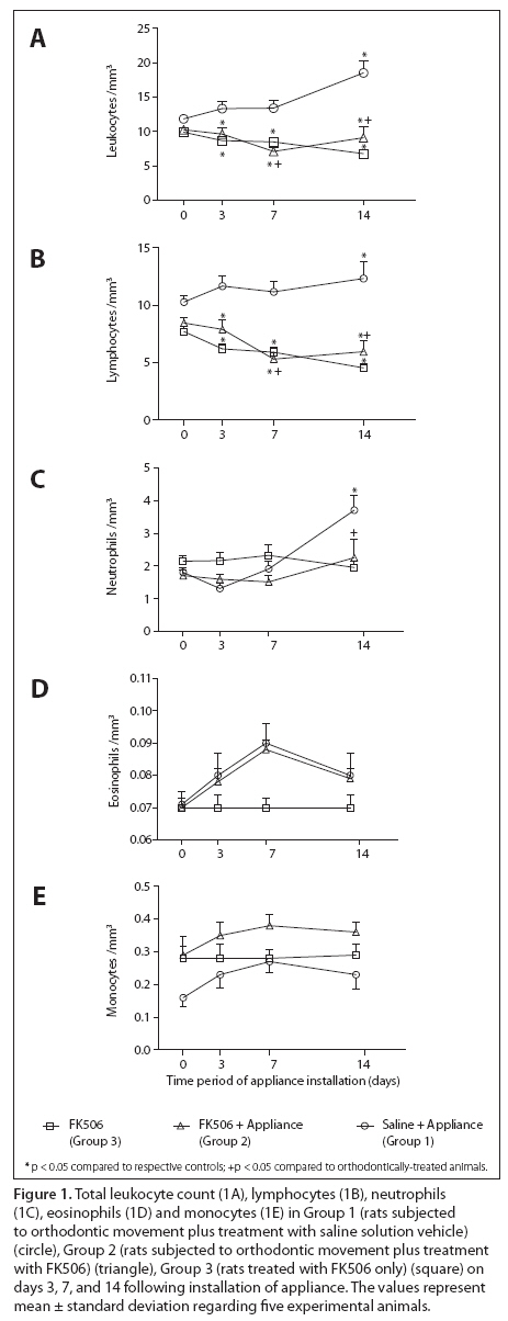
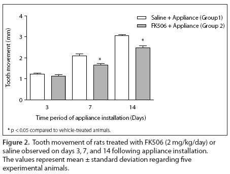
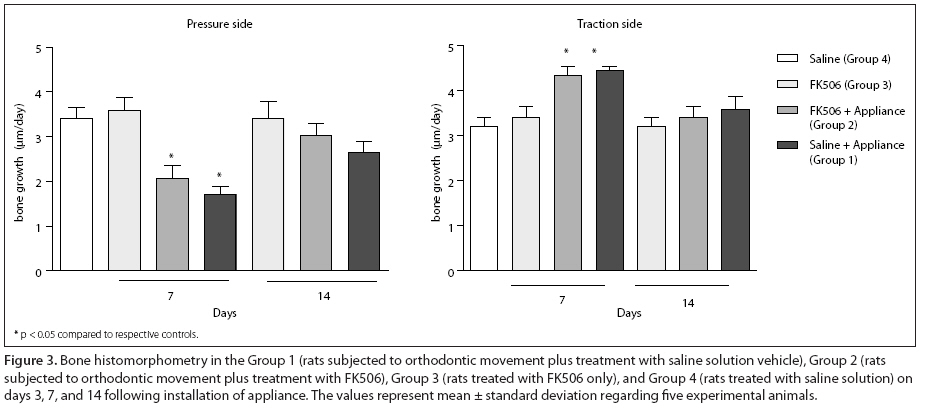
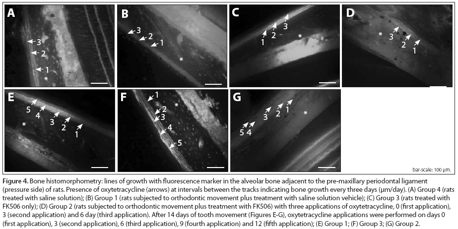
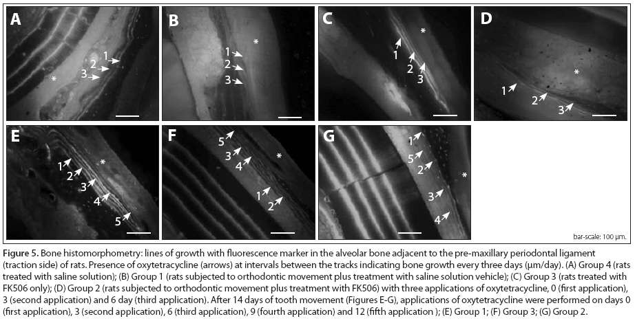
Received for publication: December 01, 2008 Code Number: os09004 Abstract Aim: Several medications affect bone metabolism and the rate of tooth movement. The objective of the present study was to test the hypothesis that treatment with immunosuppressant tacrolimus (FK506) can interfere with bone turnover, decreasing the rate of tooth movement. Methods: Sixty male Wistar rats were divided into four groups of 15 animals each: Group 1: rats subjected to orthodontic movement plus treatment with saline solution vehicle; Group 2: rats subjected to orthodontic movement plus treatment with FK506; Group 3: rats treated with FK506 only; and Group 4: rats treated with saline solution vehicle only. The FK506 dose was 2 mg/kg/day. The treatment was initiated 14 days before the appliance installation and then kept for up to 14 days. In addition to the administration of the immunosuppressive drug, 10 mg/kg of oxytetracycline were injected at intervals of three days in order to show osteoblastic activity and bone growth at a histological level. Results: Histomorphometrical measurements showed greater tooth movement in Group 1 than in Group 2 at all periods (days 3, 7 and 14), though significant difference (p < 0.05) was observed only on days 7 and 14. Conclusions: FK506 significantly influenced the rate of tooth movement in rats subjected to the application of this medication. Keywords: tooth/immunology, tooth movement, bone and bones. Introduction Tacrolimus (FK506) is an immunosuppressive agent derived from Streptomyces tsukubaensis1, which is widely used in patients subjected to organ transplantation2. Some authors3,4 have suggested that FK506 exerts its anti-inflammatory effect mainly by interfering with the activation of T-cells, by suppressing the production of cytokines, particularly TNF-α, IL-1β, IL-2, and IL-6, modulating the inflammation, and decreasing both tissue turnover and bone resorption. On the other hand, bone destruction and development of severe osteoporosis have been reported5. Studies using cell culture6,7 have demonstrated that not only messenger RNAs of NFATc1, NFATc2 and NFATc3 are present in the osteoclast-precursor cells, but also FK506 inhibits the final stages of cell cycle through osteoclastic apoptosis. Altogether, these findings are in accordance with the idea that the mechanisms by which osteoclastogenesis is inhibited and osteoclastic apoptosis is promoted are similar to those involved in inhibiting the production of transcription factor NFAT and inflammatory cytokines within T lymphocytes8. Therefore, the objective of the present study was to test the hypothesis that FK506 treatment can interfere with bone turnover, decreasing the rate of tooth movement. Material and methods Animal model A total of 60 male Wistar rats (Rattus norvegicus) aged nine weeks and weighting 220 to 280 g were housed according to the guidelines for animal research as recommended by Fundação Oswaldo Cruz (Fiocruz, Rio de Janeiro, RJ, Brazil). The immunosuppressive agent FK506 (Tacrolimus, Prograf®, Astellas, Ireland) was orally administrated at a dose of 2 mg/kg/day as a suspension containing water and 5% dextrose9. The treatment was initiated 14 days before the appliance installation and, then, kept for up to 14 days. In addition to the administration of the immunosuppressive drug, 10 mg/kg of oxytetracycline (Ouro Fino, Cravinhos, SP, Brazil) were injected at intervals of three days, in order to show osteoblastic activity10 and bone growth at a histological level. The experiments were reviewed and approved by the ethics committee on animal research of Universidade Federal do Rio de Janeiro (UFRJ). Experimental groups Four groups of 15 animals each were formed according to the treatment modality: Group 1: rats subjected to orthodontic movement plus treatment with saline solution vehicle; Group 2: rats subjected to orthodontic movement plus treatment with FK506 (2 mg/kg/day); Group 3: rats treated with FK506 only (2 mg/kg/day); and Group 4: rats treated with saline solution vehicle only. Appliance design The appliance was made according to description by Arias and Marquez-Orozco11 regarding wire diameter and installation. The initial load to be exerted by the orthodontic appliance was determined before its insertion by means of a ball dynamometer (Dentaurum 040-711, Ispringen, Germany) for measuring of 35 gf11. The rats were anesthetized with an intraperitoneal injection of sodium thiopental (Cristália, Campinas, SP, Brazil; 50 mg/kg body weight). Incisor gaps were measured in the cervical region of the tooth and recorded by two investigators using a precision caliper accurate to 0.01 mm (Starret, Itú, SP, Brazil), and the mean values were recorded when the animals were sacrificed. Blood count Total and differential leukocyte counts (lymphocytes, monocytes, eosinophils and neutrophils) were evaluated on day 0 (before appliance installation) and on sacrifice day in all groups, in order to monitor the immunosuppressive effect of FK506. Blood was collected from the tail tip of each rat and leukocyte count was realized with a light microscope (Olympus BX40; Olympus, Hamburg, Germany). Histomorphometry After 3, 7, and 14 days of orthodontic movement, the rats were sacrificed and then decapitated. Their heads were dissected and the pre-maxillae were removed, fixed in 10% formaldehyde solution for 24 hours and, thereafter, washed with PBS solution. The specimens were dehydrated and embedded in Spurr epoxy resin (Ted Pella Inc., San Jose, CA, USA), which was dissolved in ethanol at proportions of 1:3, 1:2, 1:1, 2:1, and 100% spurred every 12 hours before polymerization for 24 hours at 70 °C. The specimens were sectioned in the frontal plane at 1 mm from the palatal face of central incisors, resulting in bone blocks of 3 mm. The blocks were placed perpendicular to the diamond saw machine (Model 650, South Bay Technology, Inc., San Clemente, CA, USA), producing 40-µm-thick cross-sections, which were polished with #400-, #600-, #1200-, #2000- and #2500-grit abrasive papers (3M do Brasil, Sumaré, São Paulo, Brazil) to obtain a uniform thickness for visualization with a fluorescence microscope (Axioplan, Zeiss, Jena, Germany). This histomorphometric analysis focused on the alveolar bone located laterally to the incisors, which was divided into cervical pressure (distal) and traction (mesial) sides to the apical region following the cervical-apical axis of the root (adapted from Kale et al.)13. Three parallel sections were randomly counted without repetition, from cervical to apical regions of the tooth, regarding pressure and traction sides. A single operator blinded to the groups recorded the data on measurements of the fluorescence lines (perpendicularly interlines distance) regarding bone growth during the 3-day-interval applications of oxytetracycline. Image Tool 1.0 software (Image Tool, San Antonio, TX, USA) was used for processing the data collected and for intra and intergroup comparisons. The distance between the growth lines was divided by three in order to set the daily speed of growth (osteoblastic activity expressed as µm/day), as described by Roberts et al.12. The fields of periodontal ligament at pressure and traction sides were captured by using the AxioVision Rel 4.5 software with the fluorescence microscope at ×20 magnification. Results Effect of appliance installation and FK506 treatment on weight gain The orthodontic appliance was well tolerated by the animals during the experimental period and caused no soft tissue irritation. The weight gain regarding each period of time was calculated on the basis of the initial treatment with FK506. Both the immunosuppressant and the orthodontic appliance were found to have influenced the weight gain, since the non-treated animals had the highest gains. Group 3 had higher weight gain within 28 days (41.0 ± 1.2 g) compared to Group 1 (35.3 ± 2.0 g), though without statistical significance (p > 0.05). On the other hand, Group 2 had the lowest weight gains on days 3 (15.7 ± 2.3 g), 7 (16.7 ± 2.2 g), and 14 (21.4 ± 1.6 g). There were significant differences (p < 0.05) between Group 2 and Groups 1 and 3 in all time periods of study. Effect of appliance installation and FK506 treatment on leukocyte count Immunosuppression was observed in the animals from Groups 2 and 3 as leukocyte and lymphocyte counts were shown to have similar and decreasing values during the experiment in all time periods. Also, statistically significant differences (p < 0.05) were observed between both groups and their respective controls (day 0). There was statistically significant difference (p < 0.05) between Group 2 and Group 1 on days 7 and 14. Contrary to Groups 2 and 3, however, Group 1 (rats subjected to orthodontic movement) showed increased values of leukocytes and lymphocytes counts, i.e., there was body response to the stimulus (force) applied to the teeth. Group 1 also showed significant difference (p < 0.05) between day 0 and day 14. No statistically significant difference (p > 0.05) was found in neutrophil counts recorded in Groups 2 and 3. Group 1 showed an increased neutrophils count on day 14, which was significant different compared to the respective control (day 0) (p < 0.05). Eosinophil and monocyte counts showed similar patterns between Groups 1 and 2, with increased values on day 7 and decreased values on day 14. Group 3 had significantly greater (p < 0.05) monocyte count compared to Group 1 on day 0, which was kept for other time periods. No significant difference (p > 0.05) regarding this cell types was observed between the groups in all periods of time studied (Figure 1). A trend of increase in the number of eosinophils due to the presence of orthodontic appliance could be observed, whereas the presence of FK506 promoted an increase in the number of monocytes. Tooth movement Before installing the appliances, a lack of measurable space, between the upper incisors, was observed. Group 1 showed greater tooth movement compared to Group 2 on days 3, 7, and 14 (Figure 2), however significant difference (p < 0.05) was observed only on day 7 (2.09 ± 0.10 mm and 1.66 ± 0.09 mm) and day 14 (3.06 ± 0.05 mm and 2.48 ± 0.11 mm), respectively. Bone histomorphometry Bone growth was lower on the pressure side of the groups subjected to orthodontic movement compared to saline group, and higher in the FK506 group after seven days. There was statistically significant difference (p < 0.05) between the saline (3.40 ± 0.24 µm/day) and saline plus orthodontic appliance (1.69 ± 0.22 µm/day) groups as well as between the saline (3.40 ± 0.24 µm/day) and the FK506 plus orthodontic appliance (2.07 ± 0.27 µm/ day) groups after seven days of orthodontic movement. The traction side of the group subjected to orthodontic movement plus treatment with saline solution had higher bone growth after seven days compared to the group subjected to application of saline solution only. There was statistically significant difference (p < 0.05) between the saline (3.21 ± 0.18 µm/day) and saline plus orthodontic appliance (4.44 ± 0.11 µm/day) groups as well as between the saline (3.21 ± 0.18 µm/day) and the FK506 plus orthodontic appliance groups (4.35 ± 0.19 µm/day) after seven days of orthodontic movement. Within 14 days of tooth movement, no statistically significant differences (p > 0.05) were found among the groups on either pressure or traction sides (Figure 3, Figure 4 and Figure 5). Discussion Previous studies have demonstrated that treatment with FK506 (tacrolimus) can induce bone loss in human beings14 as well as in experimental models5,15. In cell cultures, on the other hand, FK506 can also inhibit the final phases of the life cycle of osteoclast-precursor cells through induction of apoptosis6,7. It has been suggested that FK506 inhibits the production of pro-inflammatory cytokines, particularly TNG-α, IL-1β and IL-63, both modulating the inflammation and decreasing tissue destruction and bone resorption4. Because FK506 has the capacity to alter bone metabolism, one can suggest that it can interfere with the rate of tooth movement. Investigating this possibility was the goal of the present study. Although rats have continuous tooth eruption, their periodontal structures are arranged similarly to that of human beings, with the incisors quickly responding to orthodontic forces ranging from 20 to 40 gf16. A load of 35 gf was used in the present study11. It was observed that animals subjected to orthodontics plus treatment with FK506 had a smaller amount of tooth movement. This finding was confirmed by the bone histomorphometry, which showed greater bone growth (µm/day) for the group subjected to orthodontic movement plus treatment with FK506 compared to the group subjected to orthodontic movement plus treatment with saline in the pressure side adjacent to the orthodontic movement. Bone is a tissue that grows and maintains its structural integrity by two basic physiological processes: modeling, when size and shape of the skeletal unit change, and remodeling, when there is no change in the shape of skeletal unit in previously existing bone metabolism. Histomorphometry is well recognized as an important benchmark for evaluating the lines of bone growth with vital colors, such as those obtained with oxytetracycline, alizarin, and calcein10 on fluorescence microscope. Similar bone growth was observed in the animals subjected to the application of FK506 and saline, which suggests that the immunosuppressive drug did not interfere significantly with bone growth, though an increasing trend was found in the groups treated with the immunosuppressant. As the influence of FK506 on bone metabolism is a dose-dependent event9, in the present experiment, the doses were daily calculated and adjusted according to the weight gain of each animal. Some factors such as metabolic and gastrointestinal changes17,18 caused by FK506 can also interferer with weight gain. The presence of an orthodontic appliance itself is another factor that can have an influence on weight changes as it makes feeding more difficult, which might explain why animals receiving either orthodontic treatment or FK506 treatment had low weight gain compared to animals treated with saline solution. In the present study, weight gain observed in FK506-treated animals was higher than that found in previous studies9,19, which used the same daily FK506 dosage. In fact, the use of orthodontic appliance in combination with FK506 treatment resulted in significant decrease in the weight gain, a joint influence of both factors. Nevertheless, histological data demonstrated that FK506 can significantly reduce the induction of alveolar bone loss and granulocytes infiltration4, because of its capacity to inhibit osteoclastogenesis in vivo and in vitro7 by means of osteoclastic apoptosis. Such an effect would result in inactivation of NFATc1, NFATc2, and NFATc3 (messenger RNAs) existing within osteoclast-precursor cells6. Osteoclasts are the main cells involved in the process of alveolar bone remodeling and there seems to be a direct relationship between rate of tooth movement and number of osteoclasts20. In the present study, however, the bone growth observed in the animals subjected to orthodontic movement was greater than in those animals subjected to orthodontic movement plus FK506 treatment and significantly greater compared to the control animals (treated with saline), which is in accordance with the findings of previous investigations6,7,21,22. In conclusion, the analysis of tooth movement and the histomorphometrical measurements of the alveolar bone indicate that the therapy with immunosuppressant tacrolimus (FK506) influenced significantly the bone turnover. There was a lower rate of tooth movement in the group subjected to application of the immunosuppressant. Acknowledgements The authors would like to thank the Fundação de Amparo à Pesquisa do Estado do Rio de Janeiro (Faperj), for the research funding, and the Department of Pathological Anatomy of the UFRJ, for the great help in carrying out this research. References
Copyright © 2009 - Piracicaba Dental School - UNICAMP São Paulo - Brazil The following images related to this document are available:Photo images[os09004f2.jpg] [os09004f5.jpg] [os09004f1.jpg] [os09004f4.jpg] [os09004f3.jpg] |
| |||||||||
