 |
| About Bioline | All Journals | Testimonials | Membership | News |
|
||||||
|
||||||
Brazilian Journal of Oral Sciences, Vol. 8, No. 1, Jan-Mar, 2009, pp. 25-29 Surface morphology alterations in bovine dentin exposed to different bleaching agents Juliana Nascimento Santos1, Daniel Pinto de Oliveira1, Fábio Roberto Dametto1, Brenda Paula Figueiredo de Almeida Gomes2, Alexandre Augusto Zaia2, José Flávio Affonso de Almeida2, Caio Cezar Randi Ferraz2 1 DDS, MSc, PhD, Department of Restorative Dentistry and Endodontics, Faculdade de Odontologia de Piracicaba, Universidade Estadual de Campinas (Unicamp), Piracicaba (SP), Brazil
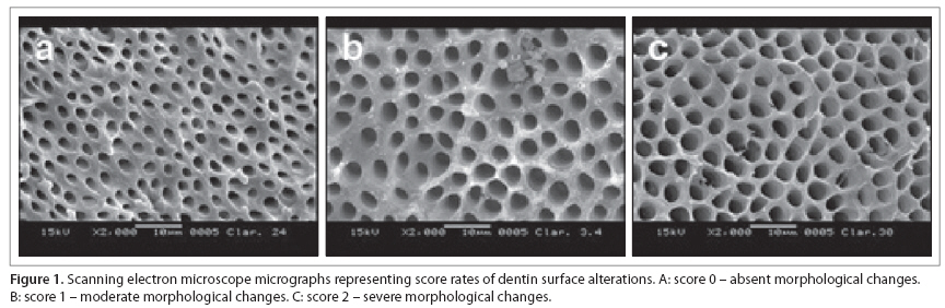
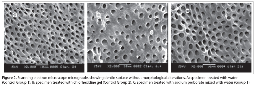
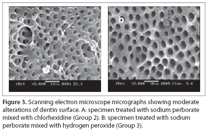
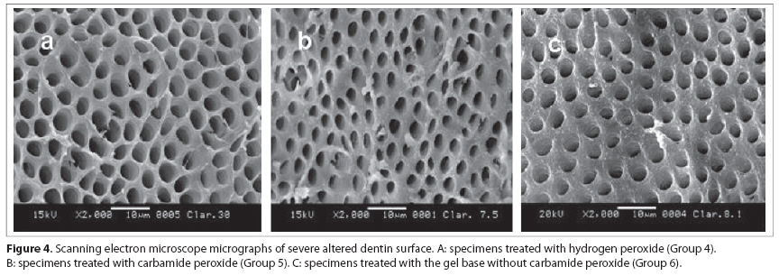
Received for publication: November 5, 2007 Code Number: os09005 Abstract Aim: This study evaluated the morphological changes caused by internal bleaching agents on dentin surface. Keywords: tooth bleaching, dentin, dental pulp cavity. Introduction Color changes may occur in teeth that had undergone endodontic treatment, consisting in an important esthetic problem. Bleaching is one of the procedures that can be used to treat discoloration, and it is commonly performed in non-vital teeth1. The walking bleach technique is considered the safest and most accepted intracoronal bleaching technique1-2. It consists on the placement of bleaching agents in the pulp chamber of root-filled teeth. Traditionally, intracoronal bleaching is achieved with the use of 30% hydrogen peroxide and sodium perborate, which can be used either separately or in combination3. Carbamide peroxide formulations, commonly used for nightguard vital bleaching, have also shown to be effective as an intracoronal bleaching agent4. Recently, a gel base containing 2% chlorhexidine was introduced as a vehicle for sodium perborate, being an alternative to water or hydrogen peroxide in order to prevent coronal microleakage during the walking bleach technique5. However, some side effects resulting from the use of internal bleaching agents have been reported. Thirty-percent hydrogen peroxide has been associated to the development of external cervical root resorption6. Chang et al. 7 found that intracoronal bleaching with 30% hydrogen peroxide and sodium perborate used, either alone or in combination, weakened dentin. A significant alteration on the levels of inorganic components of dentin8 and surface morphological changes9 were reported after treatment with carbamide peroxide. During intracoronal bleaching, the materials are placed in direct contact with dentin, and subsequently the access cavity should be readily sealed to minimize coronal leakage10. Being the major constituent of tooth structure, any changes on the surface morphology of dentin are likely to affect the outcomes of dentin bonding2. The aim of this study was to determine in vitro, the efffects of different bleaching agents on dentin surface morphology using scanning electron microscopy. Material and methods Twenty bovine mandibular incisors were obtained from slaughtered cattle at the age of one to two years, and stored in 0.2% thymol to be used within six months after extraction. The coronal portion of each tooth was cut longitudinally from mesial to distal into two equal segments with a water-cooled diamond disk (KG Sorensen, Barueri, SP, Brazil). The buccal segment was sectioned in “x” and “y” directions and serial coronal slabs were obtained. The exposed pulp chamber dentin was cleaned, by immersing the slabs in 5.25% NaOCl for ten minutes, and sequentially in 17% EDTA for the same time in order to remove the smear layer formed during sectioning11. The pooled coronary slabs were rinsed in running water for 12 hours, dried, and then randomly assigned to eight groups of five specimens each. Each experimental group was treated with one of the following bleaching agents: G1 – sodium perborate (NaBO3 .4H2 O; Proderma, Piracicaba, SP, Brazil) mixed with water, G2 – sodium perborate mixed with 2% chlorhexidine gel (Proderma), G3 – sodium perborate mixed with 30% hydrogen peroxide, G4 – 30% hydrogen peroxide, G5 – 37% carbamide peroxide (Whiteness, Porto Alegre, RS, Brazil), and G6 – gel base without carbamide (Whiteness). The following control solutions were used: C1– distilled water and C2 – 2% chlorhexidinegel. The bleaching agent was combined with the designated vehicle for each group to maintain a 2:1 ratio in solution. The specimens were immersed in the respective test material and incubated at 37 °C for seven days in 100% humidity, as previously described by Zalkind et al. 9. The samples were, then, rinsed in an ultrasonic bath during on hour, dehydrated in ascending alcohol concentrations, dried and sputter coated with gold (Denton Vaccum Desk II, Moorestown, NJ, USA)9. Dentin surface morphology was analyzed with a scanning electron microscope (JEOL – JSM 5,600 LV, Noran Instruments, Tokyo, Japan) operated at 15 KV. The fi ve most representative images from di fferent regions of each tooth segment were captured at × 2000 magnifi cation, and the surface morphological alterations were analyzed by three previously calibrated examiners. Morphological changes were classi ed as absent (score 0), moderate (score 1) and severe (score 2), according to pre-established parameters (Figure 1). Inter-examiner agreement was veri ed using the Kappa test. The rank averages obtained for the groups were subjected to Kruskal-Wallis analysis of variance at 5% significance level. Results The effects of the various bleaching agents on dentin surface morphology are expressed in ranks in Table 1. The Kappa value for inter-examiner agreement was 0.9. Most tested materials caused some morphological alteration on dentin, except for sodium perborate mixed with water (G1), the control solutions distilled water and 2% chlorhexidine gel (C1 and C2) (Figure 2). Specimens treated with sodium perborate mixed with 2% chlorhexidine gel (G2) and sodium perborate mixed with 30% hydrogen peroxide (G3) showed more evident surface alterations than Groups G1, C1 and C2, as a moderate attending of dentin surface and a mild erosion of intertubular dentinal matrix could be detected. The dentinal tubules remained opened but not widened (Figure 3). Severe morphological changes could be observed for the groups treated with 30% hydrogen peroxide (G4), 37% carbamide peroxide (G5) and the gel base without carbamide peroxide (G6), which differed signi cantly (p < 0.05) from the other groups. Theses agents gave to the dentin a flattening and an etching-like appearance on intertubular area. The dentinal tubules were wider, showing demineralization of intratubular dentin (Figure 4). Discussion The results of this study indicated that the materials commonly used for internal bleaching may have an e ffect on dentin surface morphology. The alterations observed showed a strong relationship between the morphological changes and the bleaching agent used. It is quite relevant to test the eff ects of the bleaching materials on dentin, since a close contact occurs during internal bleaching procedures. Most morphological studies have evaluated external bleaching agents and their eff ects on enamel12-13, whilst information concerning the in fluence of materials used for internal bleaching on dental tissues is rather limited. Structural changes of dentin substrate may have an important role on the performance of dental restorations. In fact, a variety of studies have shown a detrimental eff ect of the bleaching procedures on the bond strength and sealing ability of composite restorations10,14-16 to dentin, thus the authors suggest morphological alterations as one of the reasons for that. The most significant morphological changes were caused by 30% hydrogen peroxide, 37% carbamide peroxide and the gel base without carbamide peroxide. The adverse effects of hydrogen peroxide on dental hard tissues in external and internal bleaching has been previously reported on the literature and include alterations in the chemical structure of dentin17 and a reduction on the calcium/phosphorous (Ca/P) ratio8-9, which could be responsible for the alterations observed. In addition, hydrogen peroxide was found to increase dentin solubility and cause protein oxidation of organic matrix8, this may explain the severe etching-like appearance and surface attening observed in this study. Th e acidity provided by the low pH of hydrogen peroxide solutions2 might have contributed for the enlargement of dentinal tubules. The above-mentioned complications, allied to a frequent occurrence of external root resorption, made some researchers recommend avoidance of highly concentrated hydrogenperoxide solutions for intracoronal bleaching18-19. Bleaching agents containing carbamide peroxide are commonly used in the treatment of discolored vital teeth. Th ese materials started to be considered for intracoronal bleaching since satisfactory esthetic results were reached4 and no association with external root resorption was found20. However, the severe dentin surface alterations observed in this study indicate that carbamide peroxide agents may have an adverse eff ect on the organic and inorganic components of dentin. Moderate surface alterations have already been reported for 10 and 15% carbamide peroxide9,21. Since a highly concentrated (37%) formulation was used in the present study, more severe morphological changes would be expected. Another study20 showed a decrease on dentin microhardness similar to that caused by hydrogen peroxide-based solutions. As carbamide peroxide breaks down into urea and hydrogen peroxide, the previously reported adverse eff ects of hydrogen peroxide bleaching may be applicable for carbamide peroxide. Even though an acidic pH is attributed to hydrogen peroxide solutions, the pH value of carbamide peroxide is almost neutral9, indicating that the surface morphological alterations were not strictly related to pH variations among the bleaching agents. Another important consideration is the fact that the gel base depleted of carbamide peroxide also caused severe morphological changes on dentin surface. Although the eff ects of carbamide peroxide’s vehicle have not been tested before, these fi ndings indicate that other constituents of the bleaching formulation may also cause some kind of structural alterations on dental hard tissues. Specimens treated with sodium perborate mixed with water did not show any morphological changes and were similar to the Control Groups C1 and C2. Th is result confi rms a previous report of the absence of dentin alterations related to the use of sodium perborate as a bleaching agent9. In addition, it has been stated that the solubility and chemical composition of dentin remained undisturbed3, and the biomechanical properties were reduced only to a small extent7. Th ese fi ndings could be explained by the lower amount of hydrogen peroxide released from this formulation. However, signi cant surface changes were observed when sodium perborate was mixed with hydrogen peroxide, probably as a result of the increase in hydrogen peroxide concentration. The association of sodium perborate with hydrogen peroxide results in an alkalinization of the pH of the latter, which may explain why the morphological changes were less severe than those found for the specimens treated with hydrogen peroxide alone. A preparation of sodium perborate and 2% chlorhexidine gel was also tested on this experiment. Chlorhexidine digluconate in a gel base has a broad antimicrobial activity spectrum and substantivity22. It showed a good potential to increase the antimicrobial properties of the bleaching agents when used as a vehicle23, since the bleaching eff ect of sodium perborate associated with chlorhexidine gel was not decreased5. Th e morphological changes caused by this formulation were very slight and occupied an intermediary classi cation, when compared to the results obtained with the other preparations containing sodium perborate. It is likely that the use of a gel based vehicle – a hydroxyethyl cellulose – would allow a slower ionization of sodium perborate molecule than the other liquid vehicles used in the present study, thus lowering hydrogen peroxide concentration24-25. In the present study, tooth slabs were exposed to bleaching agents for seven days before the analysis of surface morphological alterations. Clinically, the bleaching procedure is usually repeated for additional seven days until achieving a satisfactory esthetic outcome. Based on the present results, it is possible to anticipate more intense alterations on dentin surface with repeated applications of the bleaching agents. Nevertheless, this hypothesis should be further investigated. This study con firmed the general concern about the hazardous eff ects of 30% hydrogen and carbamide peroxide on dental hard tissues, and indicated that the use of these materials as an intracoronal bleaching agent should be carefully considered. Sodium perborate-based formulations caused slight or even absent alterations on dentin surface morphology and seemed to be the safest agent for use in non-vital tooth bleaching. The association of a gel-based chlorhexidine with sodium perborate showed satisfactory results concerning dentin surface integrity and should; therefore, be further investigated. Acknowledgements This study was supported, in part, by grant 02/14168-6 from Fundação de Amparo à Pesquisa do Estado de São Paulo (Fapesp). References
Copyright © 2009 - Piracicaba Dental School - UNICAMP São Paulo - Brazil The following images related to this document are available:Photo images[os09005f3.jpg] [os09005t1.jpg] [os09005f1.jpg] [os09005f2.jpg] [os09005f4.jpg] |
| |||||||||
