 |
| About Bioline | All Journals | Testimonials | Membership | News |
|
||||||
|
||||||
Brazilian Journal of Oral Sciences, Vol. 8, No. 1, Jan-Mar, 2009, pp. 34-37 Shear bond strength of a new composite for orthodontic use under different situations Matheus Melo Pithon1, Rogério Lacerda dos Santos1 1 Specialist in Orthodontics, Faculdade de Odontologia, Universidade Federal de Alfenas (Unifal), Alfenas, MG, Brazil; Master and doctorate student, Universidade Federal do Rio de Janeiro (UFRJ), Rio de Janeiro, RJ, Brazil Correspondence to: Centro Odontomédico Dr. Altamirando da Costa Lima, Matheus Melo Pithon, Avenida Otávio Santos, 395 – sala 705 – Recreio, CEP 45020-750 – Vitória da Conquista (BA), Brazil E-mail: matheuspithon@bol.com.br Received for publication: March 4, 2009 Code Number: os09007 Abstract
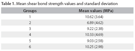
Aim: The aim of this study was to evaluate the shear bond strength of metallic orthodontic brackets bonded with Eagle Bond composite under different enamel surface conditions. Keywords: shear strength, dental bonding, orthodontic brackets. Introduction Until the 1970s, orthodontic accessories were fixed by using bands in all teeth. According to Zachrison1, such a bonding procedure had several disadvantages, which included, difficult cleaning, complexity, time-consuming clinical application and loss of esthetics. As a result, the technique for directly bonding orthodontic accessories to teeth was considered an essential advance for developing, simplifying and expanding Orthodontics. This direct bonding technique was possible only after the advent of the acid etching2, which became a routine procedure for bonding fixed appliances. The first paper known to address the direct bonding of brackets to dental surface dates back to the late 1950’s3. According to Nordenvall et al.4, such a technique brought several advantages to Orthodontics, namely absence of proximal contact5, easy bonding and debonding of accessories, shorter chair-time, esthetics, improved oral hygiene and less incidence of gingival inflammation5,6. A wide array of materials have been developed in recent years for bonding orthodontic brackets and, thus the scientific knowledge of these materials is crucial for their clinical use. These composites usually have high bond strength, hardness and dimensional stability, and they also have some disadvantages regarding viscosity, preparation time7-11 and fluoride release12. Eagle Bond composite (American Orthodontic, Sheboygan, WI, USA) is one of the composites currently introduced to the dental market, and little research has been done with this material. According to the manufacturer, Eagle Bond is easily applied, has good bond strength and moderate viscosity. The actual need of testing newly introduced materials justifies the present study, whose objective was to evaluate both the shear bond strength and the Adhesive Remnant Index (ARI) of Eagle Bond composite, applied according to the manufacturer’s instructions and under different experimental conditions. Material and methods Ninety bovine permanent lower incisors were selected, properly cleaned, stored in 10% formaldehyde solution and kept refrigerated at 6 oC. The teeth were embedded in PVC cylinders (Tigre, Joinville, Brazil) filled with acrylic resin (Clássico, São Paulo, Brazil), in such a way that only their crowns were left exposed. The buccal surfaces of the crowns were positioned perpendicular to the shearing die’s base, using a T-square to ensure that the mechanical test could be performed correctly. After resin polymerization, the specimens were stored in distilled water and maintained in refrigeration. Prior to the bonding procedures, the buccal surfaces of all teeth were subjected to prophylaxis with a slurry of extra-fine pumice (S.S. White, Juiz de Fora, MG, Brazil) and water in a rubber cup (Viking, KG Sorensen, Barueri, SP, Brazil) for 15 seconds. Next, the specimens were washed with an air/water spray for 15 seconds and dried with oil/moisture-free air streams for 15 minutes. The rubber cup was replaced after every five consecutive applications in order to keep the experimental pattern. After prophylaxis, the specimens were randomly divided into six groups (n = 15), and upper central incisor brackets (Abzil Lancer, São José do Rio Preto, SP, Brazil) with a base area of 13.8 mm2 were used in the bonding procedures. The six groups are divided as follows:
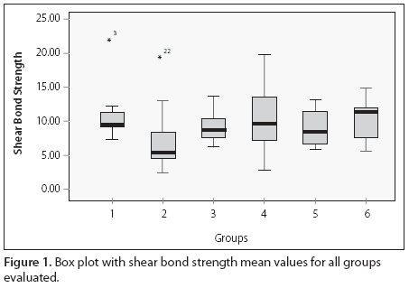
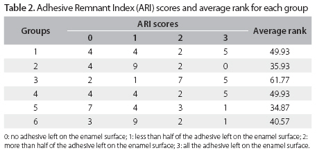
The composition of Eagle Bond is paste composed of silica, Bis-GMA, silane, N-dimethyl benzocaine, hexafluoride phosphate; primer with Bis-GMA, silane, N-dimethyl benzocaine, hexafluoride phosphate. The brackets were light-cured during 40 seconds (ten seconds for each face –mesial, distal, incisal and gingival) at a distance of 1 mm, a halogen light-curing device (XL 1,500; 3M Dental Products, Monrovia, USA) with light intensity of 450 mW/cm2 as measured with curing radiometer (Demetron, Danbury, CT, USA). After the bonding procedures, the specimens were stored in distilled water at 37 oC for 24 hours. A custom-made device was developed to hold the specimen completely stable during the mechanical test. The shear bond strength test was performed in a universal testing machine (Emic DL 10.000; São José dos Pinhais, PR, Brazil), at a crosshead speed of 0.5 mm/min through a chisel-shaped rod. The results were obtained in Kgf, converted into N and, then divided by the base area of the bracket so that values in MPa could be obtained. After the mechanical test, the buccal surface of each specimen was evaluated with a stereoscopic magnifying glass (Carl Zeiss, Goettingen, Germany), at ×8 magnification in order to quantify the ARI, according to the criteria established by Artun and Bergland7, that is, zero means no adhesive left on the enamel surface; one, less than half of the adhesive left on the enamel surface; two, more than half of the adhesive left on the enamel surface; and three, all the adhesive left on the enamel surface. Shear bond strength mean values were analyzed statistically by analysis of variance and Tukey’s test, in order to compare Group 1 (Control) to the Experimental Groups. Kruskal-Wallis test was used for assessing the ARI scores. Results No statistically significant differences were found between Group 1 (XT primer/Transbond XT composite – Control), Group 2 (Eagle Bond primer/Eagle Bond composite), Group 3 (Transbond Plus Self-Etching Primer + Eagle Bond composite), Group 4 (Eagle Bond composite without primer), Group 5 (Homogenized Eagle Bond composite), and Group 6 (Eagle Bond composite applied to saliva/blood-contaminated enamel). However, as can be seen in Table 1, Group 2 presented the highest shear bond strength numerical mean values (p > 0.05), as can be seen in Figure 1. The ARI scores in each group are presented in Table 2. Regarding Group 1, no statistically significant differences were found in relation to Group 2 (p = 0.154), Group 3 (p = 0.321), Group 4 (p = 0.999), Group 5 (p = 0.130), and Group 6 (p =0.335). The same was observed for Group 2 in relation to Groups 4 (p = 0.154), 5 (p = 0.775), and 6 (p = 0.539), as well as between Groups 4 and 5 (p = 0.130) and Groups 4 and 6 (p = 0.335). However, statistically significant differences were observed between Groups 2 and 3 (p = 0.002), Groups 3 and 5 (p = 0.006), and Groups 3 and 6 (p = 0.008). Discussion Transbond XT (Group 1 – Control), which has confirmed adhesive characteristics, was used in the present study according to the manufacturer’s instructions. No statistically significant differences were observed comparing the shear bond strengths of all groups, although Group 2 had the lowest values. These results indicate that Eagle Bond is appropriate for bonding orthodontic accessories to enamel surface, with shear bond strength ranging from 5 to 20 MPa, which is considered by Owens and Miller13 to be sufficient to resist the orthodontic forces. As mentioned above, no statistically significant difference (p > 0.05) was found between Group 2 (conventional Eagle Bond) and 3 (Eagle Bond + Transbond Plus Self Etching Primer - TPSEP), which is consistent with the findings of previous studies8,14 using Transbond XT under similar conditions. Similar results were also reported by Romano et al.15 using Transbond XT and Z 100, and by Pithon et al.16, using Orthobond composite. This finding is of clinical relevance, since TPSEP has been shown to make the bonding procedure 65% faster, according to Whyte17. Aiming at simplifying the technique proposed by the manufacturer, Eagle Bond was used for bracket bonding without the priming step (Group 4). The mean shear bond strength was higher than that obtained for Group 2 (Eagle Bond with primer), though without statistical significance. This result is of great importance since a bonding step can be eliminated, shortening the clinical chairtime, and may be due to the lower resistance of the resin without load that was applied in Group 2. However, it is important to have in mind that, although the elimination of this step reduces the clinical chairtime, the priming procedure protects the etched enamel that was not covered by the bracket after bonding. Among the innumerous questions raised by other studies, regarding composites used for bonding orthodontic accessories, the behavior of these materials when previously homogenized should also be known. Composite homogenization is justifiable for achieving a suitable distribution of the components, which would allow an improved flow during direct bonding of orthodontic brackets18,19. The bonding ability of homogenized Eagle Bond (Group 5) was compared to the bonding ability of the conventional composite (Group 2). No significant difference was found between the groups, suggesting that homogenized Eagle Bond would be a viable alternative when an improved flow is desirable. Similar results were also found by Patel et al.18, who tested the homogenization of the Superbond composite. Contamination of dental surface, with either blood or saliva during the bonding procedures, happens all the time. The interference of contamination after drying the contaminated area (Group 6) was tested. No significant difference was found between the groups, which is also consistent with the findings of Pithon et al.20, who used similar methodology with another composite. Therefore, it was demonstrated that the whole clinical sequence did not need to be repeated, provided that the contaminated area was dried before bonding the brackets. Once the enamel surface is contaminated with blood and saliva during the bonding procedure, it is necessary to dry the area to be bonded with Eagle Bond in order to obtain enough bond strength. Regarding the ARI, no statistically significant differences were found between the groups, except between Groups 2 and 3, Groups 3 and 5, and Groups 3 and 6. Such differences were due to the lower ARI scores observed in Groups 2, 5 and 6, compared to the higher ARI scores observed in Group 3, in which Transbond Plus Self Etching Primer was used. The good adhesiveness to the teeth, promoted by associating Eagle Bond with TPSEP, favored the achievement of higher shear bond strength mean values and, hence enamel protection during bracket debonding, that is, a greater amount of the adhesive material was left on the enamel surface. In Groups 2 and 5, however, the majority of fractures occurred at the enamel/composite interface following the debonding process, with ARI scores being predominantly zero or one, that is, no or lesser amount of composite adhered to enamel. Such a result may be explained by the improved flow, provided by either primer application before bonding the brackets (Group 2), or composite homogenization (Group 5). These results are favorable as far as the maintenance of enamel integrity is concerned, since enamel micro or macrofragments can be removed together with bracket and composite during debonding. The values obtained in Groups 2 and 5 are corroborated by most studies in the literature21,22. Based on the results of the present study, the following conclusions may be drawn: conventional Transbond XT and Eagle Bond systems showed good results in the shear bond strength testing, when bonded to enamel surface etched with 37% phosphoric acid; Eagle Bond composite presented good bond strength to enamel, treated with Transbond Plus Self Etching Primer; the use of Eagle Bond primer was found to be facultative, that is, it was not necessary for achieving full adhesion; and homogenization of Eagle Bond composite did not reduce the shear bond strength values, being an alternative if improved flow is desirable. References
Copyright © 2009 - Piracicaba Dental School - UNICAMP São Paulo - Brazil The following images related to this document are available:Photo images[os09007t1.jpg] [os09007f1.jpg] [os09007t2.jpg] |
| |||||||||
