 |
| About Bioline | All Journals | Testimonials | Membership | News |
|
||||||
|
||||||
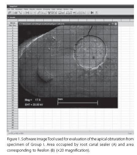
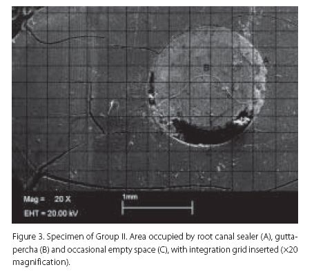
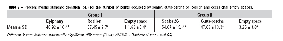
Braz J Oral Sci, Vol. 8, No. 3, July-September, 2009, pp. 132-136 Apical obturation quality of Epiphany/Resilon root canal filling system Lucas da Fonseca Roberti Garcia1, Lucas Zago Naves1, Simonides Consani2, Lourenço Correr-Sobrinho2, Fernanda de Carvalho Panzeri Pires-de-Souza3 1DDS, MSc, Department of Restorative Dentistry, Dental
Materials Area, Piracicaba Dental School, State University of Campinas, Brazil
Correspondence to: Lucas da Fonseca Roberti Garcia Rua Bernardino de Campos, 30 - apto. 1002 - Higienópolis 14015-130 - Ribeirão Preto - SP - Brasil Phone: +55 - 16 - 3964-6910 / +55 - 16 - 9796-0776 E-mail: drlucas.garcia@gmail.com Received for publication: June 4, 2009
Code Number: os09027 ABSTRACT Aim: This study analyzed by scanning electronic microscopy
(SEM) the quality of the apical obturation of a methacrylate based (polycaprolactone)
root-canal filling system (Epiphany/Resilon) compared to that of an epoxy-resin
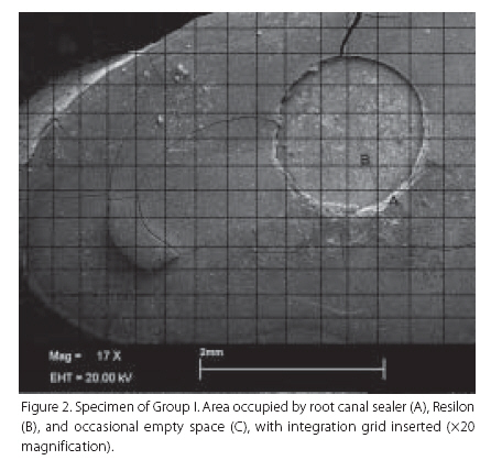
based endodontic sealer (Sealer 26/gutta-percha). Keywords: endodontics, root canal sealer, physicochemical properties, dental materials Introduction Many different root canal sealers are currently being used in combination with gutta-percha to fill the root canal system after biomechanical preparation. Gutta-percha is still the most commonly used root canal obturation material1, but resin-filling materials have steadily gained popularity and are now accepted as a root canal filling2. Epoxy resin sealers have been used because of their reduced solubility3, apical seal4 and micro-retention to root dentin5. Recently, methacrylate resin endodontic sealers have been developed6-7, but the combination of gutta-percha points and methacrylate resin sealer has shown reduced apical sealing ability compared with gutta-percha points and conventional epoxy-resin sealer6,8. Thus, self-etch primers have been used for bonding to root canal dentin9. As epoxy resin sealers do not copolymerize with methacrylate resin-based adhesives10, a dual-curable methacrylate resin sealer (Epiphany TM, Pentron Clinical Technologies, Wallingford, CT, USA), was developed with a self-etch primer, and a new thermoplastic filled polymer (Resilon™, Resilon Research LLC, Madison, CT, USA), as an alternative to gutta-percha11. Resilon is a synthetic polycaprolactone polymer, based on a polyester-based polymer that contains dimethacrylates, which can bond to methacrylate-based resin sealers, such as Epiphany, a resin-based dual-cure root canal sealer. According to the manufacturer, Resilon also contains bioactive glass, calcium hydroxide, and radiopaque filler7-12. The Resilon/Epiphany system is also followed by a self-etch primer that contains sulfonic acid-terminated functional monomer, HEMA, water, and polymerization initiator. The use of Resilon associated with the Epiphany sealer provides one of the main characteristics of this system, which has been suggested to have the capacity to produce a bonded monoblock filling, that results in improvements in the apical seal7,10. Based on recent studies involving these materials, the objective of the present study was to assess the quality of apical obturation of this new methacrylate based root-canal filling system (Epiphany/Resilon) comparatively to an epoxy-resin based endodontic sealer (Sealer 26/gutta-percha), by scanning electronic microscopy (SEM) in cross-sections at 2 mm from the apex. Material and methods Twenty extracted human maxillary canines from the Tooth Bank of Ribeirão Preto Dental School (Protocol #2008.1.336.58.0, CAAE #0031.0.138.000-08) were used in this study, in full compliance with ethical principles of the World Medical Association Declaration of Helsinki. The teeth were chosen due to their external root anatomy, avoiding roots with flattened areas in any direction and prioritizing teeth with similar root lengths, in order to facilitate biomechanical preparation as well as canal preparation. The teeth were disinfected by immersion in a 0.5% chloramine solution at 4°C for 48 h and were then washed under running water for 24 h to eliminate traces of the chloramine solution. The teeth were decoronated at the cementoenamel junction using a double-faced diamond disk coupled to a low-speed handpiece (Dabi Atlante, Ribeirão Preto, SP, Brazil), leaving an 18-mm long root remainder. Pulp tissue was removed by copious irrigation with 0.5% NaOCl. In each specimen, the actual canal length was measured by introducing a #10 K-file (Dentsply/Maillefer, Ballaigues, Switzerland) inside the root canal until its tip could be seen in the apical foramen, and the working length was determined 1 mm short of the apex, Root canal preparation was performed by the crown-down technique using tapered ProFile nickel-titanium rotary instruments (Dentsply/Maillefer) sequentially according to the manufacturer's recommendations (20 .04, 25 .04, 30 .04, 20 .06, 25 .06, 30 .06 and 40 .06). Irrigation was done with 2 mL of 2.5% NaOCl at each change of instrument followed by a final flush with 2 mL of 17% EDTA for 1 min, using a needle (Ultradent Products Inc., South Jordan, UT, USA) introduced 2 mm short of the working length. Roots were rinsed with 5 mL of sterile water to eliminate traces of NaOCl and EDTA. After drying of the root canals with absorbent paper points (Dentsply/Maillefer), the roots were randomly assigned to two groups (n=10) according to the root canal filling systems used. The composition of the tested material is shown in Table 1. Group I: Resilon/Epiphany - The primer was applied at the working length with paper points (Dentsply/Maillefer), which were also used to remove the primer excess. Subsequently, the sealer was inserted to a distance 3 mm short of the working length using a lentulo spiral (Dentsply/Maillefer). The lentulo was withdrawn slowly from the canal and the procedure was repeated at shorter length. A .04/30 master point, compatible with the root canal diameter, was coated with sealer and slowly inserted at the working length. Two smaller accessory.02/25 points were placed passively, and the sealer was photoactivated with a halogen lamp for 40 s (Ultraled; Dabi Atlante; power density: 500 mW/cm2). After trimming the Resilon points with a heated instrument, the sealer self-cured within 30 min. Group II: Sealer 26/Gutta-percha points - The root canals were obturated with Sealer 26 and gutta-percha points (Dentsply/Maillefer) according to the classic technique. A #40 gutta-percha master point and medium fine accessory points were coated with the sealer and slowly inserted at the working length, until the root canal was completely filled. A heated instrument was used to seal the filling material off at the root canal orifices. After being sealed, the roots of all groups were immersed in distilled water and incubated at 37oC for a period equivalent to three times the time required to set the sealer according to the manufacturer. Then, the roots were removed and dried. In preparation of the specimens for SEM evaluation, the apical region of each root was abraded with an Endo-Z bur (Dentsply/Maillefer) coupled to a high-speed handpiece (Dabi Atlante) until obturation exposure. Next, the apex of each root was sectioned transversally, obtaining 2-mm-thick specimens (n=10/group). The specimens were allowed to air-dry overnight, and were sputter-coated with gold (MED 010, Balzers, Liechtenstein) and examined with a scanning electron microscope (LEO 435 VP, Carl Zeiss, Jena, Germany). Representative micrographs of tested areas sites were recorded at ×20 magnification. The SEM micrographs were analyzed by an image-analysis software (Image Tool, The University of Texas, Health Science Center in San Antonio, San Antonio, TX, USA) using an integration grid (Figure 1). For each specimen, the number of points that coincided on root canal sealer (A), on gutta-percha or Resilon (B), and the number of points for eventual empty spaces (Figures 2 and 3). The percentage of space occupied by sealer, gutta-percha or Resilon, and the percentage of empty spaces were calculated by the software using a three-point scale. The data were subjected to statistical analysis (2-way ANOVA - Bonferroni test - p<0.05). Results The data for the specimens sealed with Resilon/Epiphany (Group I) and Sealer 26/gutta-percha (Group II) are shown in Table 2. The mean values revealed an adequate adaptation of the materials to the root canal walls. Resilon/Epiphany had better adaptation than Sealer26/gutta-percha, with fewer empty spaces and a larger area occupied by Resilon, when compared to the gutta-percha area, promoting a thinner cement line. However, there was no statistically significant difference (p>0.05) between the tested groups. Discussion Leakage tests are frequently performed to examine the quality of root canal fillings6-7,13-15. Leakage evaluations show great variations in studies16, and the results are often contrasting17-18. In order to avoid the disadvantages of leakage evaluations, in the present study, the quality of the root canal fillings was assessed through a histological method by SEM evaluation10,19. A cross-section height of 2 mm from the apex was also selected by other investigators20-22. Because the most critical area of the root canal preparation is the apical 2 mm23, the cross-sections of the present study were performed in this part of the roots. The hermetic sealing of root canals has a high importance in the success of endodontic therapy, and this step consists of sealing the entire root canal system with biologically tolerated materials that have good physicochemical properties and are resistant to microleakage and capable of creating an appropriate environment for tissue regeneration.4 Some researchers have associated a lesser degree of infiltration with a greater presence of gutta-percha in the canal space1,8,24-26. Others have reported that a lesser thickness of the root canal sealer promotes better seal27-28. According to De Deus et al.29, greater sealer thickness influences negatively the sealing ability of the root canal filling. Several techniques and materials have been developed with the objective of offering greater sealing ability than that of gutta-percha13,30-31. However, no currently available material has all these desired properties. Thus, to a greater or lesser extent, apical microleakage has continued to occur in endodontic seals4,17. The method of root canal obturation using core material and sealer is widely accepted. It is advantageous for the sealer to adhere firmly to both root canal wall and the core material19, which might improve the sealing ability by the elimination of fluid-permeable spaces and contribute to improve the filling stability during mechanical stress19,25. Methacrylate-based resin sealers enable obturation in a slightly moist root canal because they are hydrophilic. This hydrophilicity, combined with advanced bonding techniques facilitates the formation of deep resin tags extending into the dentinal tubules from the root canals. Deep resin tags help enhancing bonding and increasing the clinical success of the obturation25. Although sealers adhere to dentin to varying degrees, adhesion to gutta-percha is minimal26. The introduction of the Epiphany/Resilon system replaces gutta-percha with a filled polycaprolactone polymer. In theory, the methacrylate resin-based sealer of this system is able to firmly adhere to the Resilon core material13. The Epiphany/Resilon system, in essence, produces a "monoblock" effect, where the core material (Resilon), sealer (Epiphany) and dentinal tubules become a single solid structure7,13,25. Shipper et al.13 have suggested that this monoblock would be highly desirable to provide a thorough seal of the root canal system as it would be able to minimize coronal leakage in case of loss or fracture of the temporary coronal restoration. In vitro13 and in vivo31 studies have demonstrated a good resistance of the Epiphany/Resilon monoblock system to bacterial leakage. Solubility and disintegration of root canal sealers must be as small as possible so that it can promote hermetic sealing, thus favoring clinical success, because microleakage may occur from the cervical to apical third or inversely3. Sealer 26 is an epoxy resin-based sealer, with low values of solubility and disintegration3, which presents an expansion of 3.26%. This dimensional alteration could be explained by the water sorption suffered by this type of resin after its polymerization3, thus providing a hermetic seal. One of the most important properties of root canal sealers is the ability to flow enough to completely fill the root canal space. Barbizam et al.32 demonstrated that Sealer 26 failed to fill completely the lateral root canals. The number of non-filled or partially filled lateral canals was larger in the apical third and this finding is clinically important because it is well known that there are fewer lateral canals in the apical root third. Moreover, Sousa-Neto et al.4 showed the need of removing smear layer for greater adhesion of this epoxy-based sealer (Sealer 26) to dentin. The results of this study demonstrated that Epiphany/Resilon and Sealer 26/gutta-percha presented statistically similar (p>0.05) adaptation to the canal walls, confirming the findings of Herbert et al.33. Since no ideal filling material is currently available, special attention should be directed to the biomechanical preparation and the definitive restoration of the tooth after root canal filling. It is the interdependence of one treatment step on the subsequent step that must be fulfilled in order to achieve optimal results34-35. In conclusion, the two types of root canal filling systems used in this study, Epiphany/Resilon and Sealer 26/gutta-percha, had similar behavior promoting an adequate sealing of the critical apical region, with minimum presence of cement. Acknowledgements The authors are indebted to Dr. Kitajima, Dr. Tanaka and Dr. Salaroli (NAP/MEPA-ESALQ/USP) for SEM equipment support. References
Copyright 2009 - Braz J Oral Sci The following images related to this document are available:Photo images[os09027f2.jpg] [os09027f1.jpg] [os09027t2.jpg] [os09027t1.jpg] [os09027f3.jpg] |
| |||||||||
