 |
| About Bioline | All Journals | Testimonials | Membership | News |
|
||||||
|
||||||
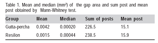
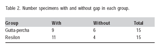
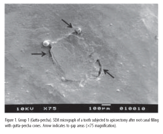
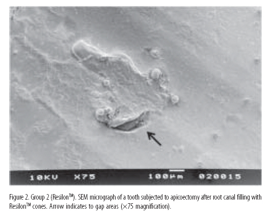
Braz J Oral Sci, Vol. 8, No. 3, July-September, 2009, pp. 141-144 Apical gaps after apicoectomy procedures performed on teeth filled with gutta-percha or ResilonTM Marco Antonio Hungaro Duarte1, Angélica Marquezim Lopes Locci2, Ivaldo Gomes de Moraes1,Juliane Maria Guerreiro Tanomaru3, Mário Tanomaru Filho3 1 DDS, MSc, PhD, Professor of Endodontics, Department
of Dentistry, Dental Materials and Endodontics, Bauru School of Dentistry,
Univeristy of São Paulo, Bauru, SP, Brazil
Correspondence to: Marco Antonio Hungaro Duarte, Departamento de Endodontia - Faculdade de Odontologia de Bauru, USP, Rua Alameda Otávio Pinheiro Brisolla, 9-75, Bauru, SP, Brasil, CEP: 17012-101 C.P. 13, Phone: +55-14-32358344, Phone/Fax: +55-14-32346147, E-mail: mhungaro@travelnet.com.br Received for publication: July 14, 2009
Code Number: os09029 Abstract Aim: This ex vivo study compared, under scanning electron
microscopy (SEM), the marginal adaptation of root canal obturation with either
ResilonTM or gutta-percha cones following root-end resection. Keywords: apicoectomy, ResilonTM, gutta-percha. Introduction Periradicular surgery is based on two goals, namely to eliminate the etiologic agents causing infection and to prevent root canal reinfection and recontamination of the periodontal tissues thereafter. Basically, the etiologic agents involved in endodontic infections may be classified as intraradicular or extraradicular microorganisms, intraradicular or extraradicular chemical substances and extraradicular physical factors1-3. The root apex surrounded by a periapical lesion presents areas of cemental resorption and harbors microorganisms and bacterial biofilm4-5. Resection of the root apical portion may be performed with either high- or low-speed rotary instruments under constant saline irrigation. It has been demonstrated that depending on its type, angulation and rotary direction, the bur used for root-end resection may create surface irregularities and expose the dentinal tubules to a greater extent. The use of surgical length fissure burs6, cross-cut fissure burs7 and diamond burs8 has been recommended for root-end resection. A previous scanning electron microscopy (SEM) study9 examined root-end resections performed using three bur configurations in both high and low-speed handpieces and observed that the smoothest surface and the least amount of gutta-percha disturbance were produced by the #57 plain fissure bur at low-speed. In addition, better fit of the filling material to the canal walls is obtained when root-end resection is performed with the handpiece moved across the tooth in a forward direction in relation to the direction of rotation of the bur10. However, the above-mentioned studies9-10 have examined gutta-percha root fillings. Although gutta-percha is universally accepted as a standard of root canal filling material, it does not have adhesion to root canal dentin and always requires association with an endodontic sealer11. Advances in adhesive technology and the search for a material with greater adhesion to the canal walls and to the sealer have resulted in a solid material named ResilonTM (Resilon Research LLC, Madison, CT, USA), which is based on a blend of synthetic thermoplastic polyester polymers and contains bioactive glass and radiopaque fillers. This material performs like gutta-percha, has the same handling properties and is usually used in combination with a dual-cure methacrylate resin-based sealer (Epiphany; Pentron Clinical Technologies, Wallingford, CT, USA) supplied with a self-etching primer12. Obturation using the ResilonTM/Epiphany system is reported to create a tight seal with the dentinal tubules within the root canal system; in essence, it is claimed to produce a "monoblock" effect, where the core material (ResilonTM), sealer and dentinal tubules become a single solid structure12-13. However, a recent study14 has found significantly lower push-out bond strength of the new obturation system to intraradicular dentin compared to gutta-percha/AH 26 sealer. The ResilonTM/Epiphany system has demonstrated good sealing properties when subjected to different leakage tests15-17, though no statistically significant difference has been found when compared to other root filling materials, like gutta-percha/AH Plus sealer18-19. Some studies20-22 have shown that ResilonTM cones have similar thermoplasticity between gutta-percha and resilon cones. Nevertheless, no study has yet evaluated ResilonTM and gutta-percha with respect to their apical fit in apicoectomized teeth. Therefore, the purpose of this in vitro study was to compare, under SEM, the apical fit of root canal obturation with either ResilonTM or gutta-percha cones after root-end resection with high-speed #170L carbide burs. Material and Methods Thirty extracted single-rooted human teeth with fully formed apices were selected for the study. The teeth were immersed in 5% sodium hypochlorite (NaOCl) for 12 h and then stored in saline until use, when they were decoronated at the cementoenamel junction with a double-faced diamond saw at low speed. A size 10 K-file (Maillefer, Ballaigues, Switzerland) was introduced into the canal until its tip was visible at the apical foramen and the working length was established 1 mm short of this length. The root canals were instrumented using the Profile rotary system (Dentsply/Maillefer, Ballaigues, Switzerland). The cervical preparation was performed with Orifice Shaper (Maillefer) number 2 (30 taper 06), number 3 (40 taper 06) and number 4 (50 taper 07). After cervical preflaring, the apical portion was prepared using the Profile 04 size 15 up to a size 45 at the working length. The canals were irrigated with 2.5% NaOCl at each change of file. When instrumentation was completed, the canals were filled with 1 mL 17% EDTA during 3 min, received a final flush with 1% NaOCl and were dried with absorbent paper points. Two groups of 15 specimens each were formed at random. In group 1, the root canals were obturated with a fitted size 45/04 gutta-percha master cone (Maillefer) and AH Plus resin-based sealer (Dentsply DeTrey Konstanz, Germany) using a lateral compaction technique. The sealer was taken to the canal using a lentulo spiral (Maillefer) before the insertion of the gutta-percha cone (Maillefer). A finger spreader was placed alongside the master cone and compaction was done to make space for up to three FF accessory gutta-percha points (Maillefer). Excess material was removed from the pulp chamber and the filling mass was vertically condensed. In group 2, the gutta-percha cone was replaced by a size 45/04 ResilonTM master cone. The endodontic sealer was taken to the canal in the same way as described for group 1 and ResilonTM accessory points were also used. The coronal portion of each root canal was sealed with IRM (Dentsply/Caulk, Milford, DE, USA). The root-filled teeth were stored in saline at 37oC during 48 h for complete setting of the sealer. After this period, the apical 3 mm of each root were resected using a plain fissure #170L carbide bur in a high-speed handpiece under constant water cooling to remove any accumulated debris and to keep the root surface moist. The cutting direction followed the direction of rotation of the bur (clockwise rotation). A new bur was used for each root-end resection and an attempt was made to produce the smoothest possible surface in all specimens. After root-end resection, the filling material was burnished against all root canal walls with a cold #33 burnisher, from the center to margins, and the resected root surfaces were washed and dried with a gentle air stream. Impressions were obtained from all faces of the resected apical segments with a condensation silicone impression material (Zeta Plus/Oranwash L; Zhermak, Badia Polesine, Rovigo, Italy). The heavy-bodied material (Zeta Plus) was first applied onto the specimen and allowed to polymerize for 7 min. Next, the light-bodied material (Oranwash L) was used to refine the impression. In both groups, each resected apical segment was paired with its respective impression. Thereafter, the impressions were replicated with epoxy resin (RD-6921; Redelease, São Paulo, SP, Brazil) with a hardening agent in positive vacuum, allowed to polymerize within 24 h. Care was taken to minimize entrapment of air bubbles. The obtained epoxy resin positive replicas were sputter-coated with gold (Hammer VI Sputtering System, Anatech Ltd., Alexandria, VA), examined with a scanning electron microscope (JSMT220A, JEOL, Tokyo, Japan) and photographed at ×75 magnification. The SEM micrographs of the epoxy resin replicas of the resected apical segments were digitized and analyzed with respect to the area (in mm2) of apical gap using ImageTool software version 3.01 (UTHSCSA, San Antonio, TX, USA). After calibration, the measurements of gap space between the obturation and the root canal walls were summed and one value (mm2) was obtained for each specimen. Data were analyzed statistically by the Mann-Whitney U-test and Fisher's exact test at 5% significance level. Results Table 1 presents the mean and median gap area (in mm2) obtained in each group and show the sum of post and mean post obtained by Mann-Whitney test. There was no statistically significant differences (p>0.05) between groups 1 and 2. Comparison of the number of specimens with and without gap between the two groups (Table 2) showed no significant differences either (p>0.05). Figures 1 and 2 show SEM micrographs of teeth subjected to apicoectomy after root canal filling with either gutta-percha or ResilonTM cones, respectively. Discussion In periradicular surgeries, curettage of the pathologic apical lesion and resection of the contaminated root apex are of paramount importance for treatment success. Even if the root canal filling is radiographically classified as adequate, the occurrence of apical gap between the obturation and the canal walls and the need for root-end cavity preparation and retrograde restoration should always be assessed after apicoectomy6. Studies have compared the action of different rotary instruments and techniques on root apex morphology after apicoectomy8, the refinement of resected root-end surfaces with finishing burs to improve root apex topography23, the use of high-power lasers for apicoectomy24-25, the use of ultrason26 and the sealing capacity of several filling materials, such as ResilonTM cones, gutta-percha cones, Epiphany sealer, AH Plus sealer16,19,20. However, to the best of our knowledge, no other study has duplicated the present experimental model to evaluate the marginal adaptation of obturations with ResilonTM and gutta-percha cones in apicoectomized teeth. The type of rotary instrument, the technique23,25-26 and the direction of rotation of the bur12 may produce an irregular surface following root-end resection and gap formation between the filling material and the root canal walls in the apical portion leading to microbial recontamination and treatment failure. In the present study, root-end resections were performed with a water-cooled high-speed #170L multifluted carbide bur because this type of rotary instrument has been shown to produce smoother surfaces9-10. The direction of root-end resection was the same as that of bur rotation in order to minimize tearing, smearing and distortion of the cones onto the root canal walls10. In the present study, comparison between the groups based on the mean values of apical gap demonstrate that the group with root canals filled with ResilonTM cones presented less gap formation (0.0015 mm2) than the group with root canals filled with gutta-percha cones (0.0042 mm2). This difference was not statistically significant, probably because the filling materials had similar thermoplasticity27. Although water-cooling was used in the present study, a temperature rise may occur during root-end resection procedures10. Adhesion of the filling material to the root canal walls after apicoectomy is another important factor. The sealer used in the present study, AH Plus, has shown better adhesion to the dentin walls when compared to other sealers28. In this sense, although ResilonTM cones have been developed for use with Epiphany sealer, in the present study AH Plus was used in both experimental groups because this sealer has demonstrated a good interaction with ResilonTM cones, and better adhesion to ResilonTM than Epihany when used with cold compaction techniques28. The use of the same sealer in both groups allowed analyzing the influence of the type of cone (gutta-percha or ResilonTM) without interference of the sealer as an additional variable. The root end was burnished prior to SEM analysis to provide a better fit of gutta-percha to the canal walls because, in a previous study29, this procedure reduced significantly the apical leakage after root end resection and glass ionomer cement retroseals. In the present study, the root canals were filled by lateral compaction because it is a widely employed obturation technique that does not require especial instruments or devices. Given that the goal of periradicular surgery is to eliminate root canal infection and prevent recontamination, apical gap of the filling material after root-end resection is an important factor that should be taken into account. In the present study, the great majority of specimens presented gap between the obturation and the root canal walls, and the type of cone used for root canal obturation (gutta-percha or Resilon) did not influence the marginal adaptation after root-end resection. This indicates that there is material able to avoid gaps at the obturation and very often the misfit is related to root canal anatomy30. In this way, the findings of this SEM evaluation reinforce the need of performing root-end cavity preparation and retrograde filling in apicoectomized teeth because the areas of apical gap observed in both groups may serve as niches for microbial recolonization invariably leading to failure of the surgical treatment. Acknowledgements The authors would like to thank Bauru Dental School, University of São Paulo, Brazil, and Mr. Edmauro de Andrade for undertaking the SEM images. References
Copyright 2009 - Braz J Oral Sci The following images related to this document are available:Photo images[os09029f2.jpg] [os09029f1.jpg] [os09029t1.jpg] [os09029t2.jpg] |
| |||||||||
