 |
| About Bioline | All Journals | Testimonials | Membership | News |
|
||||||
|
||||||
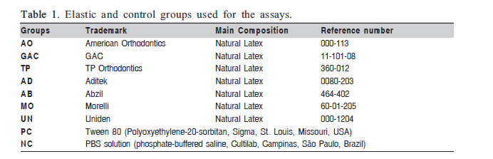
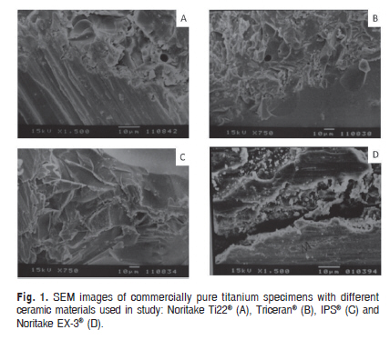
Braz J Oral Sci, Vol. 9, No. 3, July-September, 2010, pp. 366-370 Cytotoxicity of orthodontic elastics: In vitro investigation with on L929 mouse fibroblasts Rogério Lacerda dos Santos1, Matheus Melo Pithon1, Fernanda Otaviano Martins2, Maria Teresa Villela Romanos3 1Specialist in Orthodontics, Federal University of Alfenas, Minas Gerais, Brazil Master in Orthodontics, Federal University of Rio de Janeiro, Brazil PhD student in Orthodontics, Federal University of Rio de Janeiro, Brazil Correspondence to: Rogério Lacerda dos Santos Rua Ipatinga, 170, Planalto Divinópolis- MG- Brazil CEP 35501-191 E-mail: lacerdaorto@hotmail.com; lacerdaorto@bol.com.br Received for publication: April 08, 2010 Code Number: os10037 Abstract Aim: To test the hypothesis that there is no difference in the cytotoxicity among natural
latex elastics of different manufacturers using a L929 cell line
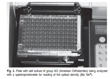
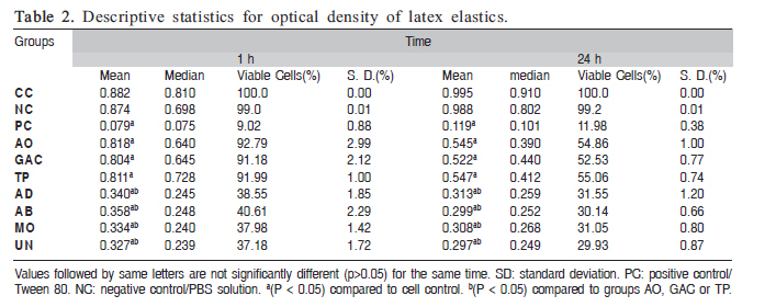
culture. Keywords: cytotoxicity, elastics, biocompatibility, orthodontics. Introduction The biocompatibility of dental materials has been subject of great speculation and uncertainty. There are, particularly in Orthodontics, several materials keeping direct contact with organic tissues for long periods. Recent studies have been concerned with the biocompatibility of different types of orthodontic materials1,2. Prevulcanized latex is produced by mixing pure natural latex3, with stabilisers such as zinc oxide and chemically vulcanized materials. The resulting mixture is then heated up to 70oC4. Although zinc is known to be neurotoxic5, the amount released by orthodontic elastics can be ingested as research studies show no evidence of harm6. Anti-ozone and anti-oxidant agents are also added to latex during the manufacture of orthodontic elastics3. This process has the advantage of producing latex with higher mechanical properties, thus increasing its strength and elasticity4,6. However, natural latex is not in the category of materials known to be entirely inoffensive7-8. Allergy caused by latex proteins has been well documented9, including immediate hypersensitivity reactions10. Amongst the allergic reactions caused by orthodontic elastics, swelling and stomatitis, erythematous oral lesions, respiratory reactions, and even anaphylactic shock, the most severe form of allergy11-12, can be cited. Latex allergy occurs in 3-17% of the cases13. The use of cell cultures for testing the toxicity of dental products is a valid way of understanding the cytotoxic behavior of such materials7. The aim of this in vitro study was to test the hypothesis that there is no difference in the cytotoxicity among natural latex elastics of different manufacturers on L929 mouse fibroblasts. Material and methods Latex intraoral elastics of different manufacturers (I.D. = 5/16", 4.5 oz.) were selected for studying their cytotoxicity in cell culture (Table 1). The samples were divided into 7 groups of 15 elastics each: Group AO (American Orthodontics, Sheboygan, WI, USA), Group GAC (GAC International, Bohemia, NY, USA), Group TP (TP Orthodontics, Lodi, CA, USA), Group AD (Aditek, Cravinhos, SP, Brazil), Group AB (Abzil, São José do Rio Preto, SP, Brazil) Group MO (Morelli, Sorocaba, SP, Brazil) and Group UN (Uniden, Sorocaba, São Paulo, Brazil) (Figure 1). The elastics used in this study belonged to the same production line for each trademark. To verify the cell response in extreme situations, 3 additional groups were included in the study: Group CC (cell control), consisting of L-929 cells not exposed to supernatants from the elastics; Group C+ (positive control), consisting of Tween 80 (Polyoxyethylene-20-sorbitan, Sigma, St. Louis, MO, USA); Group C- (negative control), consisting of phosphate-buffered saline (PBS) solution (Table 1). The cell culture model used was the monolayer containing L-929 line cells (mouse fibroblast) (American Type Culture Collection - ATCC, Rockville, MD, USA), which were maintained in Eagles' minimum essential medium (MEM; Cultilab, Campinas, SP, Brazil) by adding 0.03 mg/mL of glutamine (Sigma, St. Louis, Missouri), 50 µg/mL of garamicine (Schering Plough, Kenilworth, NJ, USA), 2.5 mg/mL of fungizone (Bristol-Myers-Squibb, New York, NY, USA.), 0.25% sodium bicarbonate solution (Merck, Darmstadt, Germany), 10 mM of HEPES (Sigma), and 10% bovine fetal serum (Cultilab) for growth medium or no bovine fetal serum for maintenance medium only. Next, the cell culture medium was incubated at 37oC for 48 h. The cells were reseeded twice a week to ensure exponential growth of the cell line. For standardization of samples, the powder coating of the elastics was removed. The elastics were washed for 15 s with deionized water by using a Milli-Q purification system (Millipore, Bedford, MA, USA) and their surfaces were slightly dried with disposable paper. Before testing, all elastics were sterilized by exposure to ultraviolet light (Labconco, Kansas, MO, USA) for 30 min for each surface14-16. The "dye-uptake"17 test was used for evaluating the cytotoxicity. This method is based on neutral red dye incorporated into live cells. It was used in this experiment only at two periods of evaluation: 1 and 24 h. These elastics are usually maintained in the oral cavity for up to 24 h. The 1-h period represents the maintenance of the elastic in the cell culture medium for 1 h after removal, whereas the 24-h period represents the maintenance of the elastic in the cell culture medium for 24 h after removal. Dye-uptake Aliquots of 100 µL of L-929 line cells were distributed into 96-well microplates. After 48 h, the growth medium was replaced with 100 µL of MEM obtained following incubation in the different types of elastics at 1 and 24 h. MEM was used because it is the same type of medium used for cell maintenance, thus not influencing the results. Positive and negative control groups consisted of culture medium put in contact with Tween 80 and PBS, respectively. After 24 h incubation, 100 µL of 0.01% neutral red dye (Sigma, St. Louis, MO, USA) were added to the culture medium in the 96-well microplates, which were incubated again for 3 h at 37oC so that the red dye could penetrate the live cells. Following this period of time, 100 µL of 4% formaldehyde solution (Vetec, Rio de Janeiro, RJ, Brazil) in PBS (130 mM of NaCl; 2 mM of KCl; 6 mM of Na2HPO4 2 H2O; 1 mM of K2HPO4 1 mM; pH 7.2) were added to promote cell attachment to the plate. After 5 min, 100 µL of 1% acetic acid (Vetec) and 50% methanol (Vetec) were added in order to remove the dye. After 20 min, a spectrophotometer (BioTek, Winooski, VT, USA) at 492 nm wavelength (l=492 nm) was used for data reading (Figure 2). Data were compared by ANOVA and Tukey's multiple-comparison test was used for identifying differences between the groups. Significance level was set at p<0.05. Results A significant difference (P < 0.05) was noted between all groups and group CC (cell control) at 1 h. Groups AD, AB, MO and UN were noticeably more cytotoxic than the other groups. Group UN produced the lowest value (37.18% ± 1.72%) and group AO produced the major viability (92.79% ± 2.99%), whereas the viability of the Tween 80 (positive cytotoxicity control) was 9.02% ± 0.88% (Table 2). After 24 h, a significant decrease in cell viability was observed in all groups. Viability ranged from 29.93% to 55.06%, relative to the cell control. The lowest viability (29.93% ± 0.87%) corresponded to group UN, whereas the viability of the Tween 80 (positive cytotoxicity control) was 11.98% ± 0.38% (Table 2). The results showed statistically significant differences between all groups tested with the group CC (cell control) (p<0.05) at 24 h. A significant difference (P < 0.05) was noted between the groups AO, GAC, TP and the groups AD, AB, MO, UN at 1 and 24 h (Table 2). Discussion The cell culture model used in the present study was the monolayer18-19. This model was used together with the dye-uptake technique17 because the cytotoxicity of the materials can be determined by spectrophotometry. The spectrophotometric assay allows rapid and reliable evidence for cell viability to be obtained based on the use of vital stain incorporated by viable cells. In this study, neutral red dye was used because it is largely employed for identification of L-929 cell viability. Dead or damaged cells cannot incorporate vital stain, thus not being recognized on optical reading. Therefore, spectrophotometry does not allow dead cells to be distinguished from the damaged ones. The amount of dye incorporated into the cells is directly proportional to the number of cells with intact membrane, which allows distinguishing the cytotoxicity of each elastic. L-929 mouse fibroblasts were used because they have results comparable to those of primary human gingival fibroblasts20-21, the cell culture results cannot be interpreted as a human response. The percentage of viable cells was obtained by comparing the mean optical density (OD) in the control group (cells with no contact with elastics) to that obtained from supernatants of cell cultures that had been in contact with elastics. As sterilization is a prerequisite for cytotoxicity essays, ultraviolet radiation14,16 was used in this study for 30 min for each elastic surface. It was observed that all elastics exhibited the same color aspect and malleability following sterilization with UV light. Because natural latex rubber has been increasingly used as dental material, many cytotoxicity issues have been reported as well15,22. A comparison was made among different latex intra-oral elastics. Preservatives such as sulfur and zinc oxide as well as antioxidants such as di-thio-carbohydrates, N-nitrosodibutylamine, and N-nitrosopiperidine are all known to be cytotoxic substances22. Holmes et al.8 have verified whether the colorants used in the fabrication of colored latex could have some toxic effect. Their results showed that these colorants exhibited low toxicity. However, such an effect is clinically inoffensive. Allergic reactions23 have been related to the use of orthodontic elastics24, which is characterized by the presence of small vesicles or acute edema and complaints of itching and burning. Allergy to natural latex occurs because of the presence of many types of proteins, and the powder covering the orthodontic elastics works as a transporter for these proteins. Therefore, the development of non-latex elastics has become increasingly important for clinical use. However, the objective of this study was to evaluate the cytotoxic effect of latex elastic in cell culture. The most serious consequence of natural rubber latex allergy commonly takes place during mucosal absorption of natural rubber latex proteins during intraoperative medical or dental procedures when health care workers or others already sensitized become patients25. The safety biocompatibility of silicone has been well proved through the use of mouth guards in dentistry26. However, Hwang and Cha22 observed that silicone rubber bands were found to exhibit a low cytotoxicity. However, in terms of the initial force level and the abrupt loss of remaining force with an increase in the extension length, great improvements in the silicone rubber band's physical properties are required. Evidence of this cytotoxic feature was shown following exposition of the elastics to cell culture medium. It was used in this experiment only two times of evaluation 1 and 24 h. These elastics are usually changed every 24 h. Natural latex elastics from Aditek, Abzil, Morelli and Uniden trademarks induced more cell lysis at 1 and 24 h compared to those from American Orthodontics, GAC and TP Orthodontics trademarks. However, all natural latex elastics were found to cause more cell lysis at 24 h. It has been shown evidence of cytotoxicity in natural latex elastics compared to the silicone elastics6,22. In this study, the elastics from TP Orthodontics and American orthodontics trademarks caused lower cell viability at 24 h compared to previous studies6,22. As the powder covering the elastics of all manufacturers was removed before performing the in vitro assays, it was not possible to know whether this powder would have produced any effect. According to Schmalz7, the great danger is that potentially cytotoxic intraoral elastics could release substances that might be ingested by the patient over time, thus causing diseases resulting from a cumulative effect. It is known that latex is not entirely biocompatible as it may interact with foods13,27 and medications 28. American Orthodontics, GAC and TP Orthodontics trademarks intraoral elastics evaluated in this study showed over 90% cell viability in the experimental period of 1 h and over 50% at 24 h. Hanson and Lobner6 evaluated latex and non-latex 3/16-inch interior lumen (medium) elastics and found cell lysis to be 50% higher for latex elastics compared to non-latex ones. However, the authors considered both types of elastics appropriate for orthodontic use. Therefore, it is suggested that elastics with cell viability less than 50% should be avoided or used with caution in order to prevent cumulative effects of the cytotoxic components released from these elastics into the organism7. Further studies assessing the mechanism of cell lysis can contribute to provide more details on the cytotoxic behavior of these materials. As these materials are widely used in clinical orthodontics, care regarding the cytotoxicity of orthodontic elastics should be taken, mainly with regard to elastics as they have a very close contact with gingiva. Thus, clinically proven biocompatible materials should be acquired whenever possible. It may be concluded that intraoral elastics from American Orthodontics, GAC and TP Orthodontics trademarks induced less cell lysis than Aditek, Abzil, Morelli and Uniden trademarks at 1 and 24 h. References
Copyright 2010 - Braz J Oral Sci The following images related to this document are available:Photo images[os10037t1.jpg] [os10037f1.jpg] [os10037t2.jpg] [os10037f2.jpg] |
| |||||||||
