 |
| About Bioline | All Journals | Testimonials | Membership | News |
|
||||||
|
||||||
Braz J Oral Sci, Vol. 9, No. 4, October-December, 2010, pp. 415-420 Pain Behavior To Electroacupuncture In Rabbit Tooth Pulp Delane Viana Gondim1, Krishnamurti de Morais Carvalho2, Mariana Lima Vale3 1MSc, Department of Clinical Medicine, Medical School, Federal University of Ceará, Fortaleza, CE, Brazil Received for publication: January 10, 2010 Accepted: November 12, 2010 Correspondence to: Delane Viana Gondim, Departamento de Medicina Clínica, Faculdade de Medicina, Universidade Federal do Ceará - UFC. R. Cel. Nunes de Melo, 1127, Rodolfo Teófilo 60430-270 - Fortaleza, CE, Brasil. Phone: +55-85-3366-8588 Fax: +55-85-3366-8333 E-mail: delanegondim@yahoo.com.br Code Number: os10047 Abstract Aim: The aim of this study was to verify the pain behavior to electroacupuncture
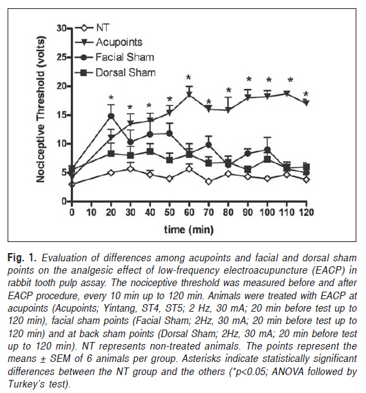
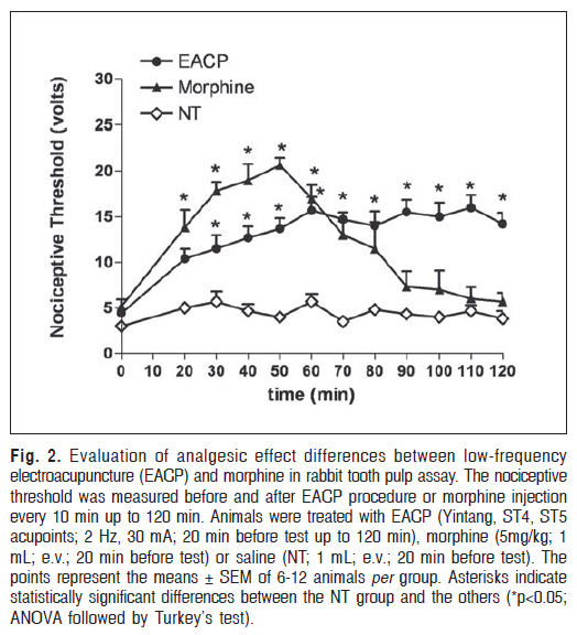
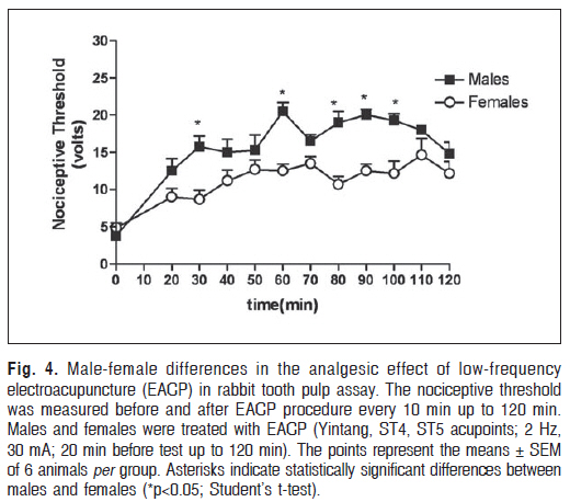
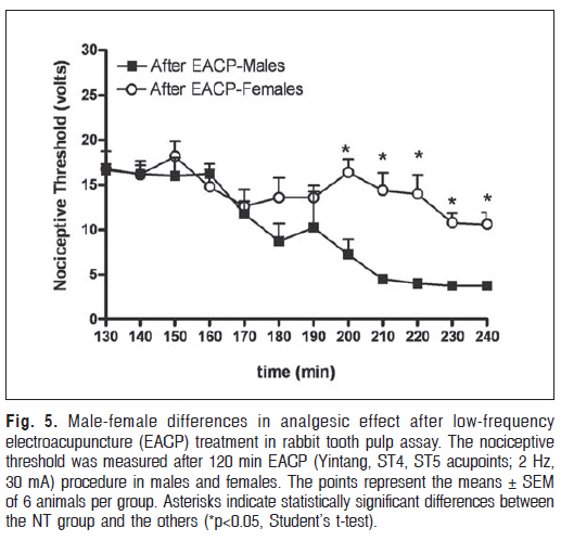
(EACP) in rabbit tooth-pulp assay. Keywords: electroacupuncture, tooth pulp, analgesic effect, endogenous opioids, pain. IntroductionAcupuncture is part of Chinese Traditional Medicine, one of the most ancient and well known human health systems, which is approximately 5,000 years old. Acupuncture is a technique for correcting reversible physiological malfunction of several parts of the body by physiological mechanisms, acting mostly through the application of skin stimuli by inserting needles in specific points called acupuncture points or acupoints1-2. Acupoints are skin spots with a rich concentration of sensory nerve terminations and they have been determined over thousands of years of medical practice3. These regions are also rich in blood vessels, muscles, and articular capsules4. Stimulation of acupoints enables direct access to the central nervous system (CNS)5. Results from some animal and clinical studies provide evidence of nervous and endocrine system involvement in the action of acupuncture6-7 . Acupuncture is also a reflex therapy, which means that a stimulus in one area acts on another(other) one(s), with this effect mainly occurring through the nociceptive system8. It has been suggested that the analgesic effect of acupuncture is the result from the saturation phenomenon that Melzack and Wall9 have called “gate control theory”. The discovery of the central endorphin system was a prominent step towards understanding the analgesic effects of acupuncture and many studies have been performed to investigate the mechanisms of acupuncture analgesia8,10-14 Electroacupuncture (EACP) is based on acupuncture and is used as a therapeutic method for general purposes. EACP stimulates acupuncture points with an electric current that enters into the body through needles rather than with manual stimulation, and it is used for disease treatment and as an analgesic method for acute and chronic pain12 Studies have demonstrated that EACP, at different frequencies, can induce the release of different neuropeptides in the CNS. Low-frequency EACP accelerates the release of encephalin and b-endorphin at supra-spinal levels, whereas high frequency EACP increases the release of dynorphin at spinal level, causing extensive physiological effects8. In this case, the antinociceptive action consists of afferent peripheral nervous stimulations through the anterolateral tract of the spinal cord, raphe nucleus, and reticular formation, thus reaching different CNS areas including various sections of the hypothalamus, thalamus, hippocampus, and hypophysis5 . The evidence found in the literature about the analgesic activity of acupuncture, acting through the release of endogenous opioid peptides, has motivated the development of this study. Thus, using the classic rabbit tooth pulp stimulation model15, the antinociceptive effects of lowfrequency EACP were investigated, assessing its duration, the significance of needle localization, and differences between males and females. Material and methods Animals Experiments were carried out with 54 albino rabbits (48 males and 6 females) weighing 1.5-2.0 kg that were housed in a temperature-controlled room with access to water and food ad libitum and 12 h of dark-light cycles. All experiments were approved by Animal Care and Use Committee of State University of Ceará (protocol number 156/06) and carried out in agreement with its Guide for the Care and Use of Laboratory Animals. Tooth cavity procedureA cavity was prepared on the distal surfaces of anterior-superior teeth close to the gingival region using a 0.5 mm round bur in a low-speed dental handpiece immediately after anesthesia16-17. Two shallow cavities were drilled until exposure of dentin18-19. Sterile saline and air spray were used to minimize thermal damage. For these procedures, animals were anaesthetized with a mixture of ketamine-xylazine hydrochlorides (90-15 mg/kg) administered by the intramuscular route, and were kept in the anesthetic condition by checking a blink reflex and tail flick by pinch. Animals were also shaved at the face or at the dorsal side. Rabbit tooth pulp assayThe nociceptive test used in this study was the rabbit tooth pulp assay, originally described by Piercey and . Schroeder (1980)15. Animals were acclimated to the immobilization procedure during 5 days before the experiment for 20 min once a day. On the 5th day, the animals were shaved (face and back) and the tooth cavity procedure was performed as described above. On the 6th day, animals were placed in boxes, where they remained for 10 min. After this . acclimation period, an electric current (voltage of 0 to 24 V from a direct-current source with an electrical resistance of 1 KÙ)20 was applied to the exposed dentin cavities via finewire steel electrodes held in each of the cavities18-19. Before the experiment, the electric stimulator equipment was tested with a voltmeter to measure and confirm the output current. The nociceptive threshold was measured by a trained observer through the observation of patterned lick-chew response associated with reflex head-jerk withdrawal movement or simple reflex muscle twitches15,21. Another observer registered the reached voltage in the nociceptive test. Mean threshold voltages were established for controls using an average of three determinations. Rabbits having control values greater than 6V were excluded from the study. Generally, non-treated animals reached a threshold of 3 to 6V. EACP and drug treatmentFor this experimental study, rabbits were subjected to EACP using stainless steel needles (0.25 x 30 mm) in predetermined acupuncture points (ST4, ST5, and Yintang)22 or in randomly chosen sham points on the face and on the back. Yintang point is located on the forehead, at the midpoint between the eyebrows (glabella). ST4 and ST5 points are located on the lateral corner of the mouth and anterior to the angle of the mandible, respectively. The facial and dorsal sham points were located near the ears and on the cervical column, respectively, and are not related to dental analgesia. Sham and acupuncture points were bilaterally stimulated with low-frequency rectangular pulses (f1=2 Hz, f2=0, recurrence time=1 sec, intensity= 3 mA) from the NKL-EL530 EACP equipment. EACP was performed 20 min before the nociceptive test14,23-24 and was applied for 120 min. In another group, the opioid antagonist Naloxone (2 mg/kg) was injected via intraperitoneal route 10 min before EACP. Morphine (5mg/kg; e.v.) was administrated 10 min before the nociceptive test in a group of rabbits not submitted to EACP procedure. The nociceptive threshold was measured every 10 min up to 120 min. After this period, EACP procedure was finished and the needles removed. After needle removal, the nociceptive threshold was still measured every 10 min until the recovery of the initial nociceptive threshold. Drugs and equipmentThe following drugs were used: morphine (Cristália, Itapira, SP, Brazil), naloxone (Rhodia Farma; São Paulo, SP, Brazil), ketamine (Francotar; Virbac do Brasil Ind. e Com. Ltda, Roseira, SP, Brazil), xilasine (Rompun; Bayer S.A. Saúde Animal, São Paulo, SP, Brazil). Sterile acupuncture needles were obtained from Wujiang Shenli Medical and Health Material Co. Ltd, São Paulo, Brazil, and electroacupuncture was applied using the NKL EL530 instrument (Bioaacus, São Paulo, SP, Brazil). Neiko AC/DC Deluxe Digital Multimeter TOL13451 (Surplus Computers, Fremont, CA, USA).The electric stimulator equipment was kindly made and donated by Dr. Roberto Brasil from the State University of Ceará - UECE, Brazil. Statistical AnalysisResults are presented as means ± S.E.M. of measurements made on at least 6 animals for each group. Differences between responses were evaluated by ANOVA followed by Tukey’s test. Statistical differences were considered to be significant at p<0.05. The Student’s t-test was used to compare two means. ResultsComparing the analgesic effect of low-frequency EACP when applied at acupuncture points versus facial and dorsal sham points, no significant differences (p>0.05) were observed between the non-treated group and the EACP group in the first 20 min after the EACP procedure. EACP at facial sham points initially showed significant nociceptive activity (148%; p<0.05), which was not maintained after 20 min of experiment. At 60, 90, and 120 min after the low-frequency EACP procedure, the EACP group showed a significant difference in nociceptive threshold (p<0.001) when compared to the untreated group (165.17%, 209.3% and 223.68%, respectively), facial sham group (111.44%, 108.43% and 170.0% respectively), and dorsal sham group (114.19%, 163.63% and 141.66%, respectively). At these times, no significant differences were observed between the untreated group and the dorsal sham group and facial sham group (Figure 1). During the evaluation of the differences between lowfrequency EACP and morphine’s antinociceptive effect, it was observed a significant difference (138%, p<0.05) between the morphine group and the untreated group that lasted 50 min, while no statistically differences was observed with EACP group. After 30 min, EACP induced a significant increase in nociceptive threshold in comparison to untreated animals (104%; p<0.01), but it remained lower than the analgesic effect observed in the morphine group (159%; p<0.001). At first there was no statistical difference between the EACP and morphine groups, but at 50 min the morphine analgesic effect was significantly different from the EACP effect (75.18%; p<0.001; Figure 2). After 50 min the morphine analgesic effect decreased and at 90 min no antinociceptive activity was observed, whereas the EACP antinociceptive activity remained throughout the whole test, up to a 180.23% difference from the non-treated group (p<0.001; Figure 2). The effect of pretreatment with naloxone on the antinociceptive activity of low-frequency EACP in the rabbit tooth pulp assay was also evaluated. Animals were pretreated with saline (1 mL; i.p.) or naloxone (2 mg/kg; i.p.) 10 min before applying EACP to acupuncture points. Naloxone pretreatment significantly inhibited the EACP antinociceptive effect up to 298.31% (p<0.001) when compared to the EACP plus saline treated group (Figure 3). When gender was assessed to evaluate differences in the antinociceptive effect of low-frequency EACP in the rabbit tooth pulp assay, statistically significant differences (p<0.05) were observed between males and females after 30 (91.56%), 60 (82%), 80 (89.62%), 90 (80%) and 100 min (70.47%) (Figure 4). When the EACP procedure was discontinued, males showed a shorter duration of the antinociceptive effect than females. The antinociceptive effects on female rabbits remained until, at least, 2 h after discontinuation of the EACP procedure (Figure 5). DiscussionAcupuncture is part of Traditional Chinese Medicine and is used in several countries all over the world. In recent years, the academic and scientific communities have had a growing interest in this area in order to find answers to nervous-anatomic-physiological problems. The effectiveness of acupuncture has always been a controversial topic for traditional medicine. Acupuncture treatment uses thermal and electrical stimulation, pressure, laser radiation, and needle insertion at specific points called acupuncture points. Results from animal and human studies provide evidence of CNS, autonomic nervous system, and endocrine system involvement in the mechanism of action of acupuncture11,14,25-26. Pain has different causes and is characterized by distinct somatosensory, visceral, affective, cultural, and cognitive qualities27. Following these patterns, the tooth pulp stimulation method can be used to test the central antinociceptive activity in rabbits15-17, and has been employed in different species18-20,28. The tooth pulp stimulation test has been successfully used to evaluate the antinociceptive effect of opiates drugs, non-opiate antiinflammatory and antipyretic drugs after intravenous and intramuscular injections15-17,21. Electric stimulation of the tooth pulp in rabbits induces typical reactions such as licking, biting, chewing and agitation of the head, which can be easily observed15,21. We have modified the original model described by Piercey and Schroeder15, since these authors used isolated pulses and pulp exposition. Other authors have also performed pulp exposure during experiments with this model11-12,16-17,2829, but we preferred not to expose the tooth pulp in order to avoid possible contamination and necrosis as done by Iwata et al. (1998)19. Instead of isolated pulses, a direct-current source with a voltage from 0 to 24 volts was used. This methodological variation was chosen to analyze EACP nociceptive activity in the presence of more aggressive stimuli and to control its effects through time. Physical and psychological stressors are known to cause a variety of behavioral and biochemical alterations, including blood pressure and norepinephrine levels. Studies have demonstrated that animals subjected to EACP with forced immobilization show a reduction in blood pressure and in plasma catecholamines24,28. Based on these facts, we have observed the EACP antinociceptive effect by tooth pulp electrical stimulation over 120 min. Higher antinociceptive activity was registered at 60, 90, 100, and 110 min. The nociceptive behavior of untreated animals and sham group did not show an increase in the nociceptive threshold, during the same period of time (120 min), when compared to the EACP group. This reinforces the theory that EACP antinociceptive activity is not related to stress in this experimental protocol. Several studies have utilized 20-40 min of EACP treatment10,12-14. However, we standardized a 120 min treatment period, with 10 min intervals, to evaluate the EACP antinociceptive effect kinetics and observe its maximum effect during all the EACP treatment period. Our results suggest that EACP, on the points Yintang, ST4, and ST5, produced an antinociceptive response at the all observed times as well as after needle removal. It was measured by the increase in the nociceptive threshold. In the Chinese Traditional Medicine, LI4 and ST44 points, which are located on the dorsal side of the forward paw (between the first and second fingers) and on the hind paw (between the second and third fingers), are essential to tooth pain22,30; however, they were not used in this study due the impossibility of paw immobilization. These points, in rabbits, could only be used with previous sedation, which could change the EACP antinociceptive response. For this reason, we used only local points, which distinguish this work from previous studies10,12. The neuronal pathway stimulated by acupuncture differs based on the frequency of the applied stimulus. We chose a low frequency (2 Hz) stimulus, which is the most common modality of EACP therapy and has been associated with less irritability. Low-frequency EACP facilitates animal manipulation, which results in a lower stress effect. Lowfrequency EACP promotes the release of encephalin and âendorphin in the brain and spinal cord, which interact with ì and ä receptors in the CNS31. This mechanism of action was demonstrated in the present work through the reversion of EACP analgesic activity by pretreatment with the nonselective opioid antagonist naloxone. In this experimental model, EACP produced a prolonged antinociceptive effect that lasted longer than the morphine antinociceptive effect. However, the early antinociceptive effect of morphine was more potent than EACP but with a shorter duration. This difference is probably related to the metabolic degradation of morphine, but more data are needed to confirm this suggestion. After the EACP procedure was finished, the antinociceptive effect was maintained for at least 2 h. This prolonged effect gives us a perspective of use of acupuncture in relieving acute pain in dentistry patients. The Traditional Chinese Acupuncture literature emphasizes the precise localization and correct combination of acupuncture points to elicit an adequate therapeutic response. Our findings agree with these data, since sham points did not produce a significant antinociceptive response when compared to acupuncture points. Regarding gender differences, female hormones have been speculated to play an important role in the development and maintenance of chronic and acute pain. Estrogen has been considered a modulator of the nociceptive afferent primary trigeminal fibers. Some authors32,33 have demonstrated that a glutamate injection into the temporomandibular joint (TMJ) can induce the jaw muscle reflex, which is more intense in female rats than in males. Gonadectomy significantly reduced the magnitude of glutamate-evoked digastric muscle activity in female rats. Treatment of gonadectomized female rats with estrogen increased the magnitude of glutamateevoked digastric muscle activity32. The results of the present study suggest male-female differences for this experimental model, since males presented a higher initial level of analgesia, but a lower capacity of maintaining the analgesic effect after termination of EACP, while females demonstrated a weaker, but longer lasting analgesic effect, even persisting after EACP was discontinued. These data need further investigation considering that female hormone cycles have effects on nociception. The findings of this study indicate that analgesia induced by EACP through the combination of stimulation at the Yintang, ST4, and ST5 sites probably occurs through endogenous opioid peptide release and the analgesic response to EACP depends on gender. Our results also suggest that precise acupuncture point localization is necessary to obtain the desired analgesic effect. However, further research is required to increase the understanding of the neuroendocrine effects of acupuncture point combination. This analgesic response could represent a promising technique for treatment of dental patients with acute and chronic pain, as well as to obtain surgical analgesia. AcknowledgementsThe authors gratefully acknowledge the technical assistance of Rosa Germana da Silva Oliveira and Rosemayre Freire for the English revision. This work was supported by grants from Fundação Cearense de Apoio ao Desenvolvimento Científico e Tecnológico (FUNCAP). We also thank Dr. Roberto Brasil for his assistance during experimental model development. References
Copyright 2010 - Braz J Oral Sci The following images related to this document are available:Photo images[os10047f2.jpg] [os10047f1.jpg] [os10047f4.jpg] [os10047f5.jpg] [os10047f3.jpg] |
| |||||||||
