 |
| About Bioline | All Journals | Testimonials | Membership | News |
|
||||||
|
||||||
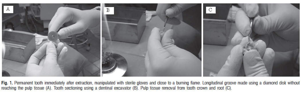
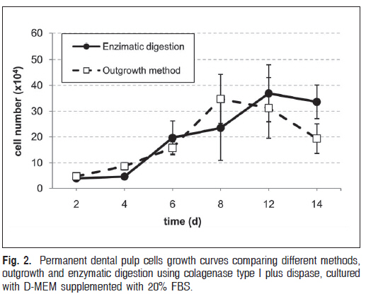
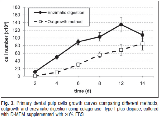
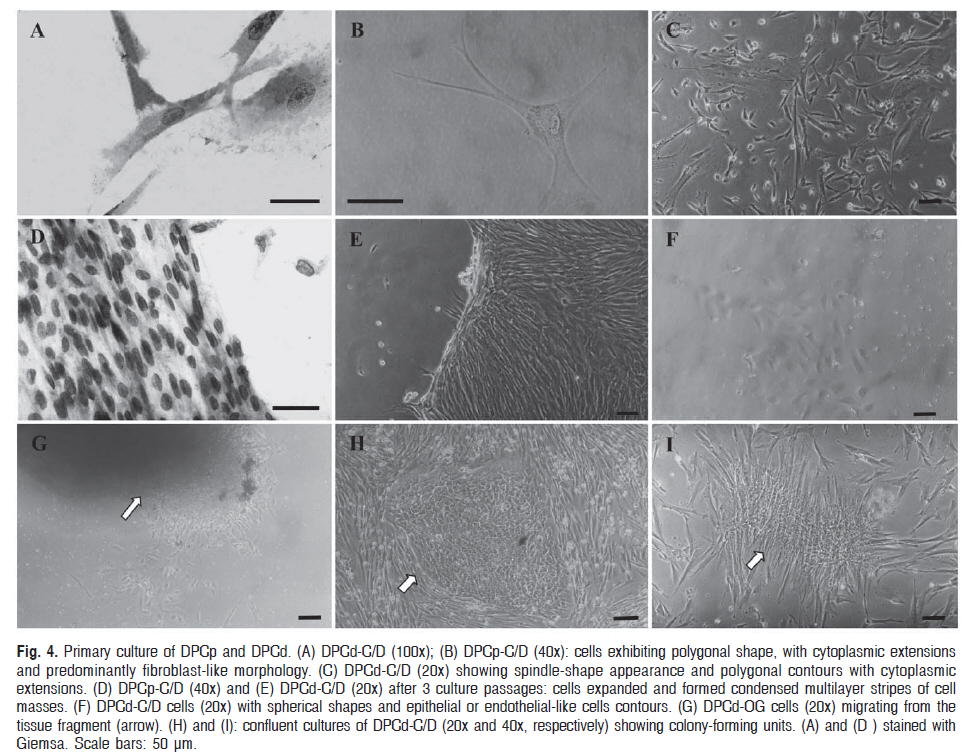
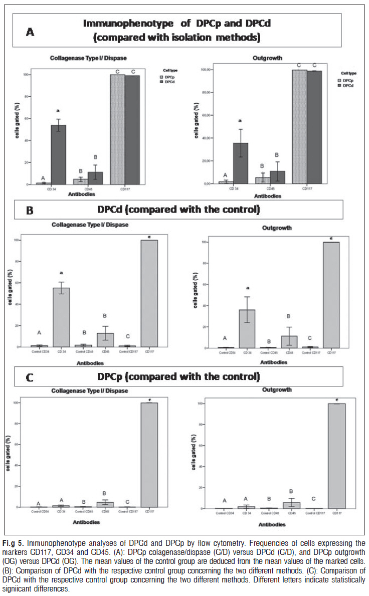
Braz J Oral Sci, Vol. 9, No. 4, October-December, 2010, pp. 427-433 Comparative isolation protocols and characterization of stem cells from human primary and permanent teeth pulp Leliane Macedo de Souza1, Juliana Duarte Bittar2, Izabel Cristina Rodrigues da Silva3, Orlando Ayrton de Toledo4, Marcelo de Macedo Brígido5, Marcio José Poças-Fonseca6 1DDS, MS, Department of Dentistry, School of Health Sciences, University of Brasilia, Brasília, DF, Brazil Correspondence to: Marcio José Poças-Fonseca Universidade de Brasília - Instituto de Ciências Biológicas - Departamento de Genética e Morfologia - Campus Universitário Darcy Ribeiro - 70910-900 Brasília - DF, Brazil. Phone: + 55 (61) 3107-3090 Fax: +55 (61) 3107-2923 E-mail: mpossas@unb.br Received for publication: March 2, 2010 Accepted: July 19, 2010 Code Number: os10049 AbstractAim: This study was developed to compare the morphological, proliferative and
immunophenotypic profiles of pulp cells from permanent and primary teeth, obtained
by two isolation methods. Keywords: stem cells, cell culture techniques, dental pulp, primary dentition, permanent dentition. Received for publication: March 02, 2010 Introduction Several studies have addressed the regeneration of pulp tissue and the induction of reparative dentin based on the biology of stem cells and tissue engineering approaches1-4. Adult mesenchymal stem cells have been identified in the pulp tissue of human permanent teeth - DPSC5-6 , in exfoliated primary teeth -SHED7-8, but also in the periodontal ligament – PDLSC9-12 They are described as multipotent stem cells, capable to self-renew and to differentiate into various cell types, such as in osteocytes, adipocytes, chondrocytes, cementoblasts, odontoblasts and neuronal cells5-6,13-14. Dental pulp cells have been isolated by enzymatic digestion5,15-17 and the outgrowth method8,14,18-19. Enzymatic digestion is a common method to obtain single cell suspensions from primary tissues and consists of exposing the tissue to enzymes for a minimal period of time in order to preserve maximum cell viability. The outgrowth method is considered simpler and faster and consists of placing pulp fragments directly into the culture plate so that cells outgrow from the pulp tissue explants. Stem cells hardly express some specific markers. In this view, they are very difficult to characterize. Moreover, there is no rigid pattern of gene expression that could be associated with the stemness state8,17,20. Few studies have addressed the possible differences in the immunophenotypic, morphological and proliferative patterns between permanent and primary teeth pulp cells as a function of different isolation protocols. In this regard, this is a pioneer study in comparing the isolation of primary teeth stem cells by enzymatic digestion and by the outgrowth method. This study aimed at performing an in vitro characterization of human dental pulp cells from primary (DPCd) and permanent (DPCp) teeth. We compared the morphological, immunophenotypic and proliferative profiles of cells isolated by enzymatic digestion (C/D) and by the outgrowth method (OG). Material and methods Primary Dental Pulp Cell Culture Human dental pulps were obtained from 10 freshly extracted impacted third molar teeth (donors aged 9-15 years) and 10 human primary teeth (donors aged 7-12 years), presenting at least two thirds of the root, extracted for orthodontics reasons at the Maxillofacial LTDA Institute Brasília (Brazil), under the approval of the Ethical Committee of the Medical School of the University of Brasília, Brazil (process number 034/2007). All experiments were undertaken after patients had signed informed consent forms according to the 196/96 Brazilian Health Department Resolution and to the World Medical Association’s Declaration of Helsinki. In the proximity of a burning flame, in order to minimize the risk of contamination, tooth surfaces were cleaned and a longitudinal groove was made using sterilized diamond discs (KGSorensen, ref.7020, Zenith Dental ApS, AgersKov, Denmark), without reaching the pulp. The pulp tissue was gently separated from the crown and root with a sterile dentinal excavator (Fig.1). The pulp tissue was transported to the laboratory in polystyrene microtubes (Corning Incorporated, Corning, NY, USA) containing Dulbecco’s modified Eagle’s medium - DMEM (GIBCOTM, Grand Island, NY, USA) supplemented with 10% fetal bovine serum (FBS) (GIBCOTM) and with 100 units/mL penicillin and 100 mg/mL streptomycin (GIBCOTM). Before culture establishment, the pulp tissue was washed three times with Hank’s Balanced Salt Solution (HyClone, Logan, UT, USA). Pulp cell cultures were established by two distinct approaches: enzyme digestion (C/D) or explant culture outgrowth (OG). For the enzyme digestion, pulp tissue was treated with a solution of 3 mg/mL type I collagenase (GIBCOTMand 4 mg/mL dispase (GIBCOTM) for 60 min at 37ºC. After enzymatic digestion, the cell suspension was washed three times by centrifugation for 10 min at 750 x g in culture medium (DMEM), and single cell suspensions (104 cells) were seeded into 6-well cell culture plates (Corning Incorporated). For the outgrowth method procedure, the explant fragments were transferred to a new plate once a week until no further outgrowth was observed (up to 5 transfers). For all methods, the culture was maintained in DMEM supplemented with 20% FBS and 100 units/mL penicillin and 100 mg/mL streptomycin (GIBCOTM) in a 80% humidity, 5% CO2 atmosphere at 37ºC. Cell proliferationIn order to compare the proliferative potential of cells obtained by enzymatic digestion and by the outgrowth method, permanent dental pulp cells (5 x 10⁴), obtained by enzymatic digestion (colagenase type I plus dispase) were seeded after 5 culture passages in 12-well plates with D-MEM supplemented with 20% FBS, 100 units/mL penicillin and 100 mg/mL streptomycin. Cell growth was assessed in triplicate by the trypan blue (Sigma Chemical Co.) exclusion assay after 2, 4, 6, 8, 12 and 14 days of culture. Culture medium was changed at every 2 days. Growth curves were performed to compare the proliferation of permanent dental pulp cells obtained by enzymatic digestion and by the outgrowth method. The same experimental approach was employed for primary dental pulp cells. To assess the time of duplication or doubling time (DT), the following equation was applied: DT = T x ln2x1 / [ln (N0 - N)], where N= final number of cells, N0= initial number of cells. This formula derives from the calculation of growth curves in N = N0 x 2t / T. Student´s t test was used to compare the proliferation rate of cells obtained by the two methods. Statistical significance was assumed at a value of p <0.05. All statistical tests were performed with MINITAB® release 14.20 (Mininc, State College, PA, USA) and the plots were obtained through the Microsoft Office Excel® software (Microsoft Corporation, Redmond, WA, USA). Morphological AnalysisFor morphological analysis by light microscopy, permanent and primary dental pulp cells (5x10⁴) were grown over microscopy cover slips (18 x 18 mm), previously treated for 30 min with poly-L-lysine (0.1 mg/mL), and placed into 6-well culture plates. After 48 h, adhered cells were fixed for 10 min with 2% paraformaldehyde in phosphate buffer saline, washed and stained with 4% Giemsa in methanol (SIGMA,) for 12 min. Another group of cells was analyzed without staining. Light microscopy analysis was performed with the Nikon Diaphot TMD M250 microscope (Kogaku, Japan). Immunophenotypic characterization by flow cytometryAfter 3 culture passages, 2x10⁶ cells from primary and permanent teeth were pellet (10 min at 750 x g) and resuspended in phosphate buffer saline (PBS) with 0.1% sodium azide and 1% bovine serum albumin (BSA) as a marking solution. Cells were then incubated for 15 min with 10 mL of antibody as indicated by the manufacturer. Stem cell markers employed were the mouse anti-human antibodies anti-CD117 or c-kit (Invitrogen Corporation, Carlsbad, CA, USA), a membrane tyrosin-kinase receptor that interacts with the Stem Cell Factor (SCF); anti-CD34 (FK-Biotec, Porto Alegre, RS, Brazil), a marker for endothelial precursors and hematopoietic stem cells; and anti-CD45 RA (FK-Biotec), a marker for immature T leukocytes. Subsequently, 104 cells were acquired and analyzed at the FACScalibur Cell Analyzer (Becton-Dickinson) running with the Cell Quest software (Becton Dickinson). Nonprobed cells were used as controls for each antibody. The statistical analysis was performed by Student’s ttest (SPSS® version 17.0; SPSS Inc., Chicago, IL, USA) and the significance level was set at 5%. The statistical analysis used for comparison of the cell types obtained with the distinct isolation methods was generated considering the normalized values. ResultsPulp cells obtainment with the two methods (Fig. 1) was effective for both crown and root tissues. Sample contamination with microorganisms was only rarely observed. Considering the proliferation of permanent dental pulp cells obtained by the two methods, growth curves (Fig. 2) showed that, on the 4th day of culture, the cell number was significantly higher for the outgrowth isolation method (p=0.015). On the other hand, on the 14th day, culture cell number was statistically higher (p=0.046) for the enzymatic digestion method. Growth curves (Fig. 3) comparing the proliferation of primary dental pulp cells obtained by the two different methods showed that cell number was statistically higher after 2 (p=0.002), 4 (p=0.000), 6 (p=0.000), 8 (p=0.004) and 12 (p=0.008) days of culture when the cells were isolated by enzymatic digestion. The calculated doubling times were: 18.54 h for DPCp-C/D; 19.61 h for DPCp-OG; 16.87 h for DPCd-C/D and 17.13 h for DPCd-OG. The primary cultures of all experimental groups were very heterogeneous at the morphological level. For both isolation methods, cells presented spindle-shape appearance and polygonal contours with cytoplasmic extensions, displaying a fibroblast-like morphology (Figures 4A, B, C). For cell cultures obtained by enzymatic digestion, cells with spherical shape and epithelial- or endothelial-like contours were also observed (Fig. 4F). After 3 weeks of culture, for both isolation methods, cells appeared aligned, presenting long extensions of cytoplasm, establishing cell connections and organizing monolayers, thus forming a tissue-like structure (Fig. 4D and E). After successive culture passages, no more spherical cells were observed for the enzymatic digestion method. In the outgrowth method, cell migration from the pulp fragment was observed throughout the culture plate. Cells closer to the fragment showed heterogeneous shapes, but a predominance of polygonal or elongated morphologies was observed (Fig. 4G). Cell cultures obtained by enzymatic digestion showed, from 7 to 15 days, the formation of clusters of cell colony-forming units, with spherical cells at the center (Fig. 4H and I). The immunophenotypic analysis (Fig. 5) showed that, in both cell groups, CD117 was the most expressed marker: DPCpC/D (100%); DPCpOG (99.69%); DPCdC/D (98.95%) and DPCdOG (98.73%), at p = 0.05. Statistical analysis showed that cells isolated by the distinct methods differently expressed CD34 (p<0.05). Results showed that DPCd also presented a high expression of CD34 (DPCdC/D=53.91%; DPCdOG=35.59%) (Fig. 5A, B). Both cell groups showed no expression of CD45, for both isolation methods. Regardless of the isolation method, the mean frequency of DPCd expressing CD117 (pC/D = 0.00; pOG = 0.00) and CD34 (pC/D = 0.01; pOG = 0:04) was different from the control group (nonproubed cells). However, the CD117marker was the only one significantly expressed in DPCp (pC/D = 0.00; pOG = 0.00) (Fig. 5C). The CD45marker was not expressed by DPCd (pC/D = 0.22; pOG = 0.33) or by DPCp (pC/D = 0.20; pOG =0.30). DiscussionIsolation and proliferation of stem cells are the first step for tissue engineering. Our objective was to compare different methods for obtainment of permanent and primary dental pulp cells aiming at the isolation of stem cells within the facilities of the University Hospital (HUB) of the University of Brasilia (UnB), Brazil. The comparison between the enzymatic digestion and the outgrowth methods had not been employed for primary teeth cells before. Although several methods for collecting pulp tissue are mentioned in different studies, they are not described in details. Some studies use carbide burs to reach the pulp tissue from extracted teeth5 or from vital teeth, removing the pulp with endodontic files (pulpectomy)17. Other studies use forceps to break extracted teeth18. Pulp heating and the increase of microbial contamination are some of the make the pulp separation difficult and increase the disadvantages of these methods. We used a diamond disk to contamination risk. create a furrow without reaching the pulp tissue in order to The primary teeth included in our study presented at reduce pulp heating, which occurs when carbide burs are least two thirds of the root. Some studies have reported the used. Tooth breakage was achieved with little pressure in use of naturally exfoliated primary teeth for the establishment the furrow using a dentinal excavator (Fig 1). We did not of pulp stem cell culture7. However, these naturally exfoliated use forceps to avoid irregular tooth breakage, which could teeth barely present pulp tissue anymore. Both the enzymatic digestion and the outgrowth methods have proved to be effective for obtaining cells. During the subsequent culture passages, cells obtained by the different methods proliferated differently. Primary dental pulp cells obtained by the enzymatic digestion method presented a higher proliferative potential when compared to the ones isolated by the outgrowth method, along the entire period of experimentation. Unpublished data from our group indicated that D-MEM supplemented with 20% FBS promoted for pulp cells a superior proliferative response, when compared to D-MEM 10 FBS%, HAM-F12 10% and 20% FBS. The doubling times calculated for primary (DPCd) and permanent (DPCp) pulp cells were similar to those observed in other studies of human dental pulp cells (hDPCs)21 and point out to a very high clonogenic capability, particularly for cells obtained from primary teeth. The major advantage of the outgrowth method is its convenience and low cost8,17. The digestion method may completely dissociate cells from the tissue, but it is technically difficult due to a certain level of cell damage and loss. Kerkis et al.8 postulated that the cultivation of pulp fragments, prior to the initial cell passage, probably prevents the stem cells premature differentiation. These authors have suggested that the outgrowth method promotes the selective proliferation of the dental pulp immature stem cells (IDPSCs). They also demonstrated that IDPSCs obtained from primary teeth expressed several mesenchymal stem cell markers, and they speculated that these cells could represent the multipotent precursors of both DPSC and SHED. Under the tested experimental conditions, few morphological differences were verified between cells obtained from primary and permanent teeth (Fig. 4). At the beginning of the culture (7 days), cells were morphologically heterogeneous, with spherical and elongated shapes. This was particularly evident for the enzymatic digestion method (Fig. 4A, B, C). After a period ranging from 15 to 22 days, cells obtained through both methods showed mainly polygonal morphology, with expansion of the cytoplasm. The nucleus was polarized, as observed in several other stem cells studies14-15,17,19. Cells with spherical morphology (Fig. 1F) have been related to epithelial or endothelial-like precursor cells22-23 . According to Sonoyama et al.24, these cells contaminate mesenchymal stem cells cultures when immunomagnetic separation is not efficient. Endothelial-like cells usually disappear after successive passages24, as it was also observed in the present experiments. Polarized cells were observed in dental pulp cultures and they are considered odontoblasts precursors19,23,25. With the enzymatic digestion method, we could identify cell clusters scattered over the culture plate (Fig. 4H, I). These clusters were described as cell colony-forming units15,25. These colonies have been identified in stem cells cultures from bone marrow and also from human dental pulp, and they are considered a prerequisite for odontoblasts differentiation14,17,26. Considering the immunophenotypic characterization, our results showed that different methods of isolation did not influence the cells immunophenotypic profiles. DPCp and DPCd showed phenotypes compatible to stem cells, with remarkable expression of CD117 (Fig. 2). Several studies have shown that stem cells from adult human dental pulp present a variety of surface antigens associated with neural crest cells precursors (CD117 or ckit)16,20,26-27. Some studies report that these primary cultures do not express hematopoietic markers like CD45, while the CD34 expression remains controversial5,8,16,24. The expression of CD117 and CD34 reinforces the immunophenotypic profile of stem cells. CD117 is an early stem cells marker, since it interacts with the stem cell factor (SCF), an important growth factor related to the proliferation, differentiation and recruitment of progenitor cells in various biological systems27 . We identified significant difference in the expression profile of CD34, an hematopoietic stem cells marker28, among the distinct cells populations. DPCp were CD117+/CD34-/ CD45-, in accordance to studies that describe dental pulp stem cells as CD34-5-6,8,15. On the other hand, DPCd were CD117+/CD34+/CD45-, in accordance to studies that describe dental pulp stem cells as CD34+ (Fig. 5)16,18,20,26. In this perspective, our data reinforce the controversy described in the literature. The CD34+ phenotype was not previously described for primary teeth cells. In conclusion, our results indicate that both isolation methods can be efficiently used aiming at the isolation of human dental pulp stem cells in a clinical environment. In addition, we could demonstrate that stem cells obtained from primary teeth also present the CD34+ phenotype, compatible to hematopoietic precursor cells. We believe that the findings of this study can contribute to the discussion of reliable and reproducible protocols for the isolation and propagation of dental pulp stem cells, aiming at future regenerative therapies not only for dental, but also for other oral structures. AcknowledgementsThis work was supported by FINEP/MCT and FUB -Brazil References
Copyright 2010 - Braz J Oral Sci The following images related to this document are available:Photo images[os10049f5.jpg] [os10049f1.jpg] [os10049f3.jpg] [os10049f2.jpg] [os10049f4.jpg] |
| |||||||||
