 |
| About Bioline | All Journals | Testimonials | Membership | News |
|
||||||
|
||||||
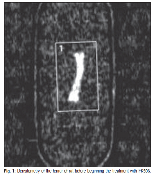
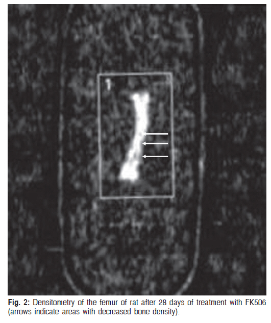
Braz J Oral Sci, Vol. 9, No. 4, October-December, 2010, pp. 459-463 Primary stability of orthodontic miniscrews inserted into femurs of osteopenic rats Rogério Lacerda dos Santos1, Matheus Melo Pithon1, Mônica Tirre de Souza Araújo2, Matilde Gonçalves da Cunha Nojima2, Lincoln Issamu Nojima2 1Specialist in Orthodontics, Federal University of Alfenas, Alfenas,
MG, Brazil; Master in Orthodontics, Federal University of Rio de Janeiro, Rio
de Janeiro, RJ, Brazil; PhD student in Orthodontics; Federal University of
Rio de Janeiro, Rio de Janeiro, RJ, Brazil Received for publication: July 15, 2010 Code Number: os10056 AbstractAim: To test the hypothesis that bone quality may affect the stability of anchorage
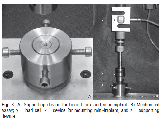
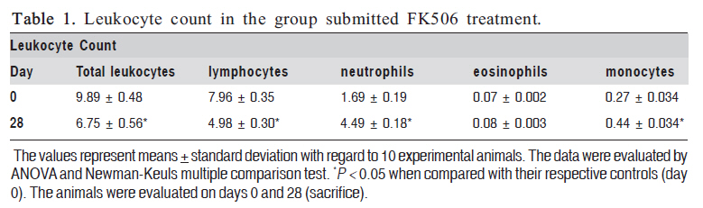
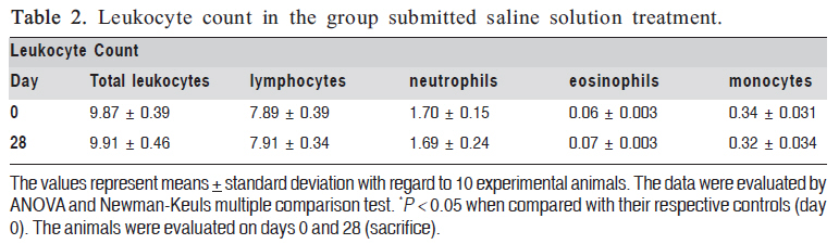
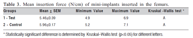
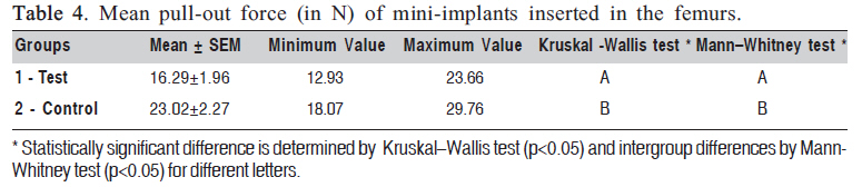
devices implanted in a rat model. Keywords: immunosuppressant, bone, miniscrew, pull-out test. IntroductionCurrently, an increasing number of young patients and adults with orthodontic needs have been submitted to different types of transplants. In addition, there has been a significant increase in the use of immunosuppressants in these patients, and questions have been raised concerning the action of these drugs on bone metabolism, causing osteopenia and osteoporosis. There are medications capable of affecting the bone metabolism and the rate of tooth movement1, such as tacrolimus, which is a calcineurin phosphatase inhibitor2. Tacrolimus (FK506) is an immunosuppressive agent derived from Streptomyces tsukubaensis3, and is widely used in patients subjected to organ transplantation4. Tacrolimus belongs to the class of macrolide immunosuppressants, a class of non-steroidal drugs that act by inhibiting the calcineurin system. Some authors5-6 have suggested that FK506 exerts its antiinflammatory effect mainly by interfering with the activation of T cells, by suppressing the production of cytokines. When the tacrolimus molecule penetrates the cytoplasm of lymphocytes, it links with the intracellular protein FKBP 12. After this the FKBP-12 molecule forms a complex with calcium, calmodulin and calcineurin, inhibiting the activity of calcineurin phosphatase. This prevents the dephosphorilation and translocation of the nuclear factor of cytoplasmatic activated T lymphocyte (NF-AT), preventing binding with its nuclear subunit. In the absence of this step, the T lymphocytes are unable to synthesize and secrete cytokines, particularly TNF-α, IL-1β, IL-2, and IL-6, by modulating the inflammation. Bone destruction and development of severe osteoporosis have been reported7 . The quality of the bone tissue is crucial for the tooth movement, including the use of several types of orthodontic anchorage devices8. Mini-implants are currently being used to help to move teeth and consequently optimize the orthodontic treatment9 . The use of mini-implants for orthodontic anchorage has revolutionized orthodontics over the years, thus allowing stable anchorage opposing the reaction forces from the orthodontic movement9-12 . Mini-implant stability can be either primary or secondary. Both involves direct contact between mini-implant and bone, but, the secondary occurs after healing13. The lack of stability may be related to osseous factors and mini-implant characteristics14, including diameter screw15 and dimension of pilot perforation16 . When the stability of a given mini-implant has to be mechanically evaluated, one of the increasingly-used methodologies is the pull-out test17-18, which is widely used in medical areas such as orthopedics, neurosurgery, cosmetic and maxillofacial surgery19, for testing the primary stability of several screw devices. The pull-out test consists of extracting the mini-implant from osseous tissue at constant speed, thus enabling assessment of the maximum force needed to remove the implanted device. Patients who need orthodontic mini-implants, but are taking medications, such as tacrolimus, which is capable of reducing the bone mineral density, can present osteopenia. In view of this, the objective of the present study was to test the hypothesis that bone quality may affect the primary stability of anchorage devices implanted in a rat model. Material and methods Animal Model A total of 20 male Wistar rats (Rattus norvegicus) aged 9 weeks old and with an average weight of 300 g were housed according to the guidelines for animal research as recommended by the Osvaldo Cruz Foundation of Rio de Janeiro (FIOCRUZ-RJ). The immunosuppressive agent FK506 (Tacrolimus, PROGRAF ®, Astellas, Ireland) was orally administered at a dose of 2 mg/kg/day as a suspension containing water and 5% dextrose20, for 28 days7. The animal experiment was approved by the Ethics Committee on Animal Research of the Federal University of Rio de Janeiro (UFRJ), Brazil. Experimental GroupsThe rats were randomly divided into 2 groups consisting of 10 animals each as follows: Group 1, rats treated with FK506 and Group 2, rats treated with saline solution vehicle. Twenty mini-implants (INP - Sistema de Implantes LTDA, São Paulo, Brazil) were used for study, all presenting their own characteristics, such as: self-drilling type, cylindrical screw design, 9 mm length, 6 mm body length, 4 mm screw length, 1.5 mm screw diameter and Ti-6AI-4V alloy. Prior to insertion, the mini-implants were characterized and measured by using a profile projector (Nikon, Model 6, Tokyo, Japan). Weight AssessmentFK506 causes metabolic and gastrointestinal alterations3 and this factor can interfere with the weight gain in the animals. The animals’ weight was measured every day in order to adjust the dosage of FK506 to be administered and to verify the influence of the immunosuppressant on weight gain. Blood CountTotal and differential (lymphocytes, monocytes, eosinophils, and neutrophils) leukocyte counts in Groups 1 and 2 were evaluated on day 0 (before immunosuppressant administration); and on sacrifice day, leukocyte counts were evaluated in Group 1, in order to monitor the immunosuppressive effect of tacrolimus. Blood was collected from the tip of each rat’s tail. The leukocyte count was performed by means of a light microscope (Olympus BX40, Olympus, Hamburg, Germany). Densitometric AnalysisWith regard to densitometry, the animals were analyzed on day 0 (before beginning the treatment with FK506) (Figure 1) and on day 28, after being sacrificed (after 28 days of treatment with FK506) (Figure 2). To avoid possible interferences in the mini-implants during the densitometric exam, densitometry was performed in the (left) femur not used for inserting the mini-implant. The bone mineral content (BMC) was measured and then divided by the area so that the bone mineral density (BMD) of the femur of each rat could be obtained by means of dualenergy x-ray absorptiometry (DXA) (Prodigy, GE/LUNAR, USA). The femur was put into a vessel of equal size and grains of rice were poured around it so that the bone was completely covered, thus simulating the surrounding soft tissues. Bone scan and data analysis were carried out by using specific software for small animals, which could be manually adjusted to analyze the region of interest. The resolution (in pixels) was 0.3 x 0.6 mm for a collimator of 0.84 mm in diameter. The scanned area was 9.9 mm wide and 11.8 mm Primary stability of orthodontic miniscrews inserted into femurs of osteopenic rats intraperitoneal injection of sodium thiopental (50 mg/kg) (Cristália, Campinas, São Paulo, Brazil) and had the femoral neck region (3 x 2 cm) was shaved. Asepsis of the operative field was done with 4% chlorhexidine digluconate (School of Pharmacy, Federal University of Rio de Janeiro, Brazil). An incision was made along the femur, and the surface of the femur was exposed from the diaphysis to the head of the femur by using a No. 15 scalpel blade mounted on a scalpel handle. The subcutaneous tissue was laterally separated using a pair of blunt-tipped scissors. All animals received one mini-implant inserted 2 mm into the femoral neck (region of compact bone) situated between the head and diaphysis. The mini-implants (self-drilling screws) were inserted into the right femur of each rat by using a torque screwdriver mounted on a digital caliper, thus allowing both insertion and torque measurements to be performed perpendicular to the femur surface. Then the animals were sacrificed and decapitated, and the femurs were dissected and bone blocks containing the mini-implant were obtained. The samples were immersed in saline and stored at -15o C for 15 days. Thereafter, the bone blocks were left at room temperature for mechanical assay of the primary stability of orthodontic mini-implants. The test was performed in a universal testing machine (Emic DL 10.000, São José dos Pinhais, Brazil). A claw-shaped device was fabricated and mounted upon the upper part of the machine so that the mini-implant could be removed. Another device served as a base for both fixing the bone block and keeping the mini-implant in a perpendicular position during the tests, thus preventing the creation of momentum (Figure 3). Mechanical assay was performed at a crosshead speed of 0.5 mm/s for removing the mini-implant from osseous bone. Load and displacement values were recorded as well as the maximum force (Fmax) (in N) for later evaluation. Statistic Analysis Experimental data were analyzed statistically with the use of SPSS software 13.0 (SPP Inc., Chicago, IL, USA). The data were submitted the non-parametric analyses with the Kruskal-Wallis and Mann-Whitney U tests to assess the torque values among different groups. Miniscrew success was defined as a complete lack of mobility and a removal torque exceeding 0. The results were found to be statistically significant at p < 0.05. The results of pull-out and bone densitometry were also submitted to the test for Pearson’s correlation at a level of significance of 1%. Other data were evaluated by ANOVA and Newman-Keuls multiple comparison test (p < 0.05). ResultsEffect of Appliance Installation and FK506 Treatment on Weight Gain The weight gain in each period of time was calculated on the basis of the initial treatment with FK506. The immunosuppressant was found to have an influence on the weight gain, since the non-treated animals (Group 2) had highest gains in 28 days (50.5±1.6 g) in comparison with Group 1 (46.0±1.7 g), though without statistically significant difference between the groups (p > 0.05). Effect of FK506 Treatment on Leukocyte CountImmunosuppression was observed in the animals submitted to FK506 treatment (Group 1) as leukocyte and lymphocyte counts were shown to have similar and decreased values during the experiment in the time periods (day 28). Moreover, statistically significant (p < 0.05) differences were observed between day 28 when compared to their respective controls (day 0). Therefore, the effect of this immunosuppressive on rats submitted to FK506 could be shown (Table 1). The number of neutrophils, eosinophils and monocytes increased after 28 days of treatment with FK506, and significant difference (p < 0.05) was observed in neutrophils and monocytes in comparison to their respective controls (day 0) (Table 1). Group 2 presented no statistically significant difference between day 0 and day 28 (p > 0.05) (Table 2). Bone DensitometryBone densitometry showed a difference in the mean bone density value on day 28, with significant difference (P < 0.05) in the animals submitted to FK506 treatment (139.2 ± 5.1 µg/cm2) in comparison to vehicle-treated animals (152.2 ± 1.8 µg/cm2). Insertion Torque and Pull-Out TestThe mean insertion force values were found to be statistically similar in both groups (P > 0.05) (Table 3). With respect to pull-out force, there was significant difference between Group 2 (control) and Group 1 (experimental) (P < 0.05) (Table 4). The mean pull-out force values between the inserted mini-implants ranged from 17.07 to 30.76 N for non-treated animals (Group 2 - Control), whereas in the animals treated with FK506 (Group 1 - Test) it ranged from 10.95 to 23.66. This finding demonstrated a strong correlation between the pull-out test and bone mineral density in the control group (0.9788) and pull-out test and low bone density in the animals submitted to treatment with FK506 (0.9850). DiscussionPrevious studies have demonstrated that the treatment with FK506 (tacrolimus) can induce bone loss in human beings21 as well as in experimental models22. It has been suggested that FK506 inhibits the production of proinflammatory cytokines, particularly TNF-±, IL-12, and IL623, all modulating inflammation24. Because FK506 has the capacity to alter bone metabolism, one can suggest that it can interfere with the primary stability of orthodontic miniimplants,which is the reason for conducting the present study. Because the influence of FK506 on bone metabolism depends mainly on the dose administered20, in the present experiment the doses were calculated and adjusted daily according to the animal’s weight gain. Some factors can interfere with weight gain, such as metabolic and gastrointestinal changes3,25 caused by FK506. In the present study, the weight gain observed in FK506-treated animals was higher than that found by Hayakawa et al.26 and Sabry et al.20, who used the same daily FK506 dosage. With regard to bone densitometry, the femurs of these animals showed different bone mineral density values on day 28, thus supporting some of the findings in the literature21-22. According to Cvetkovic et al.7, FK506 application causes imbalance in bone activity (osteoblastic/osteoclastic) due to the significant increase in the parathyroid hormone (PHT) levels, which was observed at a dosage higher than that used in the present study. On the other hand, Kirino et al.25 have reported a significant increase in PTH with maximum peak on day 21 by using a lower dose, but an increased PTH resulting from continuous FK506 administration can lead to bone loss25 . The quality of the bone tissue is crucial for the tooth movement, mainly for use of several types of orthodontic anchorage devices8. Mini-implants are currently being used to help moving teeth and consequently optimizing the orthodontic treatment9 . In view of this, the goal of the present study was to test the hypothesis that bone quality may affect the primary stability of anchorage devices implanted in a osteopenic rat model, using pull-out test. Piau pigs (Sus scropha) is an experimental animal widely used in studies on mini-implants due to the similarity between human and swine in terms of maxillary bone, a fact that is well-known and well-established in the literature27. In the present study, however, rats were used because this experimental animal is widely used for research studies on immunosuppressants28 . The pull-out test consists of extracting the mini-implant from the bone tissue perpendicular at a constant speed. This method, which is extensively used in several areas of medicine19 has been increasingly used in orthodontics since the publication of a study conducted by Huja et al.17 . Despite the non-tractional force being applied to miniimplants, the values obtained during mechanical assay show “imbrication” between the screw-part of mini-implants and the bone tissue in which they are inserted16 . As the pull-out test was performed in vitro, there is concern with regard to storage of the samples and time elapsed between sacrifice and mechanical test. Earlier studies on pull-out force demonstrated force variation over time, that is, between insertion and pull-out assay. Roe et al.29 , who tested 1-week samples stored at -20oC, reported lack of statistical differences when the test was carried out immediately after the animals were sacrificed. Another study reported a decrease in pull-out force as storage time was extended from 4 to 8 weeks. In the present study, after the animals had been sacrificed, the samples were immediately dissected and stored in saline for 15 days at -15oC. All procedures of the present study were in accordance with other studies on orthodontic mini-implants17-18 . On the 15th day, the samples were left at room temperature to gradually unfreeze. In order to fix the bone fragment during mechanical test, a metal device was made, which was mounted on a universal testing machine. The bottom part of the device was made to keep the mini-implant perpendicularly positioned on the base without having to apply resin to the osseous block, as suggested elsewhere17-18. This decision was taken because of the reduced size of the sample and the possibility of resin penetrating into the osseous tissue, which might mask the results. Studies evaluating screw pull-out performed immediately after insertion and 8 weeks later found no statistical differences30. In another study on titanium implants inserted in facial bones, only a modest gain in the pull-out strength was measured after comparing the healing periods of 1, 2, 3, 4, 6, 9, 12, and 32 weeks. Based on these studies, the secondary stability required during treatment is directly related to the primary stability achieved during mini-implant insertion. However, further studies are needed to evaluate the primary and secondary stability of mini-implants without force application. The mean pull-out values observed in this study were lower than those found elsewhere, possibly because of the different bone characteristics between the experimental animals studied17, but in general, the optimal forces required for orthodontic movement range from 0.3 to 4.0 N17. The pull-out forces found in the present study were significantly higher than those for clinical purposes, which may validate the use of mini-implants even when patients are taking immunosuppressive medications that interfere with bone metabolism. However, further research relating the amount of loading and force application direction on mini-implants is needed to validate the use devices in osteopenic patients. The cortical thickness is recognizably important in terms of primary stability of mini-implants. The fact that the medullary bone is most affected by these medications25 could explain the lack of difference in implant insertion torque between the evaluated groups. Osteoporosis may have greater influence on primary stability and the use of mini-implants in osteoporotic patients is contraindicated in such cases. In conclusion, the hypothesis tested in this study was accepted. Bone quality may affect the stability of miniimplants. Orthodontic mini-implants did not present good primary stability in a rat model with osteopenia. AcknowledgementsWe would like to thank the Rio de Janeiro State Research Support Foundation (FAPERJ) for supporting the research. References
Copyright 2010 - Braz J Oral Sci The following images related to this document are available:Photo images[os10056f3.jpg] [os10056t4.jpg] [os10056t2.jpg] [os10056f1.jpg] [os10056t3.jpg] [os10056t1.jpg] [os10056f2.jpg] |
| |||||||||
