 |
| About Bioline | All Journals | Testimonials | Membership | News |
|
||||||
|
||||||
Brazilian Journal of Oral Sciences, Vol. 10, No.1, Jan-Mar, 2011, pp. 22-26 Effect of fluoride-containing bleaching agents on bovine enamel microhardness Samira Padilha Gabasso1, Cristiane Franco Pinto2, Vanessa Cavalli3, Adriana Franco Paes-Leme4, Marcelo Giannini5 1Undergraduate Student, Department of Restorative Dentistry, Piracicaba Dental School,
University of Campinas, Piracicaba, SP, Brazil Received for publication: July 07, 2010 Correspondence to: Marcelo Giannini, Department of Restorative Dentistry - Operative Dentistry Division Piracicaba School of Dentistry, P.O. BOX 52 University of Campinas - UNICAMP Av. Limeira, 901 Areião - Piracicaba, SP Zip Code: 13414-900 - Brazil, Phone:55 19 21065340 /Fax:55 19 21065218 E-mail:giannini@fop.unicamp.br Code Number: os11005 Abstract Aim: The purpose of this study was to evaluate the effect of 10% carbamide peroxide
(10%CP) bleaching agents with different fluoride concentrations on enamel microhardness after induction
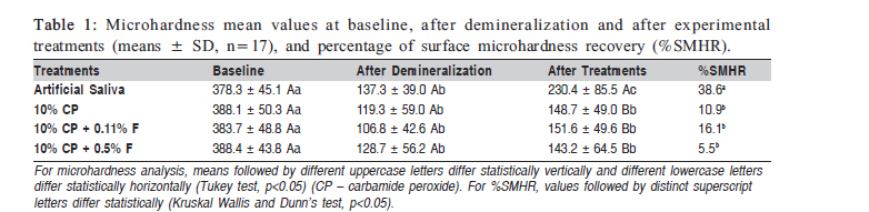
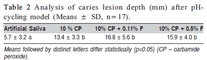
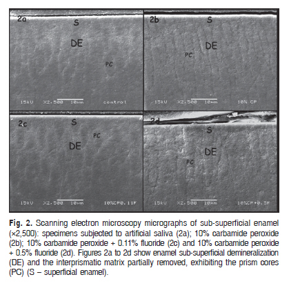
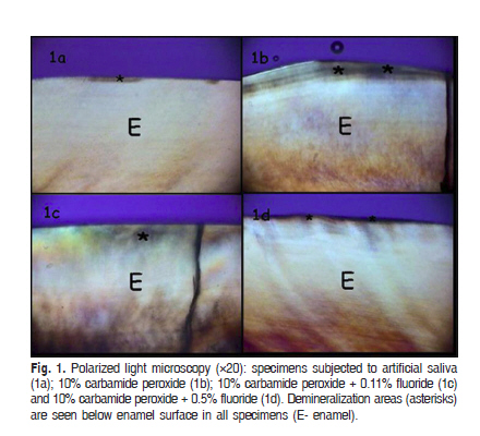
of artificial caries lesions during pH-cycling model. Keywords: dental enamel, tooth bleaching, fluoride. Introduction Carbamide peroxide is the active ingredient of most home-use tooth bleaching agents. Since the introduction of nightguard vital whitening, concern has been expressed regarding the potential effects of carbamide peroxide solution on dental hard tissues1. As the bleaching technique of vital teeth comprises the direct contact of the whitening gel on the outer enamel surface, the oxidation reaction for an extended period of time can be related to demineralization processes2-3. Various effects of carbamide peroxide bleaching on teeth have been reported. Scanning electron microscopy (SEM) investigations have shown that 10% carbamide peroxide changes enamel, promoting surface dissolution and topographical alterations4-6. The morphological changes and reduction of enamel microhardness are not limited to the enamel surface; alterations have also been detected in the subsurface enamel layer7-9. Moreover, some studies have shown alterations of the histological aspects and composition of sound dental enamel after carbamide peroxide gel application9-10. The bleaching treatment with 10% carbamide peroxide slightly increased caries susceptibility of enamel11-12. However, although most studies have shown the effects of bleaching agents on sound teeth, the effect of carbamide peroxide gels on white spots lesions and early erosions is not known, and neither is the capability of fluoridated carbamide peroxide gels in recovering the enamel microhardness. Thus, the objective of this study was to evaluate the effect of one unfluoridated and two fluoridated carbamide peroxide gels on the microhardness of enamel with caries-like lesions induced during pH-cycling model. The null hypothesis was that enamel surface microhardness recovery (SMHR) is not influenced by the type of bleaching agent used. Material and methods Three bleaching agents containing 10% carbamide peroxide were tested: one unfluoridated gel (FGM Prod. Odont. Ltda, Joinville, SC, Brazil) and two fluoridated carbamide peroxide bleaching agents (Opalescence PF with 0.11 % fluoride, Ultradent Products Inc., Salt Lake City, UT, USA and Whiteness with 0.5% fluoride, FGM Prod. Odont. Ltda, Joinville, SC, Brazil). Specimen Preparation Bovine teeth, stored in saturated thymol at 5oC for up to 1 month were used in this study. Eighty bovine dental enamel blocks (4x4x2 mm) were obtained from the buccal surface with the use of double-faced diamond discs (KG Sorensen, Barueri, SP, Brazil). The buccal enamel surfaces were wet-polished with 800-, 1000- and 1200-grit SiC paper, followed by diamond pastes (3 µm and 1 µm). Five microhardness indentations spaced 100 µm from each other were performed on the enamel surface with a microhardness tester (FM-1e, Future Tech, Tokyo, Japan), under a 50-g load for 5 s. Means of the five indentations were calculated for each block and the samples with surface Knoop hardness ranging from 430.9 to 92.6 KHN units were selected to standardize the samples among the experimental groups, which had enamel blocks with similar initial Knoop microhardness values. Half of the enamel block surface (8 mm2) was coated with an acid-resistance varnish and the exposed enamel surfaces were subjected to demineralizing solution containing 0.05 M acetate buffer, pH 5.0, 50% saturated with enamel bovine powder, for 16 h at 37oC using 2 mL of solution to each 1 mm2 of exposed area13. The aim of demineralized solution was to produce artificial caries lesion. Afterwards, the enamel blocks with surface microhardness ranging from 72.6 to 172.8 KHN units were selected. pH-Cycling Regimen and Experimental Groups The enamel blocks were randomly divided into 4 groups (n = 17) and subjected to a 12-day pH-cycling13 consisted of 5 phases: (1) 1-min soak in fluoridated dentifrice (1,100 ppm F as NaF)/water slurries three times a day to simulate daily toothbrushing; (2) between the treatments with dentifrice, samples were individually immersed in artificial saliva14 (14.2 mM sodium carboxymethylcellulose, 280 mM xylitol, 13.4 mM potassium chloride, 17.1 mM sodium chloride, 0.004 mM sodium fluoride, 0.2 mM magnesium chloride, 0.4 mM calcium chloride, 2.9 mM potassium phosphate, 0.1 mM potassium thiocyanate, pH 7.2) at 37 oC; (3) to simulate the daily acid challenge, enamel blocks were individually immersed in demineralized solution for 2 h at 37oC, with the same composition of the solution used in the initial carious lesion procedure; (4) the samples were immersed in human saliva during 2 h at 37oC before bleaching treatment to promote acquire pellicle formation; (5) to simulate daily treatment, the samples were subjected to artificial saliva (control group) or bleaching treatment with 3 whitening gels: 10% carbamide peroxide (FGM Prod. Odont. Ltda); 10% carbamide peroxide with 0.11% fluoride (Opalescence PF); and 10% carbamide peroxide with 0.5% fluoride (FGM Prod. Odont. Ltda) for 8 h (nocturnal period) at 37oC. For the bleaching procedure (5), 0.1 mL of the whitening gel was mixture with 0.05 mL of artificial saliva and this mixture was applied on enamel surface and covered with an individual tray. The mixture with artificial saliva tends to increase the decomposition of hydrogen peroxide and release more water, oxygen gas and free radicals, simulating the mouthguard bleaching technique14. During the bleaching, the specimens were placed in 100% relative humidity at 37°C and, after bleaching, the specimens were rinsed with an air/water spray for 10 s and stored in 100% humidity environmental until analysis. Surface Microhardness and Microhardness Recovery Analyses of Control and Tested Groups. Surface microhardness was determined for the enamel blocks after polishing (baseline or sound enamel), after demineralization and after experimental treatments15. The percentage of SMHR (%SMHR) was calculated as: % hardness recovery = hardness after pH cycling - hardness after demineralization x 100 / sound enamel hardness (baseline) - hardness after demineralization13. After surface microhardness measurements, all blocks were longitudinally sectioned into 2 halves with a diamond saw (Isomet 1000, Buehler, Lake Bluff, IL, USA) and one half was used to evaluate the lesion depth by polarized light microscopy (PLM) and the other half was used to examine the subsurface of treated enamel by SEM. PLM Analysis Slices of 150 ± 10 mm from specimens were polished with 600- and 1200-grit aluminum oxide disks under water (Table 1) cooling to a thickness of 100 ± 10 µm. The slabs were immersed in distilled and deionized water, mounted in glass slides and the demineralization depth was analyzed in a polarized light microscope (DM LSP, Leica Microsystems, Heerburg, Switzerland). The images were transferred to the computer via digital camera. The lesion depth was measured at five points from the enamel surface using computer software (Image-Pro Plus, 4.1 version for Windows, Media Cybernetics, Silver Spring, MD, USA) and the values were expressed in micrometers (µm) to calculate the means. SEM Analysis The surfaces from the other halves were polished with 600-, 1200- and 2000-grit SiC papers, followed by diamond pastes (6, 3, 1 and ¼ µm) and dehydrated in ascending ethanol concentrations (30, 50, 70, 90 and 100%). Afterwards, they were sputter-coated with gold (MED 010, Baltec, Balzers, Liechtenstein) and observed with a scanning electron microscope (JSM-5600, Jeol Inc., Peabody, MA, USA). Representative areas of enamel were photographed at 2,500× magnification. Statistical Analysis The %SMHR data were analyzed by the Kruskal Wallis and Dunn's test (a=0.05). The data from microhardness analysis and demineralization depth were, respectively, analyzed by two-way (bleaching and enamel treatment) repeated-measures analysis of variance and one-way analysis of variance followed by Tukey's test (a=0.05). Results The microhardness mean values and standard deviation (baseline, after demineralization and after experimental treatments) for the groups and %SMHR data are displayed in Table 1. The control and experimental groups showed significant lower microhardness (p<0.01) after deminera-lization when compared to baseline. After pH-cycling regimen, the surface microhardness after treatment was significantly higher than after demineralization but lower than baseline. The microhardness and microhardness recovery from control group was significantly higher than the experimental groups (p=0.0007). Significant differences concerning the %SMHR were not observed among the experimental groups (p values = 0.1873, 0.3544 and 0.5363). The analysis of demineralization depth by PLM (Table 2) showed superficial and sub-superficial demineralization areas in enamel for all the groups evaluated (Figs 1a, 1b, 1c and 1d). The demineralization depth for specimens immersed in artificial saliva was lower than the bleached groups (p<0.0026). SEM micrographs revealed demineralization areas located at sub-superficial region of enamel for all the experimental groups (Figs 2 a, b, c and d). However, the demineralization seemed to be milder in enamel immersed in artificial saliva (Figs 1a and 2a) than in the bleached enamel. Discussion A recent study has shown that enamel treatment with either fluoridated or unfluoridated carbamide peroxide gels, at both neutral and acidic pH, yielded enamel more susceptible to demineralization16. However, it is not known if the 10% carbamide peroxide can promote similar effects on enamel with caries lesions. The bleaching agents containing or not fluoride did not show mineral recovery when the enamel microhardness values after demineralization were compared to those after pH-cycling, which is in accordance with previous study2,17-20. Indeed, even in the presence of daily treatment with a fluoridated dentifrice during the pH-cycling, it was not able to promote remineralization. Oliveira et al.21 (2005) evaluated the effect of carbamide peroxide containing calcium (0.05% and 0.2%) or fluoride (0.2% and 0.5%) on enamel and showed the reduction of enamel surface microhardness post-bleaching treatment. Also, the bleaching of enamel with carbamide peroxide followed by fluoride treatment with 2,000 ppm fluoride solution four times during 2 min did not improve erosive resistance22. The effect of fluoridated dentifrice and saliva on remineralizing artificial caries lesions has been recognized13,23. However, the lack of fluoride effect either in the bleaching agent or in dentifrice can be explained by the fact that carbamide peroxide could have promoted erosion on enamel surface, such as open enamel prisms, increasing porosities as previously described17, which could impair an accurate surface (Fig. 1, Fig. 2) microhardness measurement. Also, Attin et al.16 (2003) showed that carbamide peroxide/fluoride mixture did lead to a fluoride uptake less than enamel samples treated with pure fluoride gel. However, the study did not evaluate whether this amount of fluoride uptake had any cariostactic effect. The present study did not show remineralization effect of fluoride-containing bleaching gel when compared to non-fluoridated gel. Conversely, Pretty et al.24 (2005) showed that tooth bleaching with carbamide peroxide did not increase the susceptibility of enamel to acid erosion or caries. Furthermore, remineralization of bleached enamel was improved by application of a high fluoride concentration (2.23% fluoride, Duraphat, Colgate, Piscataway, NJ, USA)17. In the present study, a fluoridated dentifrice was used at the same dilution that occurs in the mouth during toothbrushing, but no substantial effect was observed for the bleached groups. Another study showed that carbamide peroxide containing or not fluoride promoted mineral loss after demineralization and remineralization cycles, concluding that the treatment with either fluoridated or nonfluoridated carbamide peroxide gels (neutral and acidic gel) rendered enamel more susceptible to demineralization16. This study used bovine teeth, which can be considered a research limitation. Based on the fact that some authors reported some differences between human and bovine teeth25-26, the results speculate what would happen with human enamel in the same conditions proposed for this study. Induction of demineralization reduced the bovine enamel microhardness approximately from 384.6 (430.9 - 338.3) to 122.7 (72.6 - 172.8) KHN units. This investigation was based on previous studies, which showed similar surface microhardness after demineralization13,27. It is important to emphasize that although the caries-like lesions were superficial, it was possible to observe differences among the treatments (Table 1), i.e., the effect of the artificial saliva (unbleached control group) compared to bleaching treatments. Enamel demineralization was observed for all specimens, however, with lower mineral loss for those stored in artificial saliva. SEM observations revealed that matrix interprismatic of subsuperficial enamel was removed, exposing the prism cores for all experimental groups (Figs. 2a, b, c and d). PLM analysis also showed demineralization areas for all specimens (Figs. 1a, b, c and d). However, the demineralization seemed to be more intense for enamel treated with bleaching agents in subsuperficial part of enamel and on its surface (Figs. 1b, c and d), as a result of bleaching procedures regardless of the presence of fluoride in its composition. The non-bleached specimens were kept in artificial saliva, which favored the remineralization, according to Featherstone et al.28 (1986). Thus, artificial saliva and fluoridated dentifrice treatments during pH-cycling seemed to increase the %SMHR. If the study was performed with more periods of observation after treatments, the storage of the bleached samples for prolonged time in saliva could increase the remineralization28. The results of the present study showed that the presence of fluoride in the bleaching gels did not increase mineral recovery, suggesting that bleaching procedures on enamel with active caries-like lesions should be avoided or used with caution even with daily use of fluoridated dentifrice and fluoride-containing bleaching agent. Acknowledgements This study was supported by grants from PIBIC-CNPq, SAE/PRP-UNICAMP and 02/04135-3 from FAPESP, Brazil. References
The following images related to this document are available:Photo images[os11005t2.jpg] [os11005f1.jpg] [os11005t1.jpg] [os11005f2.jpg] |
| |||||||||
