 |
| About Bioline | All Journals | Testimonials | Membership | News |
|
||||||
|
||||||
Brazilian Journal of Oral Sciences, Vol. 10, No. 3, Jul-Sept., 2011, pp. 158-162 Use of dental dimensions estimated from personal portraits in human identification Rachel Lima Ribeiro Tinoco1, Laíse Nascimento Correia Lima1, Mário Marques Fernandes2, Luiz Francesquini Junior3, Eduardo Daruge Junior4 DDS, MSc, Department of Forensic Dentistry, Piracicaba Dental School, University of Campinas, Brazil Received: December 07, 2010 Code Number: os11031 Abstract Many cases of human identification in which traditional methods are not applicable challenge the experts’ capability and versatility. In the absence of ante-mortem records, superimposition of skull images over photographs of a possible victim arises as a possible alternative. Keywords: forensic dentistry, victims’ identification, tooth. Introduction Forensic dentistry is an important science for human identification, especially when conventional methods cannot be applied, usually due to advanced decomposition, carbonization or fragmentation of the body. In such cases, and in the absence of antemortem dental data to be compared, craniofacial image superimposition can be an accurate and reliable technique for human identification1-3 . However, before superimposing images of the skull over photographs of a possible victim, it is mandatory that they both are properly sized in the same scale4-5 . Authors that apply computer-assisted craniofacial superimposition size reach the scale through craniometric and cephalometric landmarks easily identifiable in the face and the skull6-7 , even thought this assessment is influenced by the thickness of local soft tissue - amount and disposition of fat - which can vary among different populations8 . Considering that the teeth are naturally visible throughout life, not being influenced by soft tissue, and commonly shown in smiling pictures, they are reliable size reference for comparing images of skull and personal pictures9 . Although technical advances allow the three-dimensional facial reconstruction10-12, this is not an identification method, but rather a form to disseminate a facial approximation of that subject, willing to achieve someone who knew him/her, and can provide reliable antemortem records for human identification. Human identification through craniofacial image superimposition has been tested and used by different researchers and experts. However, the proper scaling of the photographs to be compared is a precondition for an accurate evaluation. The aim of this pilot study was to test the feasibility of obtaining real size of teeth from personal photographs, using common accessories as size reference, in order to use them for the proper scaling required for image superimposition. Material and methods Sampling Pictures of 21 volunteers (8 males and 13 females) were taken by a single photographer. All the subjects were aged between 21 and 60 years, and agreed to participate of the study by signing an appropriate document. Subjects with absence or anomalies in the upper central incisors were excluded, as well as those with notorious systemic pathology. Methods From each participant the following photographs were taken:
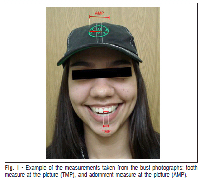
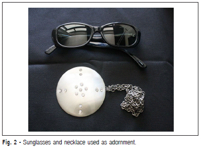
The images obtained were stored in a computer managed by Windows Vista™, and analyzed by the software Adobe Photoshop™. From the bust photographs, the mesiodistal dimensions of the upper central incisors were measured, which were called TMP (tooth measure at the picture). From the same picture, using the same zoom, the size of the adornment (sunglass, a rounded detail of the cap, or necklace) were measured, to obtain what were called AMP (adornment measure at the picture), as shown in Figure 1, with the cap. Figure 2 shows the sunglasses and the necklace. All the measurements were stored in an Excel™ file. Then, the life-size of the adornments worn by the subjects were taken, with a digital caliper (Digimess, São Paulo, Brazil), which was called adornment’s real measure (ARM). From the measures taken, tooth’s real measure (TRM) was estimated, according to algebra fundamental rules of proportion, translated by the following equation:
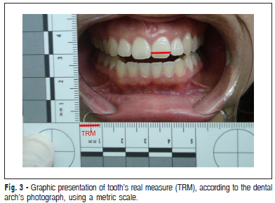
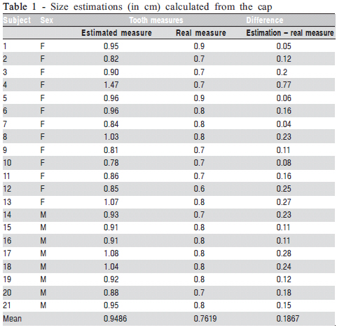
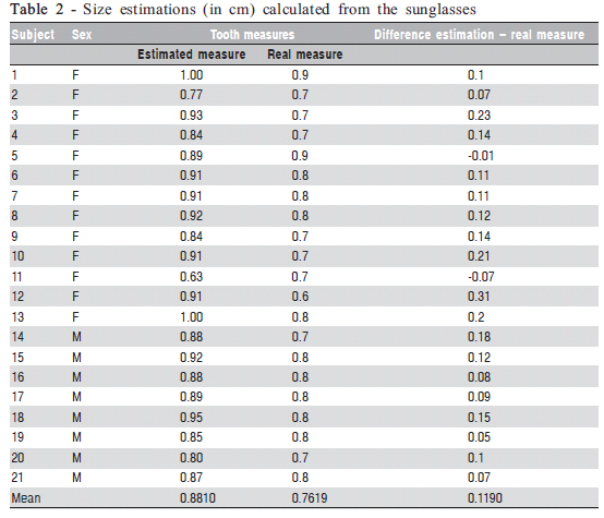
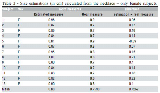
Once the estimations were concluded and stored, the pictures of the dental arches with metric scale were analyzed, and the mesiodistal dimensions of the upper central incisor were taken with a manual caliper, according to the scale at the picture, as graphically represented in Figure 3. The values obtained from this measuring procedure were then compared to the estimations. All measurements and estimations were made by a single examiner. Statistical analysis All measures – real and estimated – were divided in three independent groups, according to the adornment used as reference to the estimation: sunglasses, cap or necklace. For each group, the results were analyzed by Student’s t-test for paired samples, with the tooth’s real measure and the tooth’s estimated measure as variables. Results The dental dimensions estimated from the size of the cap were overestimated in 100% of the cases, with a mean difference of 0.1867 mm, as can be seen in Table 1. According to the Student’s t-test, the estimated dental width was statistically higher than the real width (p = 0.0000169), at a significance level of 5%. When estimating the dental width from sunglass dimensions, overestimated values were found again, except for two cases of underestimation (9.52% of the sample). The mean difference between estimated and real measurements was equal to 0.1190cm, as shown on Table 2. The values estimated from measurements of the sunglasses were considered statistically higher than the actual measurements (p = 0.0000018), with a significance level of 5%. The estimated tooth measures from the dimensional relation with the necklace were overestimated in most cases (92.31%). It is noteworthy that this type of calculation, using the necklace as a reference point, was performed only in female participants, since they have virtual exclusivity in the use of this type of adornment. The mean difference between estimated and actual measurements was equivalent to 0.1262 cm, as shown in Table 3. Tooth measures estimated from the necklace were significantly higher (p = 0.0001307) than the actual measurements, with a significance level of 5%. Discussion Since the early twentieth century, researchers have analyzed craniofacial correlations and the relationship between the face and the underlying bone morphology. On this line, are noteworthy the studies about the shape of the nose and the nasal bone, as well as variations in nose shapes of different ethnic groups13-14. These studies underlie the current principles of facial reconstruction15, which became the main target for this type of research16. As occurs in the nose, eyes also show great relationship between soft tissue and bone structure. The position of ectocanthus was studied by Whitnall in 2,000 skulls, and can be located in the so called tubercle of Whitnall. This anatomical point constitutes the major reference for sizing and positioning the image of the skull in relation to the face in superimposition methods15. The objects used as a dimensional reference in this study were selected because they frequently appear in personal pictures, in the same spatial plane of the teeth. However, the facial sizing from the teeth dimensions naturally requires the recovery of these teeth, which does not always occur, especially considering the frequency of post-mortem loss of anterior teeth. The proposed method overestimated the mesiodistal width of the central incisor, in its best result – by measuring sunglasses - 1.19 mm on average (15.62% of the real average). The worst performance was achieved by having the cap as size reference, overestimating the dental width in 1.867 mm on average. The less satisfactory result of the cap in relation to other items may be related to the position of the cap slightly oblique in relation to the plane of the central incisors. Further studies are suggested. The analysis of a personal photograph for human identification purposes requires excellent image resolution for detailed observation and accurate superimposition with the cranial image. The analysis of teeth in photographs only becomes possible under optimal conditions, close and high resolution images. Still, the use of tooth width as a size reference, given its small dimensions, creates a greater risk of failure, since fractions of a millimeter can represent important distances in the final result. Thus, when sizing images using anatomical landmarks, the larger the reference, the greater the chance of success. Although previous studies have been engaged in creating protocols and methods of sizing and positioning skull and face images for superimposition, the correct size and position are more easily achieved through successive attempts, and movement of the images3,6,1,17-19. The method evaluated in the present study is therefore inaccurate for sizing facial images for superimposition with human identification purposes. References
Copyright © 2011 - Brazilian Journal of Oral Sciences The following images related to this document are available:Photo images[os11031f1.jpg] [os11031t1.jpg] [os11031t3.jpg] [os11031f3.jpg] [os11031t2.jpg] [os11031e1.jpg] [os11031f2.jpg] |
| |||||||||

