 |
| About Bioline | All Journals | Testimonials | Membership | News |
|
||||||
|
||||||
Brazilian Journal of Oral Sciences, Vol. 10, No. 3, Jul-Sept., 2011, pp. 171-174 Comparison of tooth crown discoloration with Epiphany and AH26 sealer in terms of chroma and value: an in vitro study Fatemeh Shahrami 1, Mina Zaree 1, Arash Poorsattar Bejeh Mir2, Mojtaba Abdollahi-Armani 1, Abbas Mesgarani 3
1 DDS, MS Endodontics Department, Dentistry School, Mashad University of Medical Sciences, Iran, 2 Student Research Committee, Dental Material Research Center, Dentistry School, Babol University of Medical Sciences, Iran, 3 DDS, MS Endodontics Department, Dentistry School, Babol University of Medical Sciences, Iran
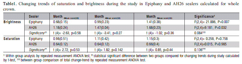
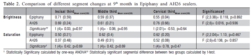
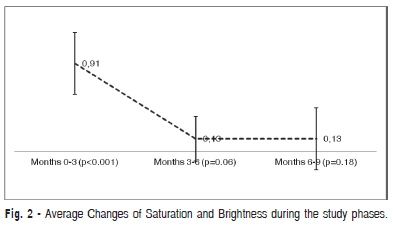
Objective: Tooth crown discoloration may possess a heavy emotional burden and esthetic concern, especially when the anterior teeth are affected. Residue of sealers within the pulp chamber is a major contributor to the occurrence of tooth discoloration. Keywords: sealer, discoloration, esthetic, Epiphany, AH26, brightness. Introduction Mainly, interaction of light with inner dentin and enamel structure reflective characteristics constitutes the color of the tooth. Hence, any change in the composition of enamel, dentin or coronal pulp may cause the modification in translation of light through the tooth, and consequently tooth discoloration1 . Actually any modification in hue, chroma (saturation, hue intensity or color) or value (brightness, hue quality) of the tooth is discussed as tooth discoloration2 . Determining the origin of tooth discoloration is an essential step for further diagnostic and remedial procedures, in order to guarantee a successful outcome. The causes of tooth discoloration are simply classified as two main categories: external or internal. There may be a combination of both in some cases3. Sedimentation of colored materials such as coffee, tea carrot, chocolate, mouthrinse or dental plaque is among the frequent reasons for external discolorations4-5. In addition, various factors, either systemic (e.g., drugs, fluorosis, cystic fibrosis, hyperbilirubinemia, dentinogenesis imperfecta) or local (e.g., pulp necrosis, pulp internal hemorrhage, pulp tissue remnant after pulpectomy, endodontic materials, crown restorative materials, aging or root resorption) may contribute to the changes in color of the tooth crown2,5. Epiphany is a recently introduced root canal sealer that has been developed specifically for use with Resilon cones. Epiphany is a composite resin sealer with dual setting action that is composed of a mixture of Bis-GMA, ethoxylated Bis-GMA, UDMA and dual hydrophilic methacrylate with calcium hydroxide fillers, barium sulfate, barium glass and silica6. To date, there are few studies that evaluated tooth discoloration after Epiphany application as an endodontic sealer. The aim of this experimental in vitro study was to evaluate of degree of crown discoloration when Epiphany is used for sealing, comparing the results with those of a traditionally used sealer, AH26. Material and methods In this in vitro experimental study, 40 intact extracted human maxillary incisors were used. Teeth were free of caries, crown restorations, cracks or any apparent coronal discoloration when selected. The teeth were cleaned with an ultrasonic device to dislodge gross debris, with additional rubber cap and pumice powder cleansing to remove remaining debris and stains from the coronal surfaces. Then teeth were randomly assigned to experimental and control groups. Randomization performed via permuted block method with 4 intervals and a final 1:1 teeth numbers. Fifteen teeth in each arm were assigned to the experimental allocations and the 5 remaining as the controls. Experimental sealers were either AH26 (Dentsply DeTrey, Switzerland) and Epiphany (SybronEndo, Orange, CA USA). Controls were filled with distilled water. Tooth preparation started with cross sectioning from the crown to 3 mm below the cementoenamel junction with the apical root discarded Pulps were extirpated and the pulp chambers were instrumented with K-files and no 2, 3 Gates Glidden drills. The canals were irrigated with 1 mL of 2.5 % sodium hypochlorite and 17% EDTA7. The experimental sealers were placed into pulp chambers via an apical access and the internal walls were thoroughly coated with the sealers. Excess sealer was removed with small pieces of cotton pellet from the orifices of the canal and chamber walls. The apical access was then sealed with a sticky wax. The prepared teeth were placed on a gray background allowed the most accurate visualization of coronal discoloration, and the digital images were captured using a digital 12 mega pixel camera (Canon Ixus, Japan) in a dark room and two 60W light sources from sides on a gray background. Thereafter, the images were saved as JPEG images. Digital images of all samples were viewed in the Photoshop software version CS8. First the full length crown of the tooth was selected. Then, the background of each sample was deleted with the tooth crown was divided into three equal segments: incisal, middle and cervical. Brightness and saturation of each segment were determined and recorded with the Eye Dropper option and the correlated histograms. Afterward, teeth were then stored in the individually vials in an incubator at 37º and 90 º humidity to simulate intra oral condition. Teeth were removed in subsequent 3, 6 and 9 months and examined in the same way. We scored (defined) brightness and saturation changes as follows: 1 for less than 5% (slight), 2 for 10% to 15% (moderate) and 3 for 15% to 20% (severe). All analyses were performed with SPSS version 17 (SPSS Inc, Chicago, IL, USA). Data were expressed as mean and standard deviation. Data calculated for final scoring of tooth discoloration were tested with repeated measures ANOVA to detect pattern of changes regarding time with sealer interaction. An ANOVA test was also applied to determine the segment differences accounted for either brightness or saturation and sealer types with their interactions at 9th month with further post-hoc between-group analyses. A two tailed p<0.05 was considered statistically significant. Results A total 15 teeth in Epiphany group and 14 teeth in AH26 group (one tooth excluded because of apparent coronal fracture after canal preparation) were evaluated as experimental group. Also ten teeth served as control group. Obtained scores were adjusted for number of teeth in these groups. Basic characteristics of calculated scores for teeth crown color changes by time in AH26 and Epiphany sealer are displayed in Table 1. Brightness was significantly Increased by time [F (2, 8) =29.16, p<0.001]. Epiphany and AH26 group was not differed in brightness changes during the 9 months of the study (p=0.084). In addition, saturation changes was not neither statistically significant [F (2, 8) =0.129, p=0.881], nor different in experimental groups (p=0.136) (Table 1). At 9th month, no differences were observed in different parts of the crown measured for Epiphany sealer [F (2, 3.38) = 0.118, p=0.892] and for AH26 sealer [F (2, 6) = 0.06, p=0.936] (Table 2). In addition, no interaction of segment*sealer (p=0.50), segment* discoloration (p=0.7), sealer*discoloration (p=0.33) and segment* discoloration* sealer (0. 83) was noticed. Changes in the control group were not discernible through the study phases. All changes in experimental groups were significantly higher than the control group (p<0.001). Changing trends of crown colors differed at different trimester intervals, with more highlighted changes occurred during first 3 months (Figure 1). Discussion In the present in vitro study, crown discoloration after filling the root canals with either Epiphany or AH26 was investigated during 9 months. Recently, Epiphany was introduced as an ideal endodontic sealer6. This is the first study that investigated the crown discoloration induced by Epiphany. Tooth crowns were deteriorated in terms of brightness in both sealer groups. However, saturation increased by time, it was not remarkable for both Epiphany (achieved power=0.48) and AH26 (achieved power=0.05). This finding is in agreement with Parson et al.6 and Partovi et al.8 researches, which concluded that tooth crown progressively discolored with application of endodontic sealers by time6,8. In addition, measured at 9th month, different crown segments were not contrasted by either saturation or brightness (achieved power=0.26). This similarity was present when compared for sealer types and interaction of either segment with discoloration or segment with sealer type. Nine-month duration was chosen since it was proposed that most severe crown discoloration may occur during the first 9-month period after sealers were used6. It is postulated that brightness or value is the most important determinant of the color. Higher values (i.e., darker shades) were observed in AH26 group when compared to Epiphany group, in all study points. Although it did not reach significant level, regarding achieved power of 25%, larger scale studies may better reveal this difference. The superiority of Epiphany with this regard may be explained by the silver (Ag) particles in AH26 sealer9-10. As one limitation of the study, we missed the immediate measurements of saturation and brightness (Figure 2). Hence, changing trends and exact amount of changes, when adjusted for the baseline scores of filled teeth, are somehow hampered by this deficit. Although not statistically significant, most prominent changes in saturation in the AH26 group occurred in the cervical third. This is in agreement with previous studies, which attributed this pattern to the thinner layer of enamel over this region6,9. Surprisingly, as another remarkable characteristic of Epiphany, it exhibited least value changes in the cervical third as compared to other regions. Many earlier investigators studied color changes of posterior teeth (premolars)6,8,10, mean while, anterior teeth concern the patients mostly with a high socio-emotional burden. Hence, we opted to examine the central teeth in the present research. Collectively, average brightness and saturation changes occurred faster during the early phase of study (months 0-3), probably due to higher tubular penetration capacity at the beginning. This finding is contrasted from the result of Parson et al.6 Visual protocol that those authors6 applied in their research, which is acceptable considering the time at which study performed, may rationalize the apparent differences from our findings. Nevertheless, our findings are in agreement with those of Partovi et al.8, who clarified more significant changes during early phase of study using a digital system. Based on our results, it is speculated that the resinbased AH26 sealer and the composite-based Epiphany sealer are similar in penetration through dentin tubules and Epiphany is even superior to the AH26. Currently methods for evaluation of tooth crown discoloration could be classified into two major categories: visual method that is a subjective method which applies a guiding color shade. This method is very difficult, since many interference variations may adversely influence the interpretation (e.g., eye fatigue and light position)7. Alternatively, color analysis with a device is a quantitative objective method that includes colorimetric and spectro-photometric analyses1. However, these techniques have their own limitations, such as limited access of clinicians to such devices. More recently, along with introduction of the computer to the dentistry, we have one ideal method for analyze tooth discoloration. Previously, a computer-aided method to discriminate chroma and value changes was used and analyzed with Photoshop software11. This may help the observer lessen the measurement bias and simple visual observation flaws. We did not calculate completecomponent-color changes using either Munsell (Nickerson formula) or CIE systems12. Using Nickerson and CIE systems, calculated I or ÄE changes of more than 5 and 3.5 is considered clinically significant, respectively. AH26 sealer was selected because it is commonly used in endodontics. According to the various previous researches on the older generations of sealer, we did not include those, to be compared with in the present study. We did not attempt to remove the smear layer. The use of 17% EDTA may lessen the inhibitory effect of smear layer for sealers to be penetrated into the adjacent dentine tubules. This may rationalize the 4day discoloration of the crown when AH26 was used and the smear layer was eliminated with 17% EDTA9. Briefly, both saturation and brightness were increased by time. Low achieved powers in our study indicate that higher sample sizes may better distinguish the corresponding changes. A larger scale study with histological examination for depth of enamel and dentin penetration is recommended. External validation with more extended reliable results may be obtained with in-vivo studies. Additively, shorter-interval observations can reveal the timely pattern of color changes more precisely. In conclusion, although it might be difficult to eliminate every trace of sealer, it is still recommended that the materials should be carefully removed from the pulp chamber immediately after filling the canal. Newer Epiphany sealer is a suitable substitute for AH26 sealer. Overly, most changes occurred during the first 3 months with minimal gradual increments for the rest of the phases of the study and brightness changes represented the main portion of these differences in the present study. References
Copyright © 2011 - Brazilian Journal of Oral Sciences The following images related to this document are available:Photo images[os11034t1.jpg] [os11034f1.jpg] [os11034f2.jpg] [os11034t2.jpg] |
| |||||||||
