 |
| About Bioline | All Journals | Testimonials | Membership | News |
|
||||||
|
||||||
Brazilian Journal of Oral Sciences, Vol. 10, No. 3, Jul-Sept., 2011, pp. 199-203 Evaluation of a digital methodology for human identification using palatal rugoscopy Karen Christine dos Santos1, Clemente Maia S. Fernandes2, Mônica da Costa Serra3
1DDS, Department of Social Dentistry, Faculty of Dentistry of Araraquara, São Paulo State University - UNESP, Brazil 2DDS, LL.B student, MSc, PhD, Post-Doctorate student, Department of Social Dentistry, Faculty of Dentistry of Araraquara, São Paulo State University - UNESP, Brazil 3DDS, LL.B, MSc, PhD, Post-Doctor, Department of Social Dentistry, Faculty of Dentistry of Araraquara, São Paulo State University - UNESP, Brazil Received: May 16, 2011 Code Number: os11040 Abstract
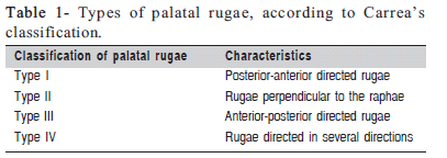
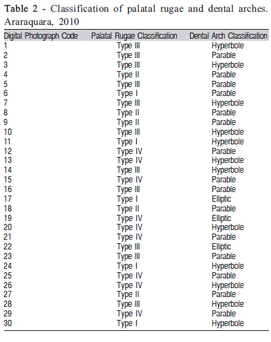
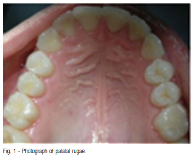
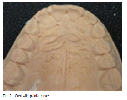
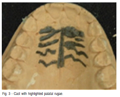
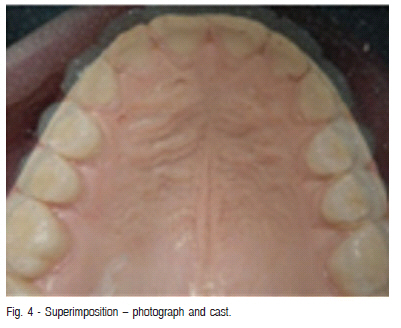
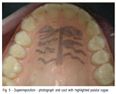
Introduction: Palatal rugoscopy, or palatoscopy, is the process by which human identification can be obtained by inspecting the transverse palatal rugae inside the mouth. Keywords: forensic sciences, forensic anthropology, forensic dentistry, human identification, palatal rugae. Introduction Human identification corresponds to the set of different procedures to identify a person or object1. Identification means demonstrating that a certain person or his/her relevant attribute, which appears in the examination at a given moment, is the same one that appeared on a previous occasion2-3. For an identification process to be applicable, five requirements need to be met: individuality, immutability, perenniality, practicality and classification4. In humans, unlike in other animals, the set formed by the rugae is asymmetric2,5. The palatal rugae are unique, unchanging, perennial and subject to classification6-10. It appears in the third month of the embryonic period, remaining for the entire life of the individual and for several days after his/her death1. When identification cannot be established by fingerprinting or by analysis of dental arches (dental records data), the palatal rugae can be considered as a source of comparative material8,11 because they are able to individualize a person, legitimizing an identification process12, even in extreme circumstances13. Illnesses, chemical injury or trauma do not seem to modify the palatal rugae structure. Muthusubramania et al.14 found that most victims of third-degree burns do not show changes in the palatal rugae, and when such changes do occur, they are less pronounced than those found on the body. The palatal rugae are better “preserved” due to the protection given by the bony scaffold, dental arches and soft structures. On the other hand, fingerprints can be easily destroyed in certain accidents or due to the action of the vast necrophagous fauna15. Palatal rugoscopy can have a major role in human identification. The cast models and antemortem intraoral photographs can be found in dental records and serve as the first protocol to perform the identification process. Furthermore, for at-risk populations (for example, aeronauts and firemen), this first record can be preventively arranged and archived for the possible need of identification. Limson and Julian11 compared the rugosities of university students by an impression with irreversible hydrocolloid and cast models in plaster type II. The rugae were highlighted with well sharpened graphite pencils. The rugae were photographed and the models were scanned, achieving a 92-97% success rate. The authors claimed that this error rate of 3-8% can be reduced by using an intraoral scanner, with a direct transfer to the computer. Some identification methods are available for edentulous victims, such as comparing the anatomy of the paranasal sinuses and comparing the bone patterns observed in radiographs. In addition to this, the victim’s own dentures can also be used, which are found inside the mouth or in their homes. Among the evidence from an edentulous victim, the palatal rugae are considered to be one of the unique morphological features16. Ohtani et al.17 studied the possibility of identifying edentulous individuals by comparing the rugae in denture molds against models obtained from impressions taken from the patients. Impressions were taken from the mucosal surfaces of complete dentures with alginate impression materials and 146 maxillary casts were made from hard dental plaster. They then compared the rugae patterns to identify the pairs (dentures and plaster models). The median percentage of correct matches was 94%, and the error rate was attributed to 3 factors: poorly demarcated eminences of rugae, change of palatal height in some cases and non-complex rugae pattern. Castellanos et al.16 reported the identification of the body of an adult woman in a city in Colombia. The woman had been declared missing by her family 15 days before the skeletonized body was found. Identification was made by comparing the palatal rugae obtained from the remainder of the body with the plaster model that was obtained from the denture that was with the family. It is often difficult to obtain dental arch impressions of corpses that arrive at the Forensic Medicine Institutes to be compared with the antemortem data of the alleged victim. As the experts or technicians are not familiar with dental impression materials and there is no dental laboratory structure, photographs are more practical. Antemortem models can be obtained from the victims’ dentists and then compared with the postmortem photographs using free image-editing software, which is more practical and economically feasible for the Brazilian Forensic Medicine Institutes. This study evaluated a digital method for human identification using palatal rugoscopy by comparing photographs of the palate with images of maxillary cast models photographed with and without the palatal rugae highlighted. Material and methods Thirty adult subjects of both genders participated in this study. Impressions were made from their upper arches with condensation silicone and their palates were photographed. These subjects agreed to participate in the survey by signing an informed consent form and the study was approved by the Ethics Committee of Araraquara Dental School, UNESP, Brazil. Casts were made of the upper dental arches of each of the 30 volunteers. In order to obtain a reliable copy of the palatal rugae positive, the first impression was prepared with heavy silicone, and the second impression with light silicone. The impressions were cast in plaster, and the corresponding models were assembled using type IV improved dental stone, in order to obtain greater strength and accuracy. The rugae of each model were classified according to Carrea18 (Table 1). The types of palatal rugae (I, II, II or IV) were determined according to their directions in relation to the raphae. The arch shapes were classified according to the method proposed by Testut. Digital photographs of the palate were taken with a 10.1 megapixel Nikon digital camera. The plaster models were photographed with the same digital camera, and then the palatal rugae were highlighted. For the design and filling of the rugae in the plaster models, a 0.3 mm mechanical pencil was used; this procedure consisted of outlining the papilla, highlighting the rugae and determining the length of the median raphe. The models with the marked palatal rugae were also photographed. Each photograph of the palate and each plaster model received a code. Each model received a different code from the code given to the corresponding photograph of the same subject. The palate photographs received Arabic numeric codes, and the models received Roman numeric codes. The images of the highlighted rugae models received the same code, with the addition of a line, which meant that it was a model with highlighted rugae, for example: II (model with no rugae highlighted) and II’ (model with highlighted rugae). The GIMP (GNU Image Manipulation Program), which is a free software raster graphics editor, was used for identification of the pairs (corresponding pictures and models)19. First, superimposition of the images from the digital photographs of the palate with the photographs of the palatal rugae of the models without highlighted rugae was performed; next, superimposition of the images from the digital photographs with the models that had the rugae highlighted by a pencil was carried out. Before being overlapped and compared, the digital images had their size standardized. The identification of the 30 pairs was performed by an examiner who had no previous contact with the photographs of the palate nor with the specimens obtained. The examiner was not aware of the previously established codes, which assured the blind nature of this experiment. The percentages of positive identifications were analyzed on the images of the models with and without highlighted palatal rugae. Results In the superimposition of the digital photographs with the photographs of the models without highlighted palatal rugae, the percentage of correct matches obtained by the examiner was 90%. Of the 30 models evaluated, one model was not correctly matched with its corresponding photograph, and two other models were not identified by the examiner, after all of photographs had been evaluated. In the superimposition of the digital photographs with the models with highlighted palatal rugae, the percentage of correct answers obtained by the examiner was 100%. The classification of the dental arches and palatal rugae of the 30 individuals is presented in Table 2. Figure 1, Figure 2, Figure 3, Figure 4 and Figure 5 show the correlation between the photographs of the palate, the images of a model without highlighted palatal rugae, a model with highlighted palatal rugae and their respective overlapped images. Discussion Palatal rugoscopy is not a recent identification method. Notwithstanding, there are few published research papers using such rugoscopy method in the international literature, and particularly using information technologies such as digital images, which does not enable to establish the comparative data found herein with several others. In this work, when the palatal rugae of the nonhighlighted models were superimposed with the digital photographs, there was a 90% rate of identification. In the overlapping of the palatal rugae of the highlighted models with the digital photographs, there was a 100% accuracy rate in the identification. These results are similar to those of Martins Filho20, who also obtained 100% identification in his study and Limson and Julian11, who achieved a success rate of 92-97% in the comparison. However, the first author compared the models with one another, not using the images from photographs of the palate. It is understood that the use of palate photographs, in a second record, represents the most practical form to be performed on a day-to-day basis at Forensic Medicine Institutes for the palatal rugoscopy identification method. The 90% rate for the non-highlighted palatal rugae suggest that, in real situations, when there may be only a digital image of the antemortem plaster model (thus, without highlighted palatal rugae) of the alleged victim to be identified, this method can be used with a high success rate for palatal rugoscopy identification. Having the plaster model physically documented, it can have the palatal rugae highlighted and can be photographed for later comparison with the digital photograph of the subject’s palate rugae, for whom an identity is sought. Regarding the classification of the arches, 50% of the arches showed a parabolic form, 40% a hyperbolic form and 10% an elliptical form. Martins Filho20 found mostly parabolic (70%) and hyperbolic arches (21%) and a lower incidence of elliptical (8%) and epsilons arches (1%). Adding the parabolic and hyperbolic arches (90%), the findings in this study are similar to those found by Martins Filho20 (91%), notwithstanding the individual results for the parabolic and hyperbolic forms. The location of the palatal rugae, protected by the anatomical structures that surround them, makes them more resistant to the action of external factors, which may be present when other structures that could serve as a basis for comparative analysis, characteristic of human identification methods, are destroyed or absent8,13,15. The findings in this study confirm the importance of using palatal rugoscopy as an identification method, not only by the traditional method, but also using information Technology, digital imaging, which can facilitate performing the technique. In individual situations or in mass disasters, when the traditional methods of identification cannot be used, such as fingerprinting or identification by data comparison from dental records, the use of palatal rugoscopy, by digital imaging, can be very useful9,11. The fact that the palatal rugae are protected by the bony scaffold, dental arches and even by the soft parts, was also reported by Castellanos et al.16, according to whom the procedure for body identifying depend on the state of preservation of the remains, that is, whether the body is complete or not, fresh, decomposed, charred, mutilated or skeletonized. These authors obtained a positive identification of an edentulous victim by using the palatal rugoscopy method. Identification by DNA analysis is also a reliable, but expensive and laborious method, in which in the absence of samples from the alleged victim, it is often necessary to obtain DNA samples from relatives. The advent of new information technologies, and the opportunity afforded by using computerized tools, generates the convenience and ease of application of the method employed in this work. The digital method assessed herein was proven efficient for human identification. The method of overlapping the photographs of the palate with the images of the upper plaster models that had the palatal rugae highlighted is more accurate than overlapping the photographs of the models without the rugae highlighted. The human identification digital method by means of the analysis of palatal rugae investigated herein is viable and within range of being implemented. It may assist and facilitate the work of forensic human identification. References
Copyright © 2011 - Brazilian Journal of Oral Sciences The following images related to this document are available:Photo images[os11040f2.jpg] [os11040t2.jpg] [os11040t1.jpg] [os11040f3.jpg] [os11040f4.jpg] [os11040f5.jpg] [os11040f1.jpg] |
| |||||||||
