 |
| About Bioline | All Journals | Testimonials | Membership | News |
|
||||||
|
||||||
Brazilian Journal of Oral Sciences, Vol. 10, No. 4, Oct-Dec., 2011, pp. 233-235 Association between dentin thickness and presence of accessory foramina in humanpermanent mandibular molars Luana de Nazaré Silva Santana¹, Luciana Brandão Freitas², Tamea Lacerda Monteiro², Thais de Mendonça Petta², Ana Cássia Reis-Costa³, Rafael Rodrigues Lima³
1Dental Surgeon and Master’s Candidate in Animal Science, Federal University of Pará, Brazil, 2Undergraduate dental students, Federal University of Pará, Brazil, 3Institute of Biological Sciences, Federal University of Pará, Brazil Received: March 14, 2011 Code Number: os11047 Abstract
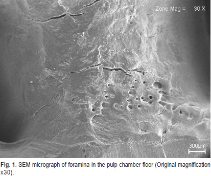
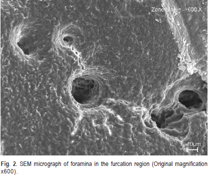
The roots and periodontal system in human dentition are closely correlated from the early stages of dental formation, maintaining this connection after teeth are established in the oral cavity through the apical foramen or other communications. Keywords: pulp chamber floor, furcation region, foramina. Introduction The anatomic relationship between dental pulp and periodontium in the furcation region trough foramina and accessory canals has prompted several research studies searching for possible pathologic consequences of this close communication1-10 . This aberrant contact between tissues in this region is the result of failed differentiation of odontoblasts due to defect formation in Hertwig’s sheath4 . Thus, these foramina can also contribute to the communication between periodontium and dental pulp. Studies with primary and permanent teeth using scanning electron microscopy (SEM)6 and topographic analyses2,9-10 found a significant incidence of foramina in the furcation area. Moreover, some studies using a simple technique (light microscopy) observed the presence of accessory canals1,3-4,7-8 . One study compared the incidence of foramina in permanent and primary teeth, and found a greater incidence in primary teeth10 . In the literature, there are few data on the influence ofpulp chamber floor surface thickness and furcation area onthe presence or absence of foramina. Therefore, the purposeof this research was to determine the presence of foramina inthe pulp chamber floor and furcation area of human permanentmandibular molars and correlate the presence of foraminawith the variation of wall thickness in these regions. Material and methods Forty sound permanent mandibular molars belongingto the Tooth Bank of the Oral Histology discipline (Opinion08/2009, Bioethics Committee of the College of Dentistry,UFPA) were examined. The crown and roots were sectionedtransversely along the tooth axis with a double-faced steeldisc 2 mm from the cementoenamel in the direction of the crown and 3 mm in the direction of the root apex to obtaina reduction in the longitudinal axis in order to facilitate thelater stages of evaluation. The samples were cleaned byimmersion in 1% sodium hypochlorite (ASFER IndústriaQuímica LTDA) for 5 min, and then in 17% EDTA (Farmácia-Escola, Federal University of Pará, Brazil) for 30 s. Sampleswere then subjected to a final rinse with distilled water in anultrasonic bath (MS 200 THORNTON, Impec Eletrônica, SãoPaulo, SP, Brazil) for 30 s and dried at room temperatureovernight. Following sample preparation, wall thicknessbetween the pulp chamber floor and the furcation area wasmeasured using a thickness caliper (JON Comércio deProdutos Odontológicos Ltda. São Paulo, SP, Brazil) accurateto the nearest 0.1 mm). Later, the samples were metalizedand subjected to scanning electron microscopy (model LEO1430/Laboratory of Scanning Electron Microscopy –LABMEV do Institute of Geosciences of UFPA), at 90 mAelectron beam current, constant acceleration voltage of 15kV and working distance of 10 mm. The SEM micrographswere analyzed under different magnifications, and thepresence/absence of foramina on the pulp chamber floor andfurcation area was recorded, divided the sample in two groups.The difference in mean thickness between the group of teethwith foramina on both surfaces and teeth without foramina on both surfaces was evaluated by Student’s t-test. Results The foramina found on both surfaces showed varied diameters and shapes and locations dispersed throughout thearea (Figure 1 and Figure 2). The results showed a mean thicknessof 2.15 ± 0.41 mm (SD) among the teeth analyzed. The datafor the sample was evaluated by the Kolmogorov-Smirnovtest, and a non-significant value of p=0.804 was obtained,confirming the normality of the sample. The study recorded a frequency of 25% of foramina onthe pulp chamber floor and 92.5% in the furcation region,with only 22.5% showing foramina on both sides. The difference in mean thickness between the group ofteeth with foramina on both surfaces and teeth without foramina on both surfaces was evaluated by Student’s t-test. There wasno statistically significant difference for the pulp chamber floor(t = -0.7587 and p = 0.4527) or for the furcation area (t = 0.5712 and p = 0.5712). Hence, there was no difference inmean thickness between the teeth with and without foramina. Discussion These anatomic communications are of great clinicalimportance with regard to the periodontal-endodonticinterrelation due to its role in the etiopathogenicity of endoperiodontal lesions11-12. Different investigations haveexamined the presence of pulpoperiodontal canals betweenthe pulp chamber floor and the furcation area and evaluatedthe possible pathological consequences of this relation5. In the literature, studies already exist showing theincidence of foramina in the furcation area and the pulp chamber floor1-3,5-9. In the present study, there were a largernumber of foramina, both in the furcation area and on thepulp chamber floor, compared with previous investigations. Kramer (2003)9 found a prevalence of 53% in the externalfurcation area and 25% in the internal furcation area usingSEM analysis9. Burch (1974)2 found 76% of foramina in the furcation area using a dissection microscope2. Vertucci (1974)3 found 46% lateral canals in the furcation area and 13% on the pulp chamber floor using a dissection microscope3. Haznedaroglu (2003)8 found a 21% incidence of patent furcalaccessory canals using a stereomicroscope8. However, in thecurrent study, we found 25% of foramina on the pulp chamberfloor and 92.5% in the furcation area, with only 22.5%showing foramina on both sides. This result is probably due to the method used, in whichorganic content and inorganic residue were removed fromthe teeth by the combination of sodium hypochlorite, EDTAand ultrasonic bath. This resulted in satisfactory cleanness,clearing the foramina and providing better visualization. Thickness was not significantly influenced by thepresence of foramina on the pulp chamber floor or in thefurcation area. Furthermore, the higher frequency of foraminain the furcation area compared to the pulp chamber floorsuggests the presence of blind foramina (accessory canalsoriginating from the pulp floor and/or periodontium andending in dentin without going on to another surface) orloop foramina (originating from the pulp floor and/orperiodontium, going through dentin, and returning to pulpchamber or periodontium), which has already been describedin another investigation13. Hence, in this study, even with22.5% of the sample showing foramina on both surfaces, itis still impossible to confirm whether there is a realcommunication between these regions even in these teeth. The dentin-pulp complex and periodontium are closelyrelated since odontogenesis, maintaining this interconnectionthrough the apical foramen. However, there are otheraccessory pathways of communication, such as lateral apicalforamina or even foramina between chamber floor and furcation areas8,10. These communications have greatrelevance in endodontic therapy, because their unsealing canresult in the maintenance of accessory ways of communication,which, in the presence of an infection process, can facilitateits spread between the periodontium and root canal system,in both directions3. From the obtained results, this study did not show acorrelation between thickness and the presence/absence offoramina. Although a higher frequency of foramina wasobserved in the furcation area compared with the pulp chamberfloor, it is not possible to infer that the frequency of foraminais associated with the frequency of communication betweenthe surfaces, which suggests the formation of blind or loopforamina. References
Copyright © 2011 - Brazilian Journal of Oral Sciences The following images related to this document are available:Photo images[os11047f2.jpg] [os11047f1.jpg] |
| |||||||||
