 |
| About Bioline | All Journals | Testimonials | Membership | News |
|
||||||
|
||||||
Prevalence and microscopic features of enamel pearls from permanent human molars Estela Kaminagakura1, Cristiane Ribeiro Salmon2, Douglas Campideli Fonseca3, Maria Cândida Almeida Lopes4, Rubens Nisie Tango5
1Professor of Stomatology, São José dos Campos Dental School, UNESP-University Estadual Paulista, Brazil, 2 PhD, Bucco-dental Biology Area, Piracicaba Dental School, University of Campinas, Brazil, 3Professor of Clinical Periodontology, Dental School, University Center of Lavras, Brazil, 4 Professor of Oral Surgery, Federal University of Piauí, Brazil, 5Professor of Dental Materials, São José dos Campos Dental School, UNESP-University Estadual Paulista, Brazil Received: August 25, 2011 Code Number: os11055 Abstract
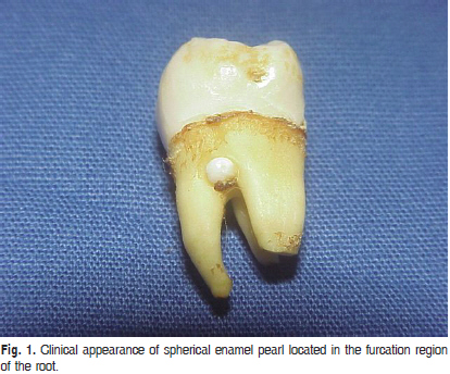
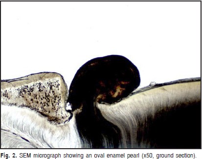
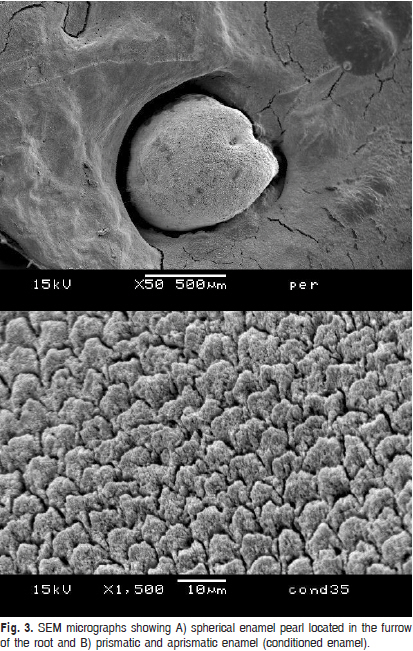
Enamel pearls are ectopic structures observed mainly on the roots of permanent teeth and could be related to periodontal diseases. Keywords: microscopy, electron, scanning, enamel, tooth abnormalities. Introduction Enamel is normally restricted to the anatomical crowns of human teeth1. However, ectopic structures, called enamel pearls, that are firmly adherent to the roots of deciduous and permanent teeth may occur1-2. Their prevalence ranges from 1.1 to 9.7% among molar teeth and the majority are detected on maxillary third molars1,3. One pearl per root is the most common occurrence, but two or more pearls have also been identified1,3-4. Microscopically, they can consist entirely of enamel, but large pearls may also contain dentin and pulp tissue. The pearls are characterized by a similar structure to that found on dental crown; however, the shape and direction of enamel prisms and dentinal tubules might be irregular5. Striae of Retzius, Hunter- Shreger bands and aprismatic enamel have been observed6 and, occasionally, enamel pearls are covered by a thin cementum layer5-6. Enamel pearls have a distinct predilection for the furcation area of molar teeth and for concavities or furrows within the root structure1,3-4. Previous studies have reported that the composition, size and topographic relation to furcation may contribute to periodontal disease1,7. The aims of this study were to evaluate the prevalence of enamel pearls in extracted molars from the UNILAVRAS Human Tooth Bank and to characterize their enamel and dentin by light microscopy (LM) and scanning electron microscopy (SEM). Material and methods The material comprised 2,201 clean and sterile extracted human permanent molars stored in the Human Tooth Bank of the University Center of Lavras, (Centro Universitário de Lavras, UNILAVRAS), Lavras, MG, Brazil. The dates of and reasons for the extractions were unknown and unavailable. The teeth were analyzed and classified according to morphological features. The presence, location and shape of the enamel pearls were investigated under a loupe at ×20 magnification. All teeth were analyzed by a single examiner. This study was approved by the local Ethics Committee (CAAE - 0002.0.189.000-05). Light microscopy (LM) Briefly, 15 molars with enamel pearls were dehydrated in an increasing series of ethanol concentrations, dried and embedded in acrylic resin (Vipi Crill®, Vipi, São Paulo, SP, Brazil) for 2 h under vacuum. Infiltration was achieved with metacrylate with 0.25% benzoyl peroxide overnight at 16oC and methacrylate with 0.5% benzoyl peroxide for 2 h under vacuum. The teeth were placed in glass test tubes and maintained at 25oC until the resin cured. Next, the tubes were broken, the resin was trimmed and the teeth were sectioned longitudinally under water coolant through enamel pearls with a diamond saw (Extec 12205; Extec Corp, Enfield, CT, USA) mounted on an automatic cutter (Model 650; South Bay Technology Inc., San Clemente, CA, USA). Sections measuring 20 µm in thickness were cut, ground and polished. The samples were ultrasonicated, dried and mounted on glass slides. The enamel pearls were analyzed by light microscope regarding composition, enamel prisms and dentin tubule organization. Scanning electron microscopy (SEM) Five enamel pearls were removed from their respective carrier teeth with a diamond saw, washed with distilled water and acid etched with 35% phosphoric acid (Dental Gel, Dentsply Ind. e Com. Ltda., Petrópolis, RJ, Brazil) for 30 s. After washing, the samples were fixed on metal stubs, dehydrated in silica for 2 h, gold sputtered and examined with scanning electron microscope (JEOL V, Tokyo, Japan). Images from the SEM were obtained using working distances (WDs) of 15 mm and 40 mm, 15kV. Results Seventy-one enamel pearls were detected on 63 permanent molars of a total of 2,201 teeth analyzed (2.86%); in 8 of these teeth (12.69%) 2 enamel pearls were observed. All enamel pearls were observed in the furcation or furrow on the root of the third molar (Figure 1). Hemispherical or spherical were the most common forms observed. The macroscopic features were the same as coronal enamel in relation to color and texture. Microscopically, two pearls were composed entirely by enamel and 13 by enamel and dentin (Figure 2). Irregularities were detected on the external surface. In most cases, the enamel observed was prismatic with an irregular course. Close to the dentinoenamel junction, the enamel appeared disorganized. In some cases, the dentinoenamel junction presented an irregular course. The number of dentinal tubules was normal and they presented curvature to continue within the root dentin of the carrier tooth. Dentinal tubules below the enamel pearls were closer to each other than normal root dentinal tubules. In the SEM analysis, the pearls showed both prismatic and aprismatic enamel similar to regular coronal enamel (Figure 3 A and B) and irregularities could be observed on the pearl surface. Discussion In this study, the prevalence of enamel pearls on extracted teeth was 2.8%, similar to previous reports8, and on about 87% of the teeth, only one pearl was observed. More than two pearls on a single tooth are rare5. All the enamel pearls were detected on third molars, in disagreement with a radiographic study that reported they are detected more frequently on the mandibular first molar, followed by the maxillary first molar9. Analyses of samples from Human Tooth Banks can lead to incomplete demographic data and clinical information, because in many cases the donor cannot be identified, as reported previously by Chrcanovic1 (2010). Additionally, third molars are the most frequent teeth available in Human Tooth Banks. The enamel pearls were easily differentiated from the cementum surface by their color and texture and the fact that they appear as discrete, glass-like globular bodies attached to the root by a sessile base3. Some authors identified these structures using the naked eye under direct light1; however, a loupe was used in the present investigation. Enamel pearls could contribute to periodontal disease onset and progression because of their composition, morphology and location1. The enamel covering the pearl can interfere in connective tissue attachment, permitting only a hemidesmosomal junction less resistant to periodontal breakdown; moreover, their morphology enhances plaque retention and protects organisms from the action of salivary enzymes and oral hygiene measures7,10. In addition, their topographic relation to the furcation can hinder treatment10, as the presence of superficial concavities filled with organic material on the enamel pearls have been described11 . In the present study, irregularities on the enamel pearl surface were observed by SEM. The majority of the enamel pearls analyzed were formed by enamel and dentin3, particularly the large pearls. Microscopically, enamel pearl structure is similar to that of a dental crown, but it shows localized defects with irregularities in the shape and course of the prisms6, which are likely related to the aberrant timing of enamel pearl formation4. The enamel identified on the roots resembles immature enamel3. It shows irregular areas of hypomineralization and the prisms do not always end on the free surface, often presenting thin layers without structure5. Occasionally, Hunter-Schreger bands and Striae of Retzius are present5 . When dentin is present, it generally appears normal. The number and course of dentinal tubules is also normal and they continue into the root dentin of the tooth without interruption6. Cavanha5 (1965), however, reported that the direction of the dentinal tubules is irregular and less mineralized. In the present study, disorganized dentin was only observed close to the dentinoenamel junction and the latter presented an irregular course, as observed by Gaspersic12 (1992). Interglobular dentin and Tome’s granular layer in large pearls and a conical-shaped core of dentin that resembles a cusp have been reported3 . Enamel pearls represent the localized activity of portions of Hertwig’s epithelial root sheath, which retain a latent potential to become functional ameloblasts and enamel organ that produces enamel deposits on the root3,12. The mechanism that permits this latent capacity remains unknown12. Genetic factors do not play a decisive role in enamel pearl formation for some authors4,6,8, but others disagree4,11. Our research group agrees with Moskow and Canut3 (1990), who suggested that enamel pearls are aberrations. The mineral contents of composite enamel and dentin from pearls are similar. The mineral-content gradient in the enamel pearl is the same as that of premolar enamel, as both have higher mineral contents at the surface. However, for pearl dentin, the opposite is true; i.e., compared with coronal dentin, it presents a reverse mineral-content gradient. This suggests that the process of mineralization in pearl dentin differs from that of permanent coronal dentin12. Another study indicated that the enamel pattern of large pearls is structurally and biochemically identical to that of the carrier tooth6. The findings of the present study do not fully support this assumption, since the dentinal tubules that continue into the root dentin of the carrier teeth were closer to each other and, because of such organization, this area seemed to be more mineralized. In conclusion, the prevalence of enamel pearls in teeth collected from the UNILAVRAS Human Tooth Bank was 2.8%; only one pearl was detected in approximately 87% of the teeth examined; the third molar was most affected tooth; and the macro and microscopic features of enamel pearl were similar to those of coronal enamel. References
Copyright © 2011 - Brazilian Journal of Oral Sciences The following images related to this document are available:Photo images[os11055f2.jpg] [os11055f1.jpg] [os11055f3.jpg] |
| |||||||||
