 |
| About Bioline | All Journals | Testimonials | Membership | News |
|
||||||
|
||||||
Brazilian Journal of Oral Sciences, Vol. 10, No. 4, Oct-Dec., 2011, pp. 272-276 Influence of calcium hydroxide on marginal leakage of endodontically treated teeth Maria Antonieta Veloso Carvalho de Oliveira1, Sara Teodoro Marra2, Paulo dos Santos Batista3, João Carlos Gabrielli Biffi4
1DS, Undergraduate student, Dental School, Federal University of Uberlândia, Brazil, 2DDS, Uberlândia, MG, Brazil, 3DDS, MS, PhD, Institute of Chemistry, Laboratory of Photochemistry, Federal University of Uberlândia, Brazil, 4DDS, MS, PhD, Professor of Endodontics, Dental School, Federal University of Uberlândia, Brazil Received: September 15, 2011 Code Number: os11056 Abstract
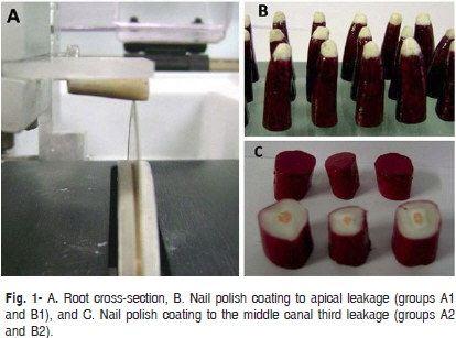
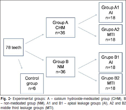
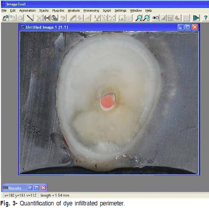
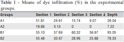
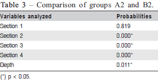
Aim: To evaluate the influence of residual calcium hydroxide (CH) intracanal medication considering two dye leakage locations (apical foramen and middle root canal third) by quantifying the diffusion of India ink in length and depth. Keywords: dental leakage, calcium hydroxide, root canal obturation. Introduction The use of the medication for root canal system disinfection has been supported to improve the treatment outcome1-2, as the complexity of the root anatomy makes more difficult their cleaning and shaping3. Intracanal medications such as calcium hydroxide (CH) are used to reduce or eliminate bacteria located in the root canal system and prevent their proliferation between sessions1-3. Regardless of the vehicle of the paste, the instrument or the solution used, CH dressing is not completely removed from the root canal, especially in the apical area1-2,4,6-14. Attempts to remove CH remnants have been made using irrigating solutions such as saline solution5,7, sodium hypochlorite1,3,5-6,8-14, EDTA1,6,10,12,14, acid citrus10, phosphoric acid8 and chlorhexidine6, alone and/or combined with manual1,3-4,6,8-9,12-13 and rotary instruments14 or ultrasound4,11-12. The remnants of CH within the radicular dentinal tubules may interfere on the properties of endodontic sealers, especially their sealing ability1,3,6,8-10,12-21. Typically, the sealing ability of filling materials is evaluated by leakage assays because they are easy to perform and do not require sophisticated materials1,3,6,8,12-13,21. However, leakage studies show a variety of assessment methods and parameters, which may be the main reason for the different results achieved with these tests1,6,8,22. Early leakage studies that evaluated the effect of CH dressing on apical seal concluded that their presence reduced the infiltration and determined a significant improvement in the quality of marginal sealing of root canal filling17,19-20. All these studies checked the infiltration of teeth after filling using methylene blue, which loses its color when in contact with some filling materials, such as CH23-24. For this reason, when the studies used the fluid transport model or Rhodamine B to quantify the infiltration of teeth with or without previous use of CH medication showed no statistically significant differences among groups1,23-24. When India ink was employed, the highest values of infiltration were found in the groups receiving the CH previous to root canal filling3,8. To date, no study has evaluated both the length and the depth of leakage after the use of CH in samples with and without the apical root canal third. Evaluation of the length and depth of leakage allows a three-dimensional analysis of dye penetration. Removal of the apical dye that can be accumulated during infiltration eliminates a variable that can affect leakage measurement within the root canal20,22. Therefore, it seemed important to conduct an in vitro leakage study to evaluate whether the presence of the remaining CH interferes with the leakage of endodontically treated teeth. Two different locations of dye leakage, apical foramen and middle root canal third, were used to quantify the diffusion of India ink in length and depth. The tested hypothesis was that the CH remnants in the root canal walls could favor dye penetration. Material and methods Seventy-eight bovine teeth of adult animals with a single straight root, apex with narrow anatomic diameter compatible with a size 40 K-file and foraminal opening coinciding with the end of the apex were selected for the study. Tooth crowns were removed by standardizing the length of the roots to 20.0 mm. All teeth were prepared, maintaining the patency of the foramen with a size 15 K-file and irrigating the canal with 2.0 mL of 1% sodium hypochlorite (Farma, Serrana, SP, Brazil) between each file throughout instrumentation. Thereafter, the root canals were dried with paper points (Tanari, Manacapuru, AM, Brazil) and two experimental groups of 36 teeth each were formed (A and B). The other 6 teeth were used as the control group. Only group A received a paste of pure CH powder (Biodinâmica, Ibiporã, PR, Brazil) mixed with normal saline solution (Ariston, São Paulo, SP, Brazil) at a powder to liquid ratio of 1:1.5. The medication paste was taken to the root canals with the aid of a plastic syringe with 27-gauge needle (Endo Eze; Ultradent Products Inc., South Jordan, UT, USA) introduced to the full working length (19.0 mm). A radiograph was taken to confirm the complete filling of the canal. Temporary sealing of the cervical region was performed with zinc oxide-eugenol sealer (IRM; Dentsply Ind. e Com. Ltda., Petrópolis, RJ, Brazil.), and thereafter the specimens were stored in 100% relative humidity at 37ºC for 7 days. Thereafter, the temporary dressing was removed with copious normal saline solution combined mild filing with memory instrument at the working length. Irrigation was performed until the reflux of irrigating solution was clear and the root canals underwent aspiration and drying with paper points. The canals in groups A and B were filled following the lateral condensation technique with zinc oxide-eugenol sealer (Endofill, Dentisply, Petrópolis, RJ, Brazil), dispensed and handled according to the manufacturer’s instructions. After placement of the temporary coronal restoration, the roots were maintained in 100% relative humidity at 37ºC for 72 h. For the marginal leakage evaluation at two different root locations, via the apical foramen and at middle third of the root canal, half of specimens from each group A and B (n = 18) was sectioned at the middle third of the root in a precision cutting machine (Buehler Ltd., Lake Bluff, IL, USA) (Figure 1 A). Then, groups were divided according to the local of dye infiltration at the root: groups A1 and B1 (n = 18 each) for apical leakage, groups A2 and B2 (n = 18 each) for middle third leakage (Figure 2). The teeth in the four groups (A1, A2, B1 and B2) were coated before the dye infiltration with two layers of nail polish (Colorama, São Paulo, SP, Brazil) (Figure 1 B and C). Specimens that had been prepared, but received no intracanal medication or filling (n = 6) served as control of the effectiveness of coated with nail polish and dye leakage. Half (n = 3) was completely coated with polish even in the apical third (negative control group) and half received no coated (positive control group). All specimens were subjected to vacuum for 10 min. After complete immersion of the roots, horizontally, in Indian ink (Faber-Castell, São Carlos, SP, Brazil) were subjected again to vacuum for 10 min. After 72 h in 100% relative humidity at 37ºC, the roots were washed; polish coating had removed and the specimens were included in self-curing resin (AM 190, Aerojet, São Paulo, SP, Brazil) to form blocks. The resin blocks glued to acrylic plates were attached to the precision cutting machine to obtain cross-sections (1.0 to 1.5 mm) from the root apex, using double-sided diamond disk (Buehler Ltd., Lake Bluff, IL, USA). The number of sections obtained in each subgroup was determined by the presence of dye leakage in root canal walls, viewed with a stereoscopic microscope. Photographs of all cross-sections were obtained with a digital camera (Nikon D60, Tokyo, Japan) to quantify dye leakage (Figure 3) in the UTHSCSA Image Tool 3.0 software (The University Texas Health Science Center, San Antonio, TX, USA). The Mann-Whitney U-test25 was used for statistical analysis. The infiltrated perimeters of the first four sections of each sample were used to estimate the length of dye leakage. The total number of sections of each sample that showed the presence of Indian ink was used to estimate the depth of infiltration. The significance level was set at 5%. Results The mean percentages of dye infiltration in length (Sections 1, 2, 3 and 4) and depth of the experimental groups are presented in Table 1. Comparison of the results of dye leakage among the groups is presented in Table 2 and Table 3. The specimens subjected to apical leakage (A1 and B1) showed statistically significant differences (p<0.05) among all comparisons of length and depth, with the highest values in the non-medicated group (B1). The groups subjected to middle third leakage (A2 and B2) showed statistically significant differences (p<0.05) between the sections 2, 3 and 4 and in depth measurements, and the highest values were found in the non-medicated group (B2). The negative control demonstrated no dye penetration, whilst the positive control showed leakage of the dye along the length and depth of the root canal. Discussion The results of the present study showed that the CH remnants influenced on leakage unlike we expected in our initial hypothesis, as less infiltration occurred in the presence of residual medication. This result has been described by Çaliskan, Türkün and Türkün21 (1998). Other studies using India ink leakage with the same goal showed contrary results to those of the present study3,8. In the studies of Kim and Kim3 (2002) and Contardo et al.8 (2007), the highest values of dye leakage were found in the groups that received the CH prior to filling. According to those authors, the presence of CH residues was not the only factor in reducing apical leakage, since the type of sealer used is also important20. By analyzing the steps of these methodologies, one can justify the confronting results due to differences in sample preparation for infiltration and/or quantification of this infiltration. Kim and Kim3 (2002) observed the India ink infiltration through cross sections by counting the number of samples with dye infiltration. Contardo et al.8 (2007), cleared the samples allowing visualization of the infiltration of India ink only in depth. In the present study, the use of cross sections allowed a three-dimensional analysis of dye penetration not only in depth, but also along the whole root length. Dye penetration was measured in millimeters along the root canal using Image Tool 3.00 software. Furthermore, the use of specimens without the apical third in groups A2 and B2 provided an assessment of dye penetration without the variation of the apical delta area20,22. It is noteworthy that the same results as the values of infiltration were found both in specimens with total length (20.0 mm) and root sections (6.0 mm). The results of this study cannot be extrapolated to the clinical situation because of the complex interrelationship between the multiple factors affecting endodontic treatment, which makes it impossible to study the correlation between in vitro dye leakage and in vivo treatment failures22 . However, the CH remnants in the root canal found in our results may represent a problem for the prognosis of endodontic therapy. CH, in the long term, can affect treatment prognosis due to leakage or to the presence the apical area resorption. The CH reacts with tissue fluids to form calcium carbonate, which is resorbable and can create space-filling in the interface wall dentin over time5,26. In addition, the properties of endodontic sealers can be affected by the presence of calcium hydroxide. Several in vitro studies have proven that the presence of CH residues within the dentinal tubules reduces the bond strength of different sealers zinc oxide and eugenol based4 and resin based6,13,15. It also reduces the apical sealing ability of zinc oxide and eugenol3, glass ionomer7 and silicone-based sealers8, and prevents the penetration of sealer into the dentinal tubules9-10 and lateral canals16. CH reacts chemically with the sealer, resulting in reduced flow capacity, shorter working and setting times, and increased sealer film thickness9. The reduction in flow and working time results in an inability to work effectively with a material, increasing the chances of creating voids1,3,9, while the thicker sealer film is believed to make sufficient condensation of the root canal filling difficult to achieve9 . However, there are studies in which incomplete removal of CH medication did not affect the adhesion or sealing ability of root canal sealers1,12 or resulted in an improvement in this ability17-21, suggesting that the effect of CH on the apical seal of the root canal filling depends on the type of sealer used 12,21 . New studies are required in order to develop vehicles for CH or auxiliary substances that chemically interact with this medication and permit its complete removal from the root canal system. Under the tested conditions, it may be concluded that the remnants of CH intracanal medication led to lower dye penetration in length and depth at the two different locations evaluated in the study. References
Copyright © 2011 - Brazilian Journal of Oral Sciences The following images related to this document are available:Photo images[os11056t3.jpg] [os11056t2.jpg] [os11056t1.jpg] [os11056f3.jpg] [os11056f2.jpg] [os11056f1.jpg] |
| |||||||||
