 |
| About Bioline | All Journals | Testimonials | Membership | News |
|
||||||
|
||||||
Evaluation of Glucose-6-Phosphate Dehydrogenase Deficiency without Hemolysis in Icteric Newborns Farzaneh Eghbalian*1, MD; Ali Reza Monsef 2, MD 1Department of Pediatrics, Hamedan University of Medical Sciences,
Hamedan, IR Iran Received: 13/7/06; Code Number: pe07007 Abstract Objective: Glucose-6- phosphate dehydrogenase (G6PD) deficiency is an inherited
deficiency that may be the cause of neonatal jaundice. Our aim was to study
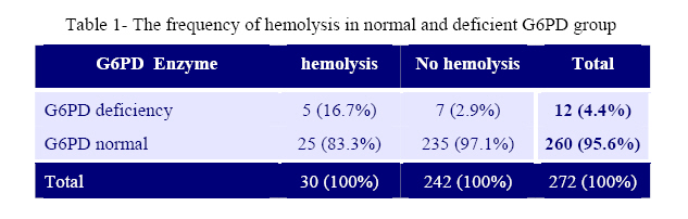
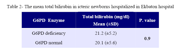
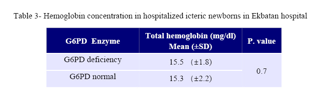
the prevalence of G6PD deficiency without hemolysis in relation to neonatal jaundice. Key Words: Glucose-6- phosphate dehydrogenase deficiency, Enzyme Deficiency, Hemolysis, Neonate, Jaundice Introduction Glucose-6-phosphate-dehydrogenase (G6PD) deficiency is the most important disorder of hexosemono-phosphate shunt in erythrocyte metabolism[1]. Worldwide, more than 200 million people are affected with this enzyme deficiency[2]. As the disease is X-linked, all males inheriting the mutant allele may potentially exhibit signs and symptoms of the disease [1]. Symptoms occur in both hemozygous males and females and they occur mildly in heterozygous females[1, 2]. Neonatal jaundice due to G6PD deficiency may occur after exposure to oxidant agents and as a consequence hemolysis occure. But quite often, there is a mutation in the promotore site of uridyle di- phosphoglucoronyl transfrase (UDPGT) which accompanies G6PD deficiency, leading to indirect hyperbilirubinemia in the abscence of hemolysis[1, 2]. One of the important manifestations of this enzyme deficiency in the neonatal period is jaundice without hemolysis. This may be so serious that it can lead to kernicterus or death and also predisposes the neonate to infection [2, 3]. Many studies conducted in the Mediterranean area and some Asian regions also confirmed that G6PD deficiency is a common cause of severe jaundice in the neonatal period [4]. A study in the USA revealed that 10% have G6PD deficiency at black neonates but severe neonatal jaundice was rare [5]. Numerous studies in Nijeria, India, Saudi Arabia, Singapore, Jamaica and Malaysia have revealed that this enzyme deficiency in icteric newborns was 40%, 12.2%, 18.4%, 1.62%, 1.57% and 3.5% respectively, in which neonatal jaundice was seen without hemolysis [6-13]. The increasing number of case of neonatal jaundice in some geographical regions known as G6PD deficiency also seem to suggest a possible correlation between the two factors, however, at this point only limited empirical studies are available that could verify this possible explanation. This study was carried out with the aim of evaluating the prevalence of G6PD deficiency, the prevalence of hemolysis with enzym deficiency and determining the severity of icterus in the hospitalized newborns in our hospital. Material & Methods This descriptive prospective cross-sectional study, was acomplished in over two years from October 2002 to september 2004 on 272 newborns hospitalized in Ekbatan Hospital, Hamedan for jaundice. The samples included in this study were: term newborns (38 weeks gestational age or more) with a total bilirubin at more than 12 mg/dL and preterm newborns (gestational age less than 38 weeks) with a total bilirubin at more than 10-14 mg/dL. The excluding criteria were; term neonates with a total bilirubin less than 12 mg /dL, preterm newborns with a total bilirubin less than 10 mg/dl and direct hyperbilirubinemia (direct to total bilirubin ratio more than 20%). In summury, we excluded cases of physiological jaundice and direct hyperbilirubinemia from the study. The criteria of hemolysis in our study was: reticulocyte count more than 3.2%±1.4 to 0.5%±0.4 (according to neonatal age in day) and hemoglobin less than 17.3± 2.3 gr/dL to 14.2±2.1 gr/dL (according to neonatal age in day) [2]. The G6PD level was measured with a fluorescent G6PD spot test of qualitative method manufactored by Kimia Pajouhan Co. Derived data included: age, sex, total, direct and indirect bilirubin, hemoglobin, reticulocyte count, blood group and Rh of mother and newborn, direct Coombs test, G6PD level and type of treatment (phototherapy, Blood exchange transfusion). The statistical Package for Social Sciences (SPSS; version 11) was used for the statistical analysis. Student's t-test was used to assess the significance between means of two groups. p≤0.05 was considered statistically significant. Findings We found that 12 neonate out of 272 (4.4%) icteric neonates were suffering from G6PD deficiency. Ten males (83.3%) and 2 females (16.7%) resulting in a male/female ratio of 5/1. The study could confirm the hemolysis criteria in 30 cases out of 272 neonates (11%), however in the 12 cases of G6PD deficiency we could confirm 5 cases (41.7%) of hemolysis and seven cases (58.3%) without hemolysis. A total of 242 neonates out of 272 neonates did not have the criteria for hemolysis. In other words, the total frequency of G6PD deficiency without hemolysis in the hospitalized icteric newborns was 2.6% (table 1). Mean total bilirubin concentration in the G6PD deficiency group compared with the normal G6PD group showed no significant difference (P=0.8) (table 2). Mean hemoglobin concentration in G6PD deficient neonates compared with the normal G6PD group also indicating no significant difference (P=0.7) (table 3). Furthermore, the study did not show a significant difference in the reticulocyte count between normal G6PD and deficient G6PD neonates (P=0.5). All G6PD deficient patients and other icteric neonates were treated with phototherapy; exchange transfusion is performed in 2 out of 12 G6PD deficient neonates (16.7%). None of the 12 G6PD deficient neonates had ABO and Rh incompatibility. Discussion The results showed that 4.4% of the study group affected by G6PD deficiency and that most of them (58/3%) were icteric without any signs of hemolysis. The results of the study confirmed the outcome of previous studies in Iran and other countries [3, 4, 6, 9, 12]. The hemolysis of G6PD is non-immune, therefore, direct Coombs test is negative, the results of our study did not show any positive Coombs test in the G6PD deficient group [2, 3]. However, some studies have pointed out the higher prevalence, for example Flym et al found that 10% of G6PD deficiency was seen in black icteric newborn in the USA [5]. Another study in Nigeria, showed that 40% of icteric newborns were suffering from G6PD deficiency, in most of them there was no without concomitant hemolysis [6]. Madan et al from India showed that in 12.2% of all icteric newborns suffered from G6PD deficiency and 48.7% of them had severe jaundice but without hemolysis [8]. A study from Saudi Arabia showed 18.4% prevalence of G6PD deficiency in icteric newborns without any signs of hemolysis [10]. Our study did not show a significant difference in the mean bilirubin level between G6PD deficient newborns and other icteric newborns. We performed exchange transfusion in 16.7% of G6PD deficient icteric neonates and 18.85% of neonates without G6PD deficiency. The results confirmed that jaundice in the G6PD deficient group is not more severe than in other causes of neonatal jaundice. A study in Saudi Arabia, showed severe jaundice without hemolysis in 18.4% of G6PD deficient neonates and in 15% of normal G6PD group neonates, they had blood exchange transfusion in 7% of G6PD deficient icteric newborns and in 8.2% of normal G6PD group. These results is compatible confirmed with the result of the present study [10]. A screening program for G6PD enzyme in Malaysia on 8900 neonates revealed that the deficiency in 100 newborns and 17 out of them had exchange transfusion (17%). This study was in accord with our study [13]. A study in Qazvin, Iran revealed that in 8% of hospitalized icteric newborns suffered from G6PD deficiency, 15.7% of them needed exchange transfusion. These results are similar with our findings [14]. Another study in Basrah, Iraq on 95 icteric newborns revealed that 51% of them suffered from G6PD deficiency and 27 out of them needed exchange transfusion [15]. These findings were not compatible with our results; from the point of higher prevalence of G6PD deficiency and more cases exchange transfusion. We did not find a meaningful difference in the mean hemoglobin level and the reticulocyte count between normal and deficient G6PD enzyme groups. This confirms that the cause of jaundice in icteric newborns is not only the hemolysis, but also the concomitant mutation, leading to a decrease of G6PD and UDPGT activity that results jaundice. The texts also confirm these findings [1, 2]. In this study the male/ female ratio was 5/1 in enzyme deficient neonates; it is predictable, because the disease is x-linked and more expressed in males. The affected females are hemozygote or heterozygote, the later unfavorable lyonization. Conclusion The results of the present study indicate that G6PD deficiency is a major cause of icterus in newborns (especially without hemolysis) in Iran. So, we recommend that G6PD activity measurement be compulsory for every icteric newborn. This should be done in spite of laboratory findings for hemolysis as a routine test and it should be done as a screening test at birth time. Early diagnosis can reduce the number of complications in icteric neonate and prevents future acute hemolytic attacks. Due to a lack of funding and inavailability of quantitative G6PD enzyme assay kit, the program was applying qualitative methods which can lead to misdiagnosis of some cases of enzyme deficiency. So screening with quantitative tests at birth for every newborn is recommended. Acknowledgment The authers would like to thank the staff of the neonatal ward and laboratory department of Ekbatan hospital for their support in this study. We would like to also thank Estefan Eghbalian (PhD) for editing the paper. References
Copyright 2007 - TUMS PUBLICATIONS The following images related to this document are available:Photo images[pe07007t2.jpg] [pe07007t3.jpg] [pe07007t1.jpg] |
| |||||||||
