 |
| About Bioline | All Journals | Testimonials | Membership | News |
|
||||||
|
||||||
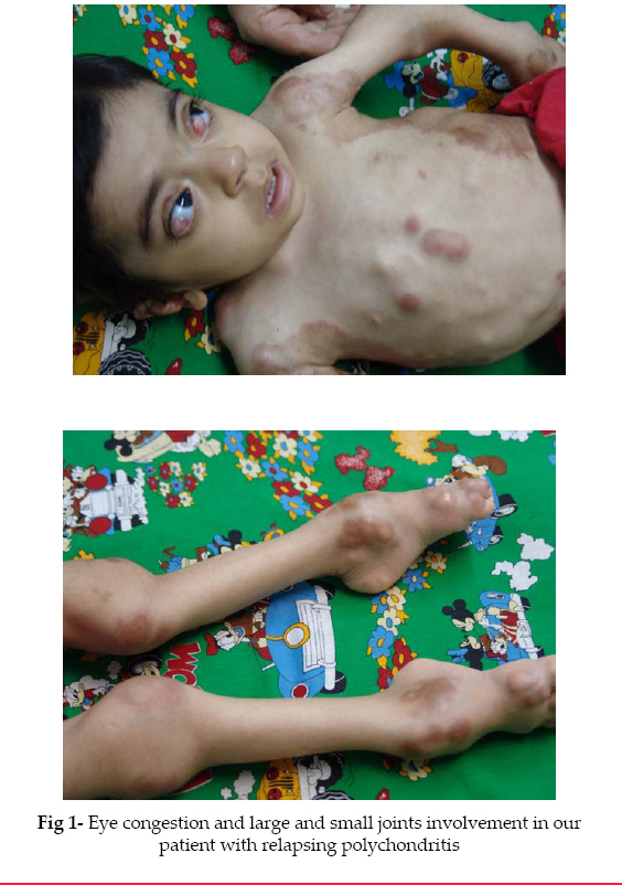
Iranian Journal of Pediatrics, Vol. 18, No. 2, June, 2008, pp. 179-182 Case Report Relapsing Polychondritis: A Case Report Yahya Aghighi1, MD, Pediatric Rheumatologist; Seyed Reza Raeeskarami1, MD, Pediatrician; Alireza Moinipur*2, MD, Resident in Pediatrics 1Department of Pediatrics, Tehran University of Medical Sciences, IR Iran Received: 26/11/07; Revised: 20/01/07; Accepted: 17/02/08 Code Number: pe08030 Abstract Objective: Relapsing Polychondritis (RP) is an uncommon inflammatory
disorder of unknown cause characterized by an episodic and progressive course
affecting predominantly the cartilage of the ears, nose and
laryngotracheobronchial tree. It has seldom been described in children. Key Words:Polychondritis, Juvenile chronic arthritis, Steroids Introduction Relapsing Polychondritis (RP) is characterized by episodic necrotizing cartilage inflammation that may be very severe[1]. Inflammation affects the cartilage of the ear or hyaline cartilage of the joints and then involves cartilage of the upper respiratory tract, including nose, trachea and bronchi[2]. It begins at the age 40 to 50 years, but may affect children and the elderly. It is found in all races, and both sexes are equally affected. No familial tendency is apparent[3]. It has seldom been described in children[4-8]. We describe a new case of RP in a 2-year old girl. Case Presentation A 2-year old girl was referred to the outpatient clinic. Initially 2 months after birth she presented with a history of several episodes of a brief, shrill cry followed by stiffness and forced expiration leading to cyanosis by moving the limbs. Hence the presumptive diagnosis was epilepsy, and treatment was initiated with oral anticonvulsant drugs. For the next 3 months, large and small joints of both upper and lower extremities began to swell and obviously ached due to active and passive motion of joints, therefore she was referred to rheumatologist. Initial blood investigation revealed an Erythrocyte Sedimentation Rate (ESR) of 110 mm, leucocytosis and thrombocytosis with mild anemia (Hb 9 gr/dl). Rheumatoid Factor (RF) was positive, the renal profile and thyroid function tests were normal. Brain CT scan and electroencephalography (EEG) was normal. Anticonvulsant drugs were discontinued. Her clinical features, laboratory studies and rule out malignancy by bone marrow biopsy, confirmed the diagnosis of Juvenile Chronic Arthritis (JCA). Treatment was initiated with non-steroidal anti-inflammatory drugs (NSAIDs) and oral steroids. The condition improved after four weeks. Six months later, the patient was brought to our outpatient clinic with hoarseness and frank stridor. Clinical examination revealed bilateral inflammatory erythematous swelling of the auricles, which was confined to cartilaginous portions. Both pinnae were tense, painful, congested and inflammatory involvement caused narrowing of external auditory meatus (Fig 1). Both eyes were congested. Bilateral conjunctivitis and episcleritis was diagnosed. In large and small joints of both upper and lower extremities, polyarticular disease relapsed. By indirect laryngoscopy laryngomalacia, swelling, inflammation and narrowing of subglotic region were observed. Rise of ESR to 130 mm, leukocytosis (16900/mm3 with 76% neutrophiles), thrombocytosis (650000/mm3) and anemia (Hb 8.5 gr/dl) were detected. Low titers of RF and antinuclear antibody tests (ANA) were observed. Her clinical features according to the clinical criteria of McAdam-Michet et al, led to final diagnosis of RP. The patient was treated with intravenous steroid pulse therapy (methyl-prednisolone 30 mg/kg/day) for three days, NSAIDs, methotrexate and oral steroids. The patient symptomatically improved and was discharged home. She is still on regular fallow up at our outpatient clinic. Discussion RP is an episodic and progressive inflammatory disease of cartilaginous structures. RP may develop with a variety of autoimmune disorders, including systemic lupus erythematosus (SLE), rheumatoidarthritis, Sjögren's syndrome and vasculitis. In most cases, these disorders antedate the appearance of RP, usually by months or years[3]. In our patient, the diagnosis of RP was made due to the appearance of criteria of McAdam-Michet et al. On this case, we can see that JCA antedates the appearance of RP by 6 months. Laboratory findings of RP are nonspecific, ranging from leukocytosis, thrombocytosis, and chronic anemia, increased ESR and elevated gamma globulin levels. Low titers of RF and ANA can be detected[9]. The onset of RP is frequently abrupt with the appearance of one or two sites of cartilaginous inflammation. Fever, fatigue and weight loss occur and may precede the clinical signs of RP by several weeks. The pattern of cartilaginous involvement and the frequency of episodes vary widely among patients. Auricular chondritis is most frequent presenting manifestation of RP and eventually affects about 85% of patients. Laryngotracheal involvement occurs in 50% of patients, symptoms include hoarseness, a non productive cough and tenderness over the larynx and proximal trachea. Mucosal edema, strictures and/or collapse of laryngeal or tracheal cartilage may cause stridor[3]. Our patient had bilateral inflammatory erythematous swelling of auricles confined to the cartilaginous portions, hoarseness, frank stridor, dys-phonia and tenderness over the larynx and proximal trachea. By indirect laryngoscopy, laryngomalacia, swelling, inflammation and narrowing of the subglotic region were observed. Both eyes were congested and bilateral conjunctivitis and episcleritis was diagnosed. The diagnosis of RP is established by the presence of 2 major criteria, or 1 major and 2 minor criteria, that suggested by McAdam- Michet et al. Biopsy and histologic examinations of affected cartilage is not required[4]. Major Criteria include: a) Inflammatory episodes involving auricular cartilage; b) Inflammatory episodes involving laryngo-tracheal cartilage; c) inflammatory episodes involving nasal cartilage, and minor criteria include: a) Ocular inflammation (conjunctivitis, keratitis, episcleritis, uveitis); b) hearing loss; c) vestibular disfunction; d) seronegative inflammatory arthritis. In our patient, recurrent bilateral chondritis of the pinna, laryngotracheal involving, bilateral conjunctivitis and episcleritis, and polyarthritis confirmed the diagnosis of RP according to the clinical criteria of McAdam-Michet et al. The etiology of RP is unknown, but the pathogenesis seems to be an immunologic reaction to type II collagen; auto antibodies against type IX and XI collagen have been found in a patient with RP[10-11]. More recently, an increase in HLA-DR4 antigen was detected in a patient with RP, although no subtype predominated[12]. Many patients respond to NSAIDs and some require corticosteroids or other immunosuppressive agents. Oral collagen has also been reported to improve symptoms. Severe, progressive and fatal disease, resulting from destruction of tracheobronchial tree and airway obstruction, is unusual in childhood[1]. Our patient symptomatically improved with intravenous steroid pulse therapy with methyl-prednisolone and was discharged home and she is still on regular fallow up at our clinic. Conclusion RP may develop with a variety of autoimmune disorders including SLE, rheumatoid arthritis and vasculitis. In most cases, these disorders antedate the appearance of RP, usually by months or years. RP should be considered in differential diagnosis of JCA. Steroids and systemic immunosuppressants can be used to treat the RP. References
© Copyright 2008 - TUMS PUBLICATIONS The following images related to this document are available:Photo images[pe08030f1.jpg] |
| |||||||||
