 |
| About Bioline | All Journals | Testimonials | Membership | News |
|
||||||
|
||||||
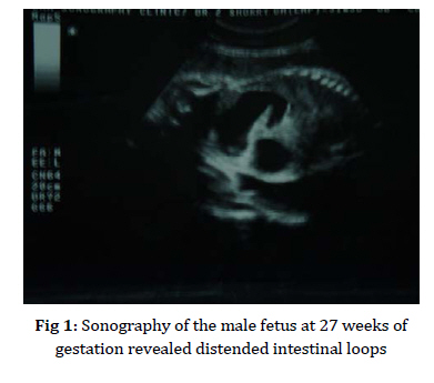
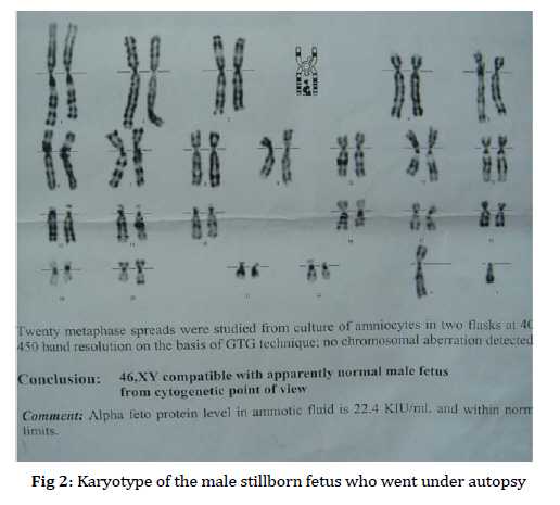
Iranian Journal of Pediatrics, Vol. 19, No. 4, 2009, pp. 435-438 Two Consecutive Stillbirths with Multiple Intestinal Atresias a in 30-Year-Old Female Fatemeh Mahjoub1, MD; Maliheh Kadivar2, MD; Behnaz Moradi3; Nargess Tabarzan3 1. Department of Pathology, Children's Medical Center, Pediatrics Center of Excellence, Tehran University of Medical Sciences, Tehran, IR Iran * Corresponding Author; Address: No 62, Children’s Medical Center, Pediatrics Center of Excellence, Dr Gharib St, Tehran, IR Iran, E-mail: fmahjoub@sina.tums.ac.ir Received: Dec 12, 2008; Final Revision: Apr 04, 2009; Accepted: May 16, 2009 Code Number: pe09056 Abstract Background: Multiple duodenal and jejuno-ileal atresias are extremely uncommon. The familial occurrence of multiple intestinal atresias is even more unusual. Also fetal death with isolated intestinal atresia is very rare, and the combination of recurrent familial intestinal atresias and intra-uterine fetal death has not been reported previously. Key Words: Multiple intestinal atresias; Repeated; Consanguineous; Marriage; Intrauterine fetal death; Familial Introduction Hereditary multiple atresias of gasterointestinal tract are an extremely rare subgroup of intestinal atresias. In the most recent article on multiple intestinal atresias in a retrospective analysis of 26 patients treated over a 20-year period between 1986– 2006, all cases were sporadic with no positive familial history[1]. We describe 2 consecutive stillborn fetuses, one male and one female with multiple intestinal atresias born to healthy consanguineous parents. both fetuses died in uterine around 32 weeks. In the autopsy of male fetus, multiple duodenal and jejuno-ileal atresias were seen, this combination is extremely uncommon[2,3]. The familial occurrence of duodenal atresia is even more unusual[4]. Also fetal death with isolated intestinal atresia is very rare[1]. To the best knowledge of the authors, the combination of recurrent familial intestinal atresias and intra-uterine fetal death has not been reported previously. Case Presentation At the time of stillbirth of the second affected fetus, the father was 37 and the mother 30 years old. The parents are cousins and both have normal karyotypes. Family history was negative. Mother has history of 5 pregnancies: two abortions in 3rd and 5th month. An 8 year old boy has speech difficulties and microcephaly. His karyotype is XYY. The 2 last pregnancies ended in 2 stillborn fetuses, on which we report in this article. Mother had no history of specific illnesses, had taken no medications during pregnancy and was exposed to no known teratogenic agents. Her routine laboratory examinations were unremarkable. Case 1 A female fetus died at 32 weeks of gestational age with intestinal atresia. Sonography at 21 weeks revealed distended intestinal loops. Repeat sonography at 31 weeks reported double bubble sign and mild polyhydramnious with normal fetal heart rate, normal physical activity, no evidence of intra uterine growth retardation (IUGR) and no other gross anomalies. Her karyotype was 46XX. Cesarean section was performed due to repeat but fetus was not sent for autopsy. Case 2 A male fetus died at 33 weeks of gestational age with multiple intestinal atresias. At 27 weeks, ultrasound study revealed multiple distended intestinal loops; upper intestinal atresia was suspected but amniotic fluid was reported as normal. At 33 weeks, repeat ultrasound revealed mild polyhydramnios and multiple distended loops (Fig 1), normal fetal heart rate, normal physical activity and no evidence of IUGR. No other gross anomalies were detectable in head, spinal cord, limbs and other internal organs. After 4 days, sonography reported dead fetus with hematoma between amniotic and chorionic sac. Cesarean section was performed and dead fetus was sent for autopsy. Autopsy revealed dilated duodenum containing coffee ground material, before complete atretic site which was located 20 cm from pylorus. Also several atretic points were seen in small intestine. Gastric and duodenal mucosa were atrophic and erythematous. Large bowel was unused with no atresia and contained few amounts of creamy material. The anus was patent and placed slightly upward. Placenta and umbilical cord were normal. Other organs were unremarkable. The karyotype of fetus was 46XY (Fig 2).
Discussion Searching all relevant articles on intestinal atresia and intra-uterine fetal death in literature, we found three articles as listed below: 1) A case of duodenal atresia complicated by massive intrauterine hemorrhage due to perforation of an umbilical cord ulceration[5]. In our case no umbilical ulcer was seen. 2) Hereditary multiple atresias of gasterointestinal tract are an extremely rare subgroup of intestinal atresias with unique features: a) Abdominal X-ray shows signs of typical large oval homogenous calcification in the abdominal cavity. b) Widespread atresias (exclusively type Ι and П) extending mostly from stomach to rectum. c) Cystic dilatation of the bile duct can be present. d) Fatal outcome is the rule[3] with a mean survival time of 50 days[6], but stillbirth was only reported in one out of 35 reported cases[3]. Also no consecutive occurrence in one family was reported. Our cases differ from this because of multiple type Ш atresias, which is never seen in hereditary multiple intestinal atresias. Also no calcification was detected in serial sonographies and autopsy in our cases. 3) In another article, two cases with normal karyotype without any other anomalies were reported. These died intrauterine at 31 and 35 weeks of gestational age[7]. Possibility of bradycardia/asystole following vagal over activity due to upper gastrointestinal obstruction was suggested as cause of death by authors, which can be a good explanation for stillbirth but could this explain two consecutive fetal deaths as in our cases? It is proposed that when extensive multiple atresias occur, a rare autosomal recessive gene is responsible for this congenital defect[8]. Due to close consanguinity of parents and two consecutive stillbirths, an autosomal recessive disorder seems to be the most probable explanation. Association of this disorder with karyotype abnormality in the living child of the family is yet unreported and prompt further attention. Conclusion Multiple and recurrent intestinal atresias are extremely uncommon and can lead to intrauterine death. We report two consecutive fetuses which had multiple atresias that led to intrauterine death in both of them. These rare cases may contribute to genetic mapping of intestinal atresias. Acknowledgment Hereby we thank Miss Zahra Omidi for her devoted cooperation. References
© Copyright 2009 - TUMS PUBLICATIONS The following images related to this document are available:Photo images[pe09056f1.jpg] [pe09056f2.jpg] |
| |||||||||
