 |
| About Bioline | All Journals | Testimonials | Membership | News |
|
||||||
|
||||||
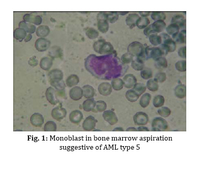
A Case of Congenital Leukemia with Nasal Hemorrhage and Jaundice Zahra Khalili Matinzadeh, MD; Zohreh Kavehmanesh*, MD; Susan Amirsalari, MD; Shahla Afsharpeyman, MD Department of Pediatrics, Baqiyatallah Medical Sciences University, Tehran, IR Iran Received: Mar 01, 2009; Accepted: Mar 15, 2010 Code Number: pe10057 Key words: Leukemia; Neonatal jaundice; Congenital; Bleeding Congenital leukemia (CL) is a very rare malignancy which occurs at estimated rate of 1 per 5 million births, and accounts for less than 1% of all childhood leukemias[1,2]. Only approximately 200 patients with congenital leukemia have been reported in the literature[3]. CL has no pathognomonic findings. The proposed diagnostic criteria for CL include: I) presentation in the first 4 weeks of life; II) proliferation of immature myeloid, lymphoid or erythroid cells; III) infiltration of these cells into nonhaematopoietict issues; IV) absence of other diseases which may explain this proliferation[5]. Most CL cases have been reported as Acute Myeloid Leukemia (AML). The acute myelomonocytic (FAB M4) and acute monocytic (FAB M5) subtypes are by far the most frequent AML respectively[4]. Herein we report a case of CL with skin lesions on his abdomen who was hospitalized because of jaundice and nasal hemorrhage. After an uneventful full-term pregnancy, a boy was born with a birth weight of 3600 gr. No maternal ante partum illness was reported. At the age of 13 days he was readmitted to the hospital because of jaundice, nasal hemorrhage and respiratory distress. Physical examination at this time revealed hepatosplenomegaly with the liver and spleen palpable 5 and 3 cm below the costal margin respectively. Multiple bluish macropapules (blue berry muffin lesion) had been scattered over his abdomen since his birth. Except for the stated signs and tachypnea, other physical examinations were normal. Laboratory tests showed: white blood cell (WBC) count 59,000 cell/mm3, Prothrombin Time (PT) >50 seconds, Partial Thromboplastin Time (PTT) >120 seconds, and total serum bilirubin 17 mg/dL with direct bilirubin not exceeding more than O.5mg/dL. AlT and AST levels were 44 and 51 IU respectively. Blood cultures were negative and serology testing was not indicative of congenital infection. Lumbar puncture did not reveal meningeal involvement. Chest X-ray showed diffuse infiltration. Cerebral ultrasound was normal. The patient was hospitalized with a primary diagnosis of sepsis, and antibiotics were initiated besides supportive therapy. Gradually all blood cells decreased: WBC count 1400 cell/mm3, hemoglobin 7 g/dl and platelet 8000/mm3. Therefore packed cells, platelet, fresh frozen plasma (FFP) and granulocyte colony-stimulating factor (G-CSF) transfusions were given. Despite treatment, his general condition did not improve and echymotic plaques began to expand over all the body. Bone marrow aspiration was occupied with >80% monoblast cells. A diagnosis of AML, French-American-British (FAB) type M5, was made based on monoblast cells (Fig. 1). Immunophenotyping showed CD33: 76%, CD64: 85% and CD45: 91% in the cell population. We recommended chemotherapy. Notwithstanding, boy's parents refused to accept it and the patient received only supportive care (by his parents’consent). Unfortunately, the boy died after 2 months due to severe and diffuse hemorrhage. There is an arbitrary difference between congenital leukemia (manifest itself within the first days of life) and neonatal leukemia (diagnosed in the first 6 weeks of life)[5]. Hypothesized factors involved in the etiology include: congenital anomalies and genetic predisposition [t (4;11) (q21;q23) is the most frequent karyotypic abnormality)][6], besides parental exposure to environmental toxins, alcohol and tobacco[5]. Skin lesions may be the first manifestation of CL. Leukemia cutis refers to the direct infiltration of cutaneous tissues by leukemic cells and occurs in approximately 25% to 30% of CL cases. They are violaceous nodules approximately 1 to 2.5 cm in diameter, accompanied by purpura, petechiae, and ecchymoses[7]. Hepatosplenomegaly, splenomegaly, manifestations of anemia and thrombocytopenia are common. Sepsis and pneumonia owing to neutropenia and respiratory distress may occur[5]. In accordance with literature, our patient presented many of these signs as well. Most common Immunophenotypes in CL are CD33, CD13, CD14, and CD15.[5,8]. The infant in this study also had a high CD33 (76%). Hematologic parameters may be normal or present profound leukocytosis (50,000 to >100,000), anemia, and thrombocytopenia as in our case. Leukemic blasts are usually found in the peripheral blood. Lactic dehydrogenase and liver function test may be abnormal. Examination of the cerebrospinal fluid frequently reveals the presence of leukemic blasts[5], although in this study it was negative. Some differential diagnosis of CL include sepsis, birth-related hypoxia, congenital infections due to TORCH, neuroblastoma, Transient Myeloproliferative Disorder and hemolytic disease secondary to Rh incompatibility[5]. In our report, the primary diagnosis was sepsis. The survival rate of congenital AML is reported to be as low as 26%[2], and to our knowledge, there are approximately 20 reported cases of congenital leukemia in which spontaneous remission has occurred[9,10]. Our patient also expired, although chemotherapy was not performed. References
Copyright 2010 - Iran Journal of Pediatrics |
