 |
search
for |
| About Bioline | All Journals | Testimonials | Membership | News |
|
||||||
|
||||||
Indian Journal of Pharmacology, Vol. 36, No. 1, Feb, 2004, pp. 34-37 Protective effect of a polyherbal drug, ambrex in ethanol induced gastric mucosal lesions in experimental rats S. Narayan, R. S. Devi, M. Jainu, K. E. Sabitha, C. S. Shyamala Devi Department of Biochemistry and Molecular Biology, University
of Madras, Guindy Campus, Chennai - 600025, India.
Received: 20.2.2003
Code Number: ph04009 Abstract Objective: To investigate the protective effect of
ambrex in ethanol-induced gastric mucosal lesions in rats.
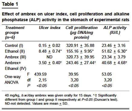
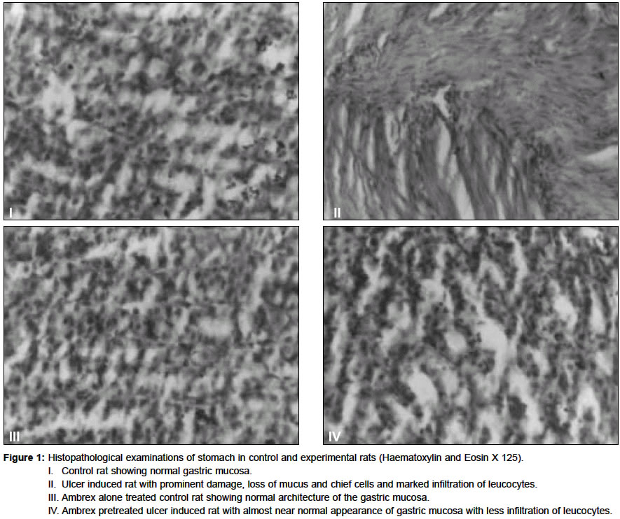
KEY WORDS: Alcohol, alkaline phosphatase, gastric ulcer. Introduction Gastric ulcers are caused due to imbalances between offensive and defensive factors of the gastric mucosa. The antiulcerogenic activity of many plant products is reported due to an increase in mucosal defensive factors rather than decrease in the offensive factors.1 A number of antiulcer drugs like gastric antisecretory drugs - H2 receptor antagonists, antimuscarinic agents, proton pump inhibitors, mucosal protective agentscarbenoxolone sodium, sucralfate and prostaglandin analogues are available which are shown to have side effects and limitations.2 There are several herbal ayurvedic preparations which have a protective effect against drug-induced gastric mucosal injury.3 The constituents of ambrex are Withania somnifera roots (6%), Orchis mascula root (10%), Cycas circinalis male cone (25%), Shorea robusta resin (10%) and Amber fossil resin (15%). The individual components have been suggested to exhibit several properties like antistress,4 antidepressant,4 antioxidant,4 immunomodulatory,4 antitumor5 (Withania somnifera), antihyperdipsia,6 (Cycas circinalis) antidiarrheal, antidysentery,7 (Orchis mascula), antibacterial,8 antiulcer9 (Shorea robusta), antiseptic and antispasmodic10 (Amber) activities. Based on the varied properties of the components of ambrex, we investigated its effect on ethanol-induced gastric mucosal injury. Material and Methods Ambrex was gifted by M/s Care and Cure Herbs Ltd, Anna Salai, Chennai. The powdered form of the drug was suspended in water and administered to rats orally. Animals Male Wistar rats weighing 120-180 g were fed with standard pellet diet (M/s. Hindustan Lever Foods, Bangalore, India) and were provided water ad libitum. Animals were housed under standard environmental conditions. All animal experiments were carried out according to the guidelines of the Institutional Animals Ethics Committee. The experimental rats were divided into four groups with six animals in each group. Group I served as control. In Group II the ulcer was induced as described below. Group III animals were pretreated with ambrex (40 mg/kg body weight for a period of 15 days) alone and Group IV animals were pretreated with ambrex (40 mg /kg body weight for a period of 15 days) before ulcer induction. Dosage fixation Ambrex was administered orally in graded doses of 10, 20, 30 and 40 mg/kg/day for 7 and 15 days. The last dose of ambrex was given on the 7th and 15th day respectively, one hour prior to ethanol administration. From the preliminary study, a dose of 40 mg/kg (ED50)/day for 15 days was selected as it showed maximum antiulcer activity. Gastric ulcer induction in rats One ml of 80% ethanol was used orally to induce gastric ulcer.11 Aqueous suspension of ambrex was administered by oral intubation at a dose of 40 mg/kg/day and was given for a period of 15 days. After the experimental period of 15 days, prior to sacrifice, animals were fasted for 24 h. For Group II and Group IV animals, 1 ml of 80% ethanol was administered orally. One hour after the ethanol administration the animals were sacrificed by cervical dislocation under ether anesthesia and the abdomen was incised and irrigated with normal saline. Subsequently, the stomach was incised along the greater curvature and washed gently in running tap water. It was placed on the watch glass and examined for severity of ulceration12 according to the following scale: 0 = normal gray colored stomach, 0.5 = pink to red coloration of stomach, 1 = spot ulcer, 1.5 = hemorrhagic streak, 2 = number of ulcers <5, 3 = number of ulcers >5, 4 = ulcers with bleeding. Ulcer index was calculated by adding the total number of ulcers plus the severity of ulcer. The mucosal tissue was scrapped from the stomach and was used for the estimation of total protein, DNA and the glandular portion of the stomach was immersed in 4 ml of carbonate-bicarbonate buffer solution (pH10) for alkaline phosphatase activity. Total protein was determined by the method of Lowry et al.13 Mucosal DNA was estimated following the method of Bregman14 which is a reliable index of cell proliferation. Estimation of alkaline phosphatase (ALP) activity The tissue immersed in 4 ml of buffer solution was ground in a mortar and centrifuged for 10 min. 3 ml of the supernatant solution was pipetted out and the marker enzyme ALP was measured using the method of Kind and King15 and expressed as IU/L. Histopathological examination The tissue samples were fixed in 10% buffered formalin and processed with paraffin wax. For histopathological examination, 5 µm sections were stained with hematoxylin and eosin. The extent and depth of ulceration and hemorrhage were evaluated.16 Statistical analysis This was carried out using one-way ANOVA followed by Duncan's multiple comparison test. All the results obtained in the study were compared with each group. P values <0.05 were considered statistically significant. Results Table 1 shows the effect of ambrex on ulcer index and the extent of cell proliferation in the experimental animals. In Group III animals, pretreatment with ambrex showed no ulcer in the gastric mucosa. In Group IV animals, the intensity of the lesions and hemorrhage was significantly reduced upon pretreatment, revealing the protective action of ambrex. The DNA content in the gastric mucosa was reduced upon ulcer induction in Group II animals whereas in Group IV animals there was a significant increase in the levels of DNA. In Group III animals, the proliferation index was near normal. The activity of ALP was assayed. Upon ulcer induction, the activity of ALP had increased significantly in Group II animals. Group IV animals showed a significant reduction in the ALP activity when compared to Group II animals. In Group III animals the activity of ALP was maintained at near normal. Microscopic examination of the lesions (Figure 1) of the gastric mucosa showed that Group II animals had total mucosal ulceration, hemorrhage and segmental mucosal necrosis of the gastric epithelium when compared to Group I animals. In Group III animals the mucosal epithelium showed maintenance of normal architecture. Group IV animals showed less mucosal epithelial loss indicating the protective effect of ambrex. Discussion Ethanol serves as a most common ulcerogenic agent and when given intragastrically to rats it produces severe gastric hemorrhagic erosions.3 The genesis of ethanol-induced gastric lesions is multifactorial with the depletion of gastric wall mucus content as one of the involved factors17 and this damage induced by ethanol may be due to mucosal leukotriene release.18 Mucosal blood flow has also been attributed to be an important factor in the damage caused by alcohol and is modulated by prostaglandin.19 Submucosal venular constriction by ethanol and eventual injury is caused due to perturbations of superficial mucosal cells,18 notably the mucosal mast cells leading to release of vasoactive mediators including histamine, that cause damage to gastric mucosa.20 Ethanol-induced damage to the gastric mucosa is associated with a significant production of free radicals leading to an increased lipid peroxidation and damage to the cell and cell membranes. Accumulation of activated neutrophils in the gastric mucosa may be a source of free radicals.18 Ethanol treatment caused a significant increase in the ulcer index whereas in ambrex pretreated rats there was a significant reduction in the ethanol effect. There are extensive experimental evidences which indicate that free radical scavengers protect the gastric mucosa.21 The reduction in the nucleic acid concentrations of the stomach in the ethanol-induced rats might be due to the accumulation of free radicals as free radicals induce significant damage to DNA.22 Ambrex pretreatment offered protection against the action of ethanol on nucleic acid content showing that the presence of some antioxidant phytoconstituents might have protected the gastric mucosa from free radical-induced damage. The effective scavenging of free radicals by Withania somnifera4 may have reduced progressive ulceration. Perturbation produced by ethanol induces histamine release from the mast cells20 which also results in the accumulation of free radicals. Shorea robusta contains flavonoid23 which reduces gastric tissue histamine content.24 This in turn may have inhibited gastric secretions, thus resulting in the reduction of the ulcer index. Increased ALP activity results from damage to tissues and the release of this enzyme has been suggested to have a role in tissue necrosis associated with various models of gastrointestinal ulceration.25 The present results concur with the previous study,25 as ALP activity was significantly increased following mucosal damage. The decrease in the activity of ALP after ambrex treatment implicates its biochemical basis as an antiulcerogenic. Also, the histopathlogical observations showed that, upon ambrex pretreatment, the mucosal epithelium had normal architecture and it had less hemorrhage as against the ethanol-induced damages in the mucosal epithelium. These observations on the cytoprotective nature of ambrex against ethanol-induced gastric ulcers prove its antiulcer activity. Acknowledgements Author, Dr. K. E. Sabitha, acknowledges the financial assistance rendered by the Council of Scientific and Industrial Research (CSIR), New Delhi. References
Copyright 2004 - Medknow Publications on behalf of the Indian Pharmacological Society. Free, full-text articles also available from http://www.ijp-online.com The following images related to this document are available:Photo images[ph04009t1.jpg] [ph04009f1.jpg] |
| |||||||||
