 |
| About Bioline | All Journals | Testimonials | Membership | News |
|
||||||
|
||||||
Indian Journal of Pharmacology, Vol. 36, No. 1, Feb, 2004, pp. 38-40 Antitumor activity of Indigofera aspalathoides on Ehrlich ascites carcinoma in mice B. Rajkapoor, B. Jayakar, N. Murugesh* Department of Pharmacology, Vinayaka Mission's College of Pharmacy,
Yercaud Road, Salem - 636008, India; and *Institute of Pharmacology, Madurai
Medical College, Madurai - 625020, India.
Received: 19.5.2003
Code Number: ph04010 Abstract Objective: To evaluate the antitumor activity of the
ethanol extract of Indigofera aspalathoides (EIA) in mice.
KEY WORDS: Cancer, flavonoids, solid tumor. Introduction A number of natural products have been studied for anticancer activity on various experimental models. This has resulted in the availability of nearly 30 effective anticancer drugs.1 Indigofera aspalathoides Vahl (Family: Papilionaceae) is a low undershrub with wide distribution, mostly found in South India and Sri Lanka. The stem is traditionally used for various skin disorders and cancer.2 The plant is popularly known as Sivanar vembu in Tamil. The aim of the present study was to evaluate the antitumour activity of the ethanol extract of Indigofera aspalathoides (EIA) against Ehrlich ascites carcinoma (EAC) in mice. Material and Methods Collection and extraction Animals Adult Swiss male albino mice (20-25 g) were procured from Perundurai Medical College, Perundurai, Tamil Nadu and used throughout the study. They were housed in microlon boxes in a controlled environment (temperature 25+20C and 12 h dark/light cycle) with standard laboratory diet and water ad libitum. The study was conducted after obtaining Institutional animal ethical committee clearance. Cells EAC cells were obtained through the courtesy of Amala Cancer Research Center, Thrissur. They were maintained by weekly intraperitoneal inoculation of 106 cells/mouse.4 Effect of EIA on survival time5 Animals were inoculated with 1 X 10⁶ cells/mouse on day `0' and treatment with EIA started 24 h after inoculation, at a dose of 250 mg/kg/day, p.o. The control group was treated with the same volume of 0.9% sodium chloride solution. All the treatments were given for nine days. The median survival time (MST) of each group, consisting of 10 mice was noted. The antitumor efficacy of EIA was compared with that of 5-fluorouracil (Dabur Pharmaceutical Ltd, India; 5-FU, 20 mg/kg/day, i.p. for 9 days). The MST of the treated groups was compared with that of the control group using the following calculation:
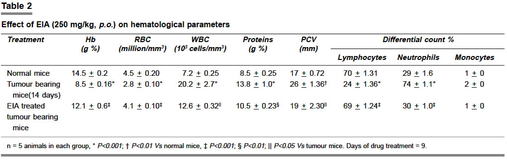
Where T = number of days the treated animals survived and C = number of days control animals survived. Effect of EIA on normal peritoneal cells5 Three groups of normal mice (n= 5) were used for the study. One group was treated with 250 mg/kg, p.o. of EIA only once for a single day and the second group received the same treatment for two consecutive days. The untreated third group was used as control. Peritoneal exudate cells were collected after 24 h treatment by repeated intraperitoneal wash with normal saline and counted in each of the treated groups and compared with those of the untreated group. Effect of EIA on hematological parameters5 In order to detect the influence of EIA on the hematological status of EAC-bearing mice, a comparison was made among three groups (n= 5) of mice on the 14th day after inoculation. The groups comprised of (1) tumor-bearing mice (2) tumor-bearing mice treated with EIA (250 mg/kg/day, p.o. for the first 9 days) and (3) control mice (normal). Blood was drawn from each mouse by the retro orbital plexus method and the white blood cell count (WBC), red blood cell count (RBC), hemoglobin, protein and packed cell volume (PCV) were determined.6-8 Effect of EIA on solid tumor9 Mice were divided into two groups (n=8). Tumor cells (1 X 10⁶ cells/mice) were injected into the right hind limb (thigh) of all the animals intramuscularly. The mice of Group I were tumor control. Group II received EIA (250 mg/kg) orally for 5 alternate days. The dose was selected based on toxicity studies which showed no toxicity up to 5 g/kg (p.o.). Tumor mass was measured from the 11th day of tumor induction. The measurement was carried out every 5th day for a period of 30 days. The volume of tumor mass was calculated using the formula V = 4/3pr2 where r is the mean of r1 and r2 which are two independent radii of the tumor mass.10 Effect of EIA on in vitro cytotoxicity Short-term cytotoxicity was assessed by incubating 1 X 10⁶ EAC cells in 1 ml phosphate buffer saline with varying concentrations of the EIA at 370C for 3 h in CO2 atmosphere ensured using a McIntosh field jar. The viability of the cells was determined by the trypan blue exclusion method.11 Statistical analysis All values were expressed as mean+SEM. The data were statistically analyzed by one-way ANOVA followed by Dunnett's test, the data of hematological parameters were analyzed using ANOVA followed by Tukey multiple comparison test and data of solid tumor were analyzed using Student's ` t ' test. P values <0.05 were considered significant. Results The effect of EIA on the survival of tumor-bearing mice is shown in Table 1. The MST for the control group was 21+1.20 days, whereas it was 33+1.20 days and 40+2.10 days for the groups treated with EIA (250 mg/kg/day, p.o.) and 5-FU (20 mg/kg/day, i.p.) respectively. The increase in the lifespan of tumor-bearing mice treated with EIA and 5-FU was found to be 57.14% and 90.47% respectively (P< 0.01) as compared to the control group. The average number of peritoneal exudate cells per normal mouse was found to be 5.8+0.4 X 10⁶. Single treatment with EIA (250 mg/kg) enhanced peritoneal cells to 8.9+0.9 X 10⁶, while two consecutive treatments enhanced the number to 9.8+1.1 X 10⁶ (P< 0.001). Hematological parameters of tumor-bearing mice on Day 14 showed significant changes when compared with the normal mice (Table 2). The total WBC count, proteins and PCV were found to increase with a reduction in the hemoglobin content of RBC. The differential count of WBC showed that the percentage of neutrophils increased (P<0.001) while that of lymphocytes decreased (P<0.001). At the same time interval, EIA (250 mg/kg/day, p.o.) treatment could change these altered parameters to near normal. There was reduction in the tumor volume of mice treated with EIA (P<0.001). Tumor volume of control animals was 2.96+0.12 ml, whereas for the extract-treated group it was 1.54+0.05 ml. The in vitro cytotoxicity study showed the IC50 of EIA to be 500+11.54 mg/ml. Discussion The reliable criteria for judging the value of any anticancer drug are prolongation of lifespan and decrease of WBC from blood.12,13 The results of the present study show an antitumor effect of EIA against EAC in Swiss albino mice. A significant enhancement of MST and peritoneal cell count was observed. The effect of EIA treatment on the peritoneal exudate cells of normal mice is an indirect method of evaluating its inhibitory effect on tumor cell growth. Normally, a mouse contains about 5 X 10⁶ peritoneal cells, 50% of which are macrophages. EIA treatment was found to enhance peritoneal cells count. These results demonstrate the indirect inhibitory effect of EIA on EAC cells, which is probably mediated by the enhancement and activation of either macrophage or cytokine production. The analysis of the hematological parameters showed mini mum toxic effect in mice treated with EIA. After 14 days of transplantation, EIA was able to reverse the changes in the hematological parameters consequent to tumor inoculation. The present study reveals that the extract was cytotoxic towards EAC. Preliminary phytochemical screening indicated the presence of alkaloids and flavonoids in EIA. Flavonoids have been shown to possess antimutagenic and antimalignant effects.14-15 Moreover, flavonoids have a chemopreventive role in cancer through their effects on signal transduction in cell proliferation16 and angiogenesis.17 The cytotoxic and antitumor properties of the extract may be due to these compounds. The present study points to the potential anticancer activity of Indigofera aspalathoides. Further studies to characterize the active principles and elucidate the mechanism of the action of EIA are in progress. References
Copyright 2004 - Medknow Publications on behalf of the Indian Pharmacological Society. Free, full-text articles also available from http://www.ijp-online.com The following images related to this document are available:Photo images[ph04010t2.jpg] [ph04010t1.jpg] |
| |||||||||
