 |
| About Bioline | All Journals | Testimonials | Membership | News |
|
||||||
|
||||||
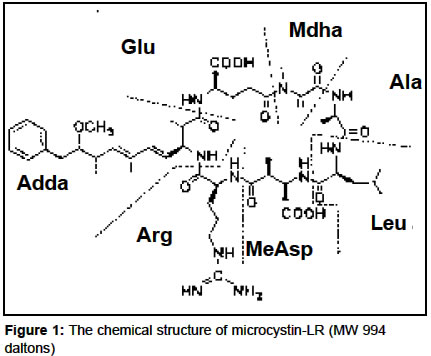
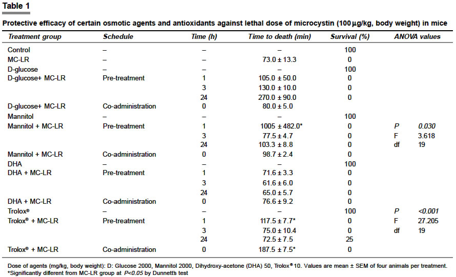
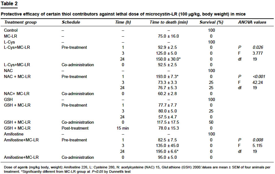
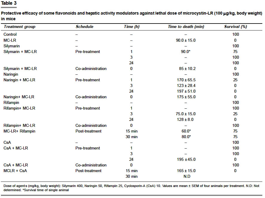
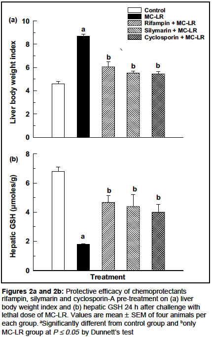
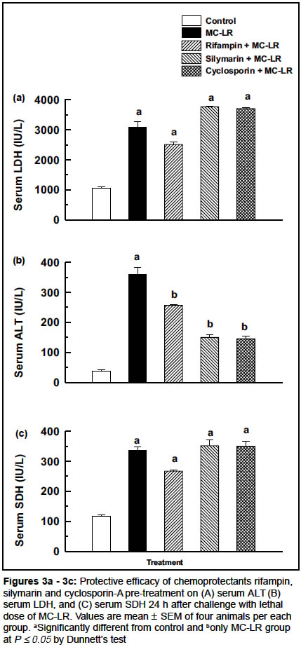
Indian Journal of Pharmacology, Vol. 36, No. 2, April, 2004, pp. 87-92 Research Paper Screening of certain chemoprotectants against cyclic peptide toxin microcystin-LR Lakshmana Rao PV, Gupta N, Jayaraj R Division of Pharmacology and Toxicology, Defence Research and Development Establishment, Jhansi Road, Gwalior - 474002, MP Code Number: ph04032 ABSTRACT Objective: To evaluate the protective efficacy of certain chemoprotectants against cyclic peptide hepatotoxic microcystin-LR in mice. Material and Methods: Swiss albino female mice were used in all experiments for screening antidotes against the lethal dose of microcystin-LR (100 mg/kg body weight, i.p.). The agents, D-glucose, mannitol, dihydroxyacetone, Trolox®, L-cysteine, N-acetylcysteine, amifostine, glutathione, silymarin, naringin, rifampin and cyclosporin-a were administered as either pre-treatment (1, 3 and 24 h), co-administration or post-treatment. Percent survival and time to death were monitored. The biochemical profile of protected animals was monitored 24 h post-treatment. Results: D-glucose, mannitol, L-cysteine, naringin and amifostine extended the survival time of animals but offered no protection against lethality. N-acetylcysteine, glutathione and Trolox® gave partial protection (25-50%) on pretreatment or co-administration. Complete protection was observed with rifampin (25 mg/kg), cyclosporin-A (10 mg/kg) and silymarin (400 mg/kg) when given as pre-treatment. In addition, rifampin and cyclosprin-A gave complete protection when co-administered with microcystin-LR. Rifampin was the only agent which gave protection at 15 min post-treatment. The biochemical profile of surviving animals 24 h after treatment showed increased liver body weight index and levels of hepatic enzymes viz. LDH, ALT, SDH in serum. Conclusion: Chemoprotectants that can inhibit the specific uptake of toxin into the liver can be promising antidotes against lethal poisoning by microcystin-LR in mice.INTRODUCTION Microcystins are a family of structurally related cyclic peptide toxins that are produced by a variety of cyanobacteria but most often by those belonging to the genus Microcystis. The general structure of microcystins is cyclo (D-Ala-X-D-MeAsp-Y-Adda-D-Glu-Mdha-) in which X and Y are variable amino acids, D-MeAsp is D-erythro-b-methylaspartic acid, Adda is 3-amino-9-methoxy-2,6,8-trimethyl-10-phenyldeca-4,6-dienoic acid, and Mdha is N-methyldehydroalanine [Figure - 1]. Over 60 microcystin variants have been reported so far. The most common and toxic among them is microcystin-LR (MC-LR, MW 994), in which the variable amino acids are leucine (L) and arginine (R). Microcystins are known to be potent inhibitors of protein phosphatases 1 and 2A as well as skin and liver tumor promoters in animals.[1] These peptide toxins are also suspected to be involved in the promotion of primary liver cancer in humans exposed to long-term doses through drinking water.[2],[3] Acute illnesses and deaths in both humans and animals following exposure to microcystin-contaminated water sources have been reported worldwide.[4],[5] The clinical, pathological, and toxicological evidence points to microcystins having been the major cause of the death of 60 hemodialysis patients at Carauru, Brazil.[6],[7] Because of the rapid, irreversible and severe damage to the liver caused by microcystins, therapy is likely to have little or no value, and effective prophylaxis is critical. In spite of the potential economic loss due to livestock poisoning and possible human hazards associated with this toxin, very little work has been done on the development of suitable chemoprotectants against these toxins. In the present study a number of chemicals were investigated for their efficacy to antagonize microcystin poisoning in mice. MATERIAL AND METHODS ChemicalsMicrocystin-LR (MC-LR) was purified from laboratory cultures of Microcystis aeruginosa (PCC 7820) and confirmed with authentic MC-LR standard obtained from Prof. W.W. Carmichael, Wright State University, Ohio, USA. Silymarin, rifampin, naringin, N-acetyl cysteine (NAC), L-cysteine, D-glucose, mannitol, dihydroxy acetone (DHA) were obtained from Sigma Chemical Co. USA. Trolox ® (a water soluble vitamin E derivative; 6-hydroxy-2,5,7,8- tetramethyl-chroman-2-carboxylic acid) was obtained from Sigma-Aldrich (Aldrich, USA), cyclosporin-A (CsA) from Fluka, and GSH from Across, Belgium. Animals Treatment regimen Assessment of toxicity Statistical analysis RESULTS Microcystin-LR at 100 mg/kg consistently produced 100% lethality and the mean time to death varied from 1 to 2 h. Previous studies have shown that mice that did not exhibit signs of toxicity within 24 h after treatment with MC-LR would not develop adverse effects thereafter.[9],[10] Therefore, survival to 24 h was chosen as an acceptable end-point for studies using potential chemoprotectants. [Table - 1] summarizes the results on the protective efficacy of certain osmotic agents and antioxidant/free radical scavengers. D-glucose at 2 g/kg after 1, 3 and 24 h pre-treatment extended survival time, but offered no protection from lethality. Similarly, mannitol (2 g/kg) and DHA (50 mg/kg) had no protective effect at all the pre-treatment time points. Pre-treatment with mannitol 1 h prior to the administration of microcystin-LR extended mouse survival time by 15-20 h. None of the agents protected the animals when co-administered with MC-LR. Trolox ® at 24 h pre-treatment protected 25% of the animals. Co-administration with MC-LR significantly extended survival time (187.5 ± 7.5 min) but could not prevent lethality. The protective efficacy of thiol contributors is shown in [Table - 2]. Pre-treatment with L-cysteine for 1, 3 h and co-administration offered no protection. But 24 h pre-treatment significantly extended survival time. One hour pre-treatment with NAC 15 mg/kg significantly extended survival time (193 ± 7.2 min). At 3 and 24 h pre-treatment 25% of the animals survived but co-administration was not effective. No protection was observed with amifostine pre-treatment at 1, 3 and 24 h though a marginal increase in survival time was observed. GSH at 2 g/kg concentration protected 25% of the mice at 3 h pre-treatment but no protection was observed at 1 and 24 h pre-treatment. Co-administration protected 50% of the animals. Post-treatment with GSH did not give any protection. The protective efficacy of certain flavonoid compounds and hepatic activity modulators screened against a lethal dose of MC-LR is shown in [Table - 3]. Silymarin at 400 mg/kg protected 75% of the animals after 1 h pre-treatment and 100% protection was observed with 3 and 24 h pre-treatment. But it offered no protective effect when co-administered with MC-LR. Post-treatment did not offer protection. Naringin, the grape fruit flavonoid was evaluated for its protective efficacy against MC-LR. At 50 mg/kg, naringin protected 25% of the animals after 1 h pre-treatment. The mean time to death was also extended in the other three animals. At 3 and 24 h pre-treatment, no protection was observed but survival time was marginally extended. Rifampin at 25 mg/kg pre-treatment 1 h prior to the dose of MC-LR completely protected all the mice. However, 3 h and 24 h pre-treatment study showed 75 and 100% mortality. Co-administration of rifampin gave 100% protection. Rifampin when administered 15 and 30 min after MC-LR could still protect 75% of the animals. CsA pre-treatment at 10 mg/kg for 1 and 3 h showed 100% protection as against 100% mortality in 24 h pre-treated mice with extended survival time. Similar to rifampin, co-administration of CsA with MC-LR gave 100% protection against lethality. But unlike rifampin, post-treatment for 15 min CsA could not prevent MC-LR induced lethality. In order to evaluate the recovery of the surviving animals after rifampin, CsA or silymarin treatment, some biochemical variables were estimated in surviving animals 24 h post-treatment after challenge with the lethal dose of MC-LR. Animals that survived after protection with all three agents still showed an enlarged liver indicated by higher LBI after 24 h. The LBI was significantly higher than the control group but less than the MC-LR only group [Figure:2a]. MC-LR treated mice showed three-fold depletion of GSH levels as compared to the control group. Though animals protected with the three agents showed GSH augmentation, the GSH levels were still lower than the controls [Figure:2b]. Serum enzymes of LDH, ALT and SDH showed elevated levels after 24 h in all the three antidote-treated groups as compared to control animals [Figure:3a] - [Figure:3c]. DISCUSSION Microcystins are the most predominantly distributed among the cyanobacterial toxins. The consequence of acute poisoning by these compounds is rapid disorganization of the hepatic architecture, breakdown of sinusoidal structures and, in mammals, pooling of blood in the liver.[11],[12] The mode of action of the microcystins at the cellular level is the specific inhibition of the activity of protein phosphatase (PP1 and PP2A) activity.[13] Studies aimed at finding antidotes for microcystin toxicity have shown in vivo protection in mice by a variety of chemically unrelated compounds. Adams et al[14] investigated chemicals known to affect macrophage function as potential prophylactic agents. Hermansky et al[9] in an empirical study performed prior to the publication of the protein phosphatase-inhibitory properties of microcystins, examined a variety of chemoprotectants against microcystin-LR. These compounds included antioxidants, enzyme inducers, calcium channel blockers, free-radical scavengers and hepatic activity modulators. Earlier studies indicate a possible role for free-radical scavengers in antagonizing microcystin-induced toxicity.[15],[16] However, of the free-radical scavengers / osmotic agents used in the study, D-glucose and mannitol extended only survival time (possibly due to the inactivation or dilution of the toxin in the peritoneal cavity) but could not prevent lethality.[9],[10] Trolox ® that penetrates bio membranes and protects mammalian cells from oxidative damage gave only a partial protection. Among thiol modulators / contributors no protection from lethality was observed with L-cysteine, and amifostine though they extended survival time. Partial protection was observed with NAC and GSH. It is speculated that the physical inhibition of absorption similar to that suggested for mannitol and glucose may be responsible for the protective effect of GSH. Additionally, GSH may react with MC-LR in the peritoneal cavity preventing sufficient absorption of the toxin to produce toxicity. Recent studies show that microcystin could conjugate with GSH and cysteine in both cell-free systems.[17],[18] Keeping in view their biological activities, two flavonoids, silymarin and naringin were evaluated. Silymarin pre-treatment completely protected mice from lethality but was not effective when co-administered. Silymarin has been shown to increase the levels of glutathione and reduce the production of malondialdehyde, as a measure of lipid peroxidation in livers of rats acutely intoxicated with ethanol.[19] The antagonistic effect of silymarin could be similar to that of dithioerythritol by stabilizing protein-thiol, which may be important to the structure of liver cells. Rifampin and CsA pre-treatment and co-administration gave 100% protection. Both rifampin and CsA are known for their immunosuppressant activities. However, the dose used in the present study may not be high enough to produce immunosuppression. Both rifampin and CsA are known to block the hepatocellular uptake of bile acids.[20],[21] CsA has also been shown to protect the isolated hepatocytes from phalloidin, a cyclic heptapeptide toxin that is similar to microcystin-LR in its uptake and mode of action. Thus, the protective effects of rifampin and CsA may be related to blocked cellular uptake of MC-LR. Screening of various compounds has shown that only rifampin, silymarin and CsA could provide complete protection against MC-LR-induced lethality. The biochemical profile of surviving animals still showed elevated levels of various enzymes and LBI indicating the persistent toxic effect of MC-LR. Microcystin-induced toxicity is mediated through the inhibition of cellular protein phosphatase 1 and 2A.[22],[23],[24] To date, there are no known inhibitors of the interaction of microcystin with protein phosphatases. Any potential chemoprotectant should have the ability to either prevent microcystin-protein phosphatase interaction or inhibit the uptake of the toxin into hepatocytes. In conclusion, rifampin, cyclosprin-A and silymarin pre-treatment could prevent microcystin-induced lethality possibly by preventing the uptake and intracellular transport of toxin into the liver. Further studies are required to identify and characterize effective antidotes against this environmentally important class of toxins. ACKNOWLEDGEMENTS The authors thank Dr. R. Vijayaraghavan, Head, Division of Pharmacology and Toxicology and Mr. K. Sekhar, Director, DRDE for their support and keen interest. Nidhi Gupta is thankful to the Defence Research and Development Establishment for the award of research fellowship. REFERENCES
Copyright 2004 - Indian Journal of Pharmacology The following images related to this document are available:Photo images[ph04032t2.jpg] [ph04032f1.jpg] [ph04032t1.jpg] [ph04032f3.jpg] [ph04032f2.jpg] [ph04032t3.jpg] |
| |||||||||
