 |
| About Bioline | All Journals | Testimonials | Membership | News |
|
||||||
|
||||||
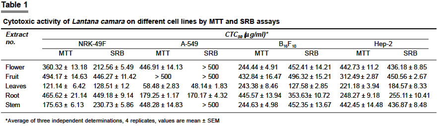
Indian Journal of Pharmacology, Vol. 36, No. 2, April, 2004, pp. 94-95 Research Letter In vitro cytotoxic activity of Lantana camara Linn Raghu C, Ashok G, Dhanaraj SA, Suresh B, Vijayan P J. S. S. College of Pharmacy, Rocklands, Ootacamund - 643001 Code Number: ph04034 Lantana camara Linn. is a large evergreen strong-smelling herb, native of tropical America, but now naturalized in many parts of India. All the parts of this plant have been used traditionally for several ailments throughout the world.[1] The leaves are used as a bechic, antitumoral, antibacterial and antihypertensive agent.[2] The root of this plant is used for the treatment of malaria, rheumatism and skin rashes.[3] Several tri-terpenoids, napthaquinones, flavonoids, alkaloids and glycosides isolated from this plant are known to exert diverse biological activities including cytotoxic and anticancer properties.[4] The crude methanolic extract of different parts of this plant was studied for its in vitro cytotoxic potential. Natural products provide an inexhaustible source of anticancer drugs in terms of both variety and mechanism of action. Hence, in continuation of our studies to identify potent natural products for antitumor activities,[5] investigation on the in vitro cytotoxic properties of the crude extracts of different parts of Lantana camara was taken up against four cancerous cell lines viz. HEp-2, B16F10, A-549 and DLA and a normal NRK-49F cell line using standard procedures. The fresh flowers (30 g), fruits (50 g), leaves (57 g), root (55 g) and stem (45 g) of Lantana camara were collected from the forests of The Nilgiris, Tamil Nadu, India, in the month of June 2001. Mr. Rajan, Survey of Medicinal Plants and Collection Unit, Government Arts College, Ootacamund, India authenticated the plant. The plant materials were coarsely powdered and extracted in a Soxhlet apparatus with methanol (1:5 w/v) for 18 h. The extracts were then concentrated to dryness under reduced pressure and controlled temperature. The yields obtained were 12.12, 8.64, 12.15, 9.67 and 12.75% respectively, for flowers, fruits, leaves, root and stem. Each extract was separately dissolved in distilled dimethyl sulphoxide (DMSO) and the volume was made up to 10 ml with Dulbecco′s Modified Eagle′s medium (DMEM), pH 7.4, supplemented with 2% inactivated newborn calf serum (Maintenance medium, PAA Laboratories, Austria), to obtain a stock solution of 1 mg/ml concentration, sterilized by filtration and stored at -20°C till use. NRK-49F (normal rat kidney) and B16F10 (mouse melanoma) cell cultures were obtained from the National Centre for Cell Sciences, Pune, India. HEp-2 (caucasian male larynx epithelium carcinoma) cell line was obtained from the Pasteur Institute of India, Coonoor, India. A-549 (small cell lung carcinoma) cell line was obtained from the Christian Medical College, Vellore, India. Dalton′s lymphoma ascites (DLA) cells were obtained from the Amala Cancer Institute, Trissur, India. The cultures of A-549, B16F10, HEp-2 and NRK-49F were propagated in DMEM, pH 7.4 supplemented with 10% inactivated newborn calf serum, penicillin (100 IU/ml), streptomycin (100 µg/ml) and amphotericin B (5 µg/ml), and maintained in a humidified atmosphere of 5% CO2 at 37°C until confluent. The cells were dissociated with 0.2% trypsin, 0.02% EDTA in phosphate buffer saline solution (TPVG). The stock culture was grown in 25 cm2 tissue culture flasks (Tarsons India Pvt. Ltd., Kolkata, India) and all cytotoxicity experiments were carried out in 96 well microtitre plates (Tarsons India Pvt. Ltd., Kolkata, India). DLA cells used were propagated and maintained in the peritoneal cavity of Swiss albino mice. Cell lines in exponential growth phase were washed, trypsinized and resuspended in DMEM medium with 10% inactivated newborn calf serum. Cells were plated at 10,000 cells/well in 96 well microtitre plate and incubated for 24 h at 37°C, 5% CO2 in a humidified atmosphere during which period a partial monolayer was formed. The cells were then exposed to different concentrations (1000 µg/ml to 15.6 µg/ml, prepared by serial two-fold dilution using maintenance medium from the stock solution) of the test extracts in quadruplicate. Control wells received only maintenance medium. The cells were incubated at 37°C in a humidified incubator with 5% CO2 for a period of 72 h. Morphological changes of the cell cultures were examined using an inverted tissue culture microscope (Olympus, Japan, Model 1X70) at 24 h time intervals and compared with the control. At the end of 72 h, cellular viability was determined using standard 3-(4, 5-Dimethylthiazol-2-yl)-2, 5-diphenyl tetrazolium bromide (MTT)[6] and Sulphorhodamine B (SRB)[7] assays. The short-term toxicity studies[5] were carried out on DLA cells employing Trypan blue dye exclusion technique[5] and the CTC50 value (concentration of the sample required to kill 50% of the cells) was calculated. Of the five methanol extracts obtained from different parts of Lantana camara, the leaf extract exhibited comparatively more cytotoxic activity against all the five cell lines tested [Table - 1]. The human lung carcinoma cell line, A-549 was found to be more susceptible with a CTC50 value of 48.1 - 58.5 mg/ml extract. The other four extracts showed less activity as indicated by the relatively high CTC50 values. In the short-term toxicity studies, the methanol extracts of the root with 191.5 ± 5.1 µg/ml and leaf with 219.5 ± 8.4 µg/ml, showed moderate activity against DLA cells after 3 h of exposure. The extracts of the stem, fruit and flowers of Lantana camara, showed less activity with CTC50 values, 268.7 ± 10.2, 492.7 ± 14.4 and > 1000 µg/ml respectively.' The results obtained from the present study show that the extract of the leaf of Lantana camara is cytotoxic in nature and may possess antitumor activity. The study also supports the ethnomedical data provided in an earlier study.[2] The cytotoxic activity may be due to the presence of toxic lantanoids and alkaloids from this plant.[4] The plant merits further investigation to identify the active principles and the nature of the antitumor activity in animal models. REFERENCES
Copyright 2004 - Indian Journal of Pharmacology The following images related to this document are available:Photo images[ph04034t1.jpg] |
| |||||||||
