 |
| About Bioline | All Journals | Testimonials | Membership | News |
|
||||||
|
||||||
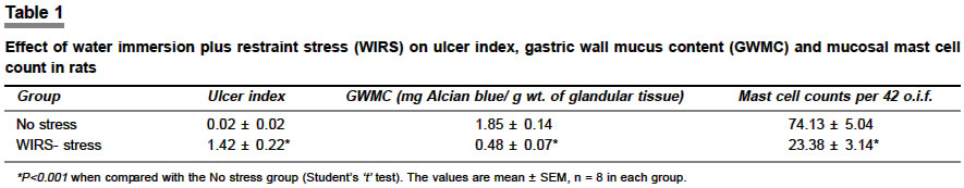
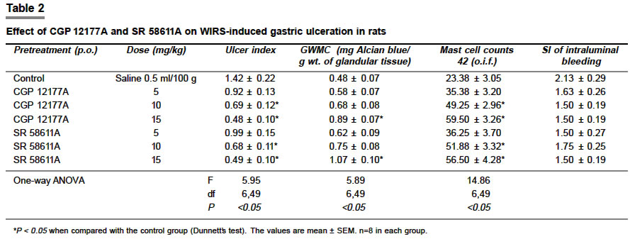
Indian Journal of Pharmacology, Vol. 36, No. 3, June, 2004, pp. 151-154 Research Paper Gastroprotective effects of b3-adrenoceptor agonists on water immersion plus restraint stress-induced gastric ulcer in rats Paul A, Goswami S, Santani D Lachoo Memorial College of Science and Technology (Pharmacy Wing), Shastri Nagar, Jodhpur - 342 003 Code Number: ph04052 ABSTRACT OBJECTIVE: To evaluate the gastroprotective effects of b3-adrenoceptor agonists CGP 12177A and SR 58611A, on water immersion plus restraint stress (WIRS)-induced gastric ulceration in rats. MATERIAL AND METHODS: Drugs were administered (5, 10 and 15 mg/kg, p.o.) 30 min prior to the ulcerogenic procedure. Ulcer index and the score for intensity of intraluminal bleeding were determined. Gastric wall mucus content (GWMC) and mast cell counts were determined in the glandular portion of the stomach. RESULTS: A dose-dependent reduction in the ulcer index was observed with both the drugs. A significant rise in the GWMC in the glandular tissue at 15 mg/kg dose was caused by the b3-adrenoceptors agonists. In the glandular tissue the mast cell count was significantly decreased at 10 and 15 mg/kg dose with both drugs. CONCLUSION: The present study shows the gastroprotective effect of b3-adrenoceptor agonists CGP 12177A and SR 58611A against WIRS-induced gastric ulceration in rats. The gastroprotective effect may be mediated by the enhancement of mucin activity and the decrease in mast cell degranulation. INTRODUCTION Recent localization of b3-adrenoceptors using immunohistochemical studies has confirmed the presence of b3-adrenoceptors in human vascular and non-vascular smooth muscles of the gastrointestinal tract.[1] The presence of b3-adrenoceptors has been well described in mast cells and basophils and the stimulation of these receptors results in the inhibition of immune-stimulated histamine release.[2],[3] Espluges et al [4] observed significant antiulcer activity with b-adrenergic drugs such as salbutamol, salmeterol and isoprenaline against polymixin-B-induced gastric ulcers involving histamine release. However, propranolol could only partially antagonize the isoprenaline-induced inhibitory effect on histamine release. This shows that besides b2-adrenoceptor stimulation, these agonists inhibit histamine release through some additional mechanism other than beta-receptor stimulation. The involvement of b1-adrenoceptors in the histamine release mechanism was ruled out by these studies. Isoprenaline was reported to be as potent as SR 58611A, a b3-adrenoceptor agonist, in stimulating b3-adrenoceptors in isolated rat colon.[5] b3-adrenoceptor agonists inhibit gastric ulcer-induced by indomethacin,[5],[6] pylorus ligation and ethanol[7] in rats. Hence, this study was undertaken to evaluate the antiulcer effect of b3-adrenoceptor agonists on water immersion plus restraint stress (WIRS)- induced gastric ulcer in rats. The study was also directed towards the elucidation of the mechanism of the antiulcer activity of b3-adrenoceptor agonists. MATERIAL AND METHODS Wistar albino rats of either sex weighing 200-250 g were selected. Rats were fed with standard chow diet and water ad libitum till the end of the experimental period. Distributions of the animals in-group, sequence of trials and treatment aspects were randomized. This experiment complied with the guidelines of our laboratory for animal experimentation. The Animal Ethics Committee of the institute cleared the experimental protocols. CGP 12177A [(±)-4-(3-t-butylamino-2-hydroxy-propoxy) benzimidazol-2-one] and SR58611A [ethyl {(7S)-7-[(2R)-2-(3-chlorophenyl)-2-hydroxyethylamino] 5,6,7,8-tetrahydro-napthalene-2-yloxy} acetate hydrochloride] were obtained as gift samples from Novartis, Switzerland and Sanofi Recherche, France respectively. Drugs dissolved in distilled water were administered orally to rats in doses of 5, 10 and 15 mg/kg. Saline treated (0.5 ml/100 g, p.o.) rats served as controls. The dose of b3-adrenoceptor agonists were selected on the basis of ED50 values of BRL 35135, a b3-adrenoceptor agonist on indomethacin-induced ulceration and total acid-output in pylorus-ligated rats.[5] Water immersion plus restraint stress-induced gastric ulceration Measurement of gastric wall mucus Examination of mast cells Statistical analysis RESULTS Severe hemorrhagic gastric glandular mucosal ulcers were observed in stress-induced control animals [Table - 1]. Significant change in the ulcer index, GWMC and mast cell count were observed in WIRS-stress as compared with non-stressed controls [Table - 1]. Both the b3-adrenoceptor agonists (CGP 12177A and SR 58611A) reduced the ulcer index in a dose-dependent manner (10 and 15 mg/kg, [Table - 2]. ED50 values for antiulcer activity (ulcer index) of CGP 12177A and SR 58611A in WIRS- induced gastric ulcer model were found to be 10.25 and 10.48 mg/kg respectively. Gastric wall mucus content was significantly higher in the CGP 12177A and SR 58611A treated group at 15 mg/kg dose as compared to controls. At 10 and 15 mg/kg doses, a significant rise in the mast cell count was observed with both the compounds [Table - 2]. DISCUSSION The experimental stress ulcer may be considered equivalent to clinical stress ulcer which occurs after surgery, head injury or shock. An acute gastric hemorrhagic lesion in the glandular stomach characterizes a stress ulcer.[13] The present study shows anti-ulcer activity of b3-adrenoceptor agonists (CGP 12177A and SR 58611A) which was evident from a significant decrease in the ulcer index at 10 and 15 mg/kg doses in a WIRS-induced gastric ulcer model. The centrally-induced vascular disturbance of mucosal capillaries is being implicated in restraint-induced gastric bleeding[13]. b3-adrenoceptor agonists can cause enhancement in antral gastric mucosal blood flow (GMBF) in rats.[5] The insignificant decrease in score of intensity of intraluminal bleeding caused by b3-adrenoceptor agonists in the present study can be partly attributed to their ability to enhance antral GMBF. The specific pathophysiologic mechanism involved in stress- induced ulcers could be ultimate multifactorial impairment of mucosal defense system. An increase in gastric acid secretion, reduction of gastric mucus and alteration in the microvasculature of the gastric mucosa play a major role in the pathogenesis of stress- induced ulcers.[14],[15] Our earlier study has shown that the mechanism of the anti-ulcer action of b3-adrenoceptor agonists in the pylorus ligation model is partly attributed to a decrease in acid secretion. b-adrenoceptor agonists are known to inhibit the release of histamine.[2] Histamine has been known to induce gastric acid secretion mainly through H2-receptor activation.[16] Gastrin-stimulated and cholinergically-mediated acid secretions require a background release of histamine from mast cells for their maximal effects. Thus any agent that reduces the release of histamine from mast cells should suppress acid secretion.[17] Therefore, the effect of b3-adrenoceptor agonists on mucin activity and mast cell counts was also studied in the present study. In the WIRS-induced gastric ulcer model, enhanced mucin activity (GWMC) and increase in mast cell counts (i.e., decrease in mast cell degranulation) caused by CGP 12177A and SR 58611A may explain the antiulcer action of b3-adrenoceptor agonists. In conclusion, our study shows significant gastroprotective activity of b3-adrenoceptor agonists against WIRS-induced gastric ulcer model. The mechanism for antiulcer action is attributed to the enhancement of mucin activity and a decrease in mast cell degranulation. ACKNOWLEDGEMENTS The authors are thankful to M/s Novartis, Switzerland and Sanofi Recherche, France for supplying gift samples of CGP 12177A and SR 58611A. REFERENCES
Copyright 2004 - Indian Journal of Pharmacology The following images related to this document are available:Photo images[ph04052t2.jpg] [ph04052t1.jpg] |
| |||||||||
